
We aim to gain a fundamental understanding of how microtubule nucleation is spatio-temporally regulated in the context of multi-cellular animals.
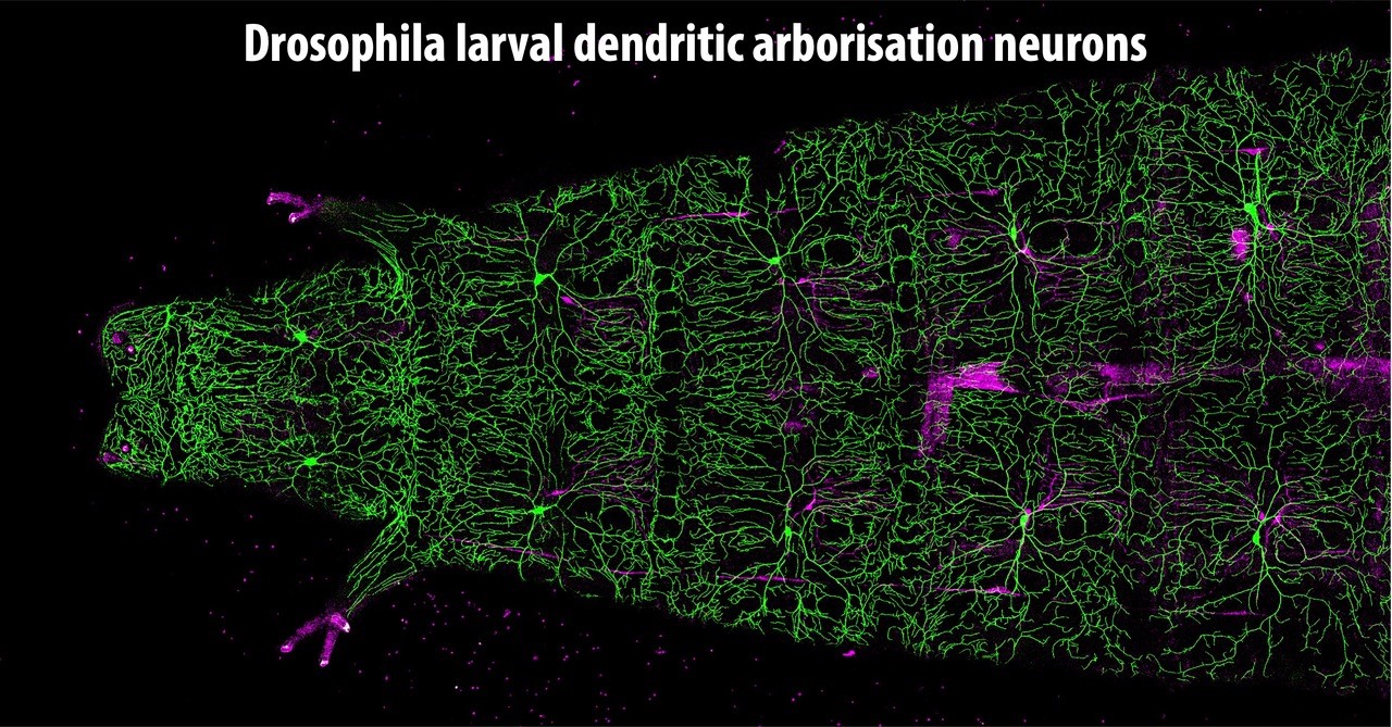
We are particularly interested in how the mechanisms regulating microtubule formation and organization vary between cell types, including dividing cells and neurons.
Keywords: Microtubules, cell division, neurons, gamma-tubulin, g-TuRC, MTOC, centrosome, Drosophila
+33 (0)1 57 27 80 95 Contact @pconduit.bsky.social
Our Vision
We want to gain a fundamental understanding of how different cells establish their highly specific microtubule networks, with a particular focus on the regulation of microtubule nucleation.
Background
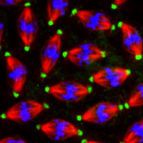
Microtubules are dynamic polymers that form a crucial part of the cellular cytoskeleton. They assemble into a spectacular variety of arrays across different cell types and developmental stages. For example, during cell division, microtubules organize into the mitotic spindle to separate duplicated chromosomes equally between daughter cells. In mature neurons, however, they form polarized networks that span axons and dendrites, essential for structural support, neurite growth, and intracellular transport. Because all cells use the same fundamental machinery to generate and organize microtubules, a central question remains: how do distinct cell types form such vastly different microtubule arrays?
Research Programme
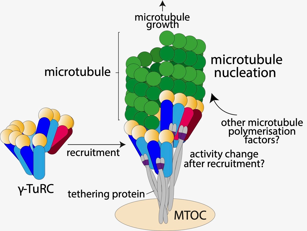
We address this question principally by studying the molecular regulation of multi-protein gamma-tubulin ring complexes (g-TuRCs), which template de novo microtubule assembly. g-TuRCs are recruited and activated at specific subcellular sites to generate new microtubules at the exact right place and time. These sites include microtubule organizing centers (MTOCs), such as centrosomes during mitosis, the nuclear envelope in muscle cells, or the Golgi apparatus in migrating cells.
Once generated, molecular motors can slide these microtubules against one another (or against the cell cortex) to guide network formation. Microtubules are also stabilized by post-translational modifications and specialized binding proteins. While these post-nucleation processes are not the primary focus of our lab, we investigate them to understand how they collectively contribute to proper microtubule array formation.
Methodology
We predominantly use Drosophila as an in vivo multicellular model system. Our approach combines precise genome manipulation, advanced fixed and live-cell imaging, and biochemical assays to probe the molecular regulation of microtubules—and the consequences of their misregulation. We also collaborate closely with structural biologists to resolve the atomic-level details of the proteins and complexes driving microtubule nucleation.

To contact a member of the team by e-mail: name.surname@ijm.fr
Publications
- Impulscience, Fondation Bettencourt-Schueller
- ANR project grant
- IDex, Université Parix Cité
21.11.2023 : Impulscience Prize 2023
We are always looking for talented postdocs that have a keen interest in microtubule regulation. If you want to join the team, email Paul Conduit with a CV and cover letter explaining why you would like to join and naming 2 referees.