Cellular Spatial Organization


NICOLAS MINC


Our team aims to understand the basis of cellular organization, by addressing questions including:

  • How do cells determine their functional morphologies?
  • How might they probe their own size and shape?
  • How do these geometrical properties contribute to regulate cell division, growth and tissue architecture?

Keywords: Cell division, Cytoskeleton, Morphogenesis, Embryo development.

+33 (0)157278052     nicolas.minc(at)ijm.fr     @MincLab    http://www.minclab.fr/

The spatial organization of cells is key to regulate many fundamental biological processes, including, cell migration, division, embryo development and tissue repair. Defects in cellular organization are often associated with the emergence of developmental defects, and severe diseases like cancer.

Our team aims to understand the basis of cellular organization, by addressing questions including: How do cells determine their functional morphologies? How might they probe their own size and shape? How do these geometrical properties contribute to regulate cell division, growth and tissue architecture?

To reach these goals we combine expertise from different fields including chemistry, biology and physics. We integrate and develop state of the art quantitative approaches, such as micro-fabrication, mathematical modeling and image analysis tools, with more traditional genetics and biochemistry approaches. We aim to establish quantitative rules that regulate morphogenesis in unicellular and multicellular organisms. 

Cells come with stereotypical shapes and sizes which are often associated with their function. Neurons grow in highly elongated morphologies to build neuronal networks, while red blood cells have discoid-like deformable morphologies to squeeze within tiny blood vessels. Questions that fascinate our team are to understand how cells define their particular shape, and how information in cell geometry may be conveyed into cell function or in organizing the cell interior. To address these fundamental questions, we integrate original methods at the frontier between molecular biology, genetics and physics and mathematics. One of our ultimate goal is to capture biological behavior with mathematical models. The current main research area of our team include:


1-     Division plane positioning and early embryo development

The proper positioning of the site of cell division is crucial not only for the survival of all cells, but also for the development of multi-cellular tissues. The lab is fascinated by the process of early embryogenesis during which the egg becomes fertilized and follows a stereotypical choreography of successive division, called cleavage patterns. Our goal is to understand, predict and establish quantitative laws that explain the patterning of cell divisions in early embryos. To this end, we combine mathematical modelling, imaging and biophysics approaches (Figure 1). Our model experimental system is the sea urchin embryo, which integrates many advantages for the study of early development. To extend our findings to other multicellular embryos and tissues, we also have collaboration with other labs, working on flies, frogs, fishes and ascidians, as well as organoids.

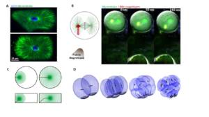
Figure 1: A. In sea urchin zygotes, like in many cells, microtubule asters allow to probe cell shape to orient the nucleus and the subsequent division axis with respect to cell shape (Minc et al. 2011). B. Use of magnetic tweezers to apply forces on mitotic spindles in intact cells, and to control division plane orientation (Xie et al. 2021). C. Numerical simulation of the forces exerted by microtubules to center the nucleus at fertilization (Tanimoto et al. 2016). D. Simulation of the successive division geometries that mark the cleavage stage of early sea urchin embryos (Pierre et al. 2016).


Video1

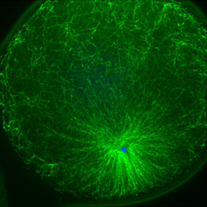
Legend : Developping sea urchin embryos where DNA is labelled with a fluorescent dye. 


2-    Cell shape and polarity

The morphology of single cells as well as cells embedded in tissues underlie many of their function. We seek to address how cells develop their particular shape and internal organization. For instance, many cells only grow at a given location, a process called polarized growth, which involves specific biochemical reactions needed to cluster a local domain of growth activity (Vidéo 2). To study these aspects we use a unicellular fungi called fission yeast. These cells have rod-shapes and are genetically tractable, with many mutants defective in morphogenesis. These cells are encased in a stiff cell wall and are turgid, which provides them with particular mechanical properties (Figure 2). One of our ultimate goal is to link biochemical and biomechanical signals which serve to shape cells.

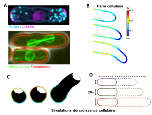
Figure 2: A. Fluorescent images of yeast cells expressing fluorescent proteins to visualize the cytoskeleton, polarity, growth and cell division (Bonazzi et al. 2015). B. Super-resolution images of cell wall thickness dynamics in a growin hyphae of the fungus aspergillus nidulans (Chevallier et al).    C. Numerical simulation of a yeast spore and its cell wall (colors) and poalrity domain (white). D. Simulation of growth speed f yeast cells.


Video2

Legend: Growth and division of yeast cells expressing fluorescent markers of the nuclear envelop and cell wall synthesis.  

Responsable :

Nicolas MINC
Téléphone : +33 (0)157278052
Email : nicolas.minc (at) ijm.fr


Membres de l’équipe :

Serge DMITRIEFF Researcher
Amir KHOSRAVANIZADEH Postdoc
Javad NAJAFI Postdoc
Yannis REIGNIER AI
Charlotte MALLART Postdoc
Célia MUNICIO-DIAZ IR
Maria Isabel ARJONA HIDALGO Postdoc
Camille BOUCHER Postdoc
Aude NOMMICK Postdoc
Jeremy SALLÉ Researcher
Laura RUIZ Ingénieur d’étude
Elyn MANSOUR ROBIC Master 2
Jiawei XU Master 2
  1. Xie J, Najafi J, Le Borgne R, Verbavatz J-M, Durieu C, Sallé J, and Minc N (2022) “Contribution of cytoplasm viscoelastic properties to mitotic spindle positioning” , Proc Natl Acad Sci U S A, 119 (8) e2115593119.
  2. Neeli-Venkata R, Municio Diaz C, Celador R, Sanchez Y, Minc N (2021) “Detection of surface forces by the cell-wall mechanosensor Wsc1 in yeast” , Developmental Cell 56, 1–15
  3. Palenzuela H, Lacroix B, Sallé J, Minami K, Shima T, Jegou A, Romet-Lemonne#G and Minc# N (2020) “In Vitro Reconstitution of Dynein Force Exertion in a Bulk Viscous Medium” , Current Biology, 30, 1–7
  4. Davi V, Chevalier L, Guo H, Tanimoto H, Barrett K, Couturier E, Boudaoud#A, and Minc# N, (2019) “Systematic mapping of cell wall mechanics in the regulation of cell morphogenesis” , Proc. Acad. Sci. USA, 116(28):13833-13838
  5. Sallé J, Xie J, Ershov D, Lacassin M, Dmitrieff S and Minc N, (2019) “Asymmetric division through a reduction of microtubule centering forces” J Cell Biol. 218(3):771-782 .
  6. Tanimoto H, Sallé J, Dodin L and Minc N (2018) “Physical forces determining the persistency and centring precision of microtubule asters” Nature Physics, 114, 848–854.
  7. Davì V, Tanimoto H, Ershov D, Haupt A, De Belly H, Le Borgne R, Couturier E, Boudaoud#A and Minc#, (2018) “Mechanosensation Dynamically Coordinates Polar Growth and Cell Wall Assembly to Promote Cell Survival” Developmental Cell, 45, 2, p170–182.
  8. Pierre A, Sallé J, Wühr M, Minc N., (2016) “Generic Theoretical Models to Predict Division Patterns of Cleaving Embryos.” Developmental Cell. 39(6):667-682
  9. Bonazzi* D, Julien* JD, Seddiki R, Romao M, Piel M, Boudaoud#A, Minc# N, (2014) “Symmetry breaking in spore germination relies on an interplay between polar cap stability and spore wall mechanics.” Developmental Cell, 28 (5):534-546
  10. Minc#, Burgess, D. and Chang F. (2011), “Influence of cell geometry on division plane positioning” Cell., 144 (3): 414-426.

Publications

Mallart, C., Netter, S., Chalvet, F., Claret, S., Guichet, A., Montagne, J., Pret, A.-M., & Malartre, M. (2024). JAK-STAT-dependent contact between follicle cells and the oocyte controls Drosophila anterior-posterior polarity and germline development. Nature Communications, 15(1), 1627. https://doi.org/10.1038/s41467-024-45963-z
Arjona, M. I., Najafi, J., & Minc, N. (2023). Cytoplasm mechanics and cellular organization. Current Opinion in Cell Biology, 85, 102278. https://doi.org/10.1016/j.ceb.2023.102278
Saleh, J., Fardin, M.-A., Barai, A., Soleilhac, M., Frenoy, O., Gaston, C., Cui, H., Dang, T., Gaudin, N., Vincent, A., Minc, N., & Delacour, D. (2023). Length limitation of astral microtubules orients cell divisions in murine intestinal crypts. Developmental Cell. https://doi.org/10.1016/j.devcel.2023.06.004
Guevara-Garcia, A., Soleilhac, M., Minc, N., & Delacour, D. (2023). Regulation and functions of cell division in the intestinal tissue. Seminars in Cell & Developmental Biology, S1084-9521(23)00004-6. https://doi.org/10.1016/j.semcdb.2023.01.004
Jasnin, M., Hervy, J., Balor, S., Bouissou, A., Proag, A., Voituriez, R., Schneider, J., Mangeat, T., Maridonneau-Parini, I., Baumeister, W., Dmitrieff, S., & Poincloux, R. (2022). Elasticity of podosome actin networks produces nanonewton protrusive forces. Nature Communications, 13(1), 3842. https://doi.org/10.1038/s41467-022-30652-6
Letort, G., Eichmuller, A., Da Silva, C., Nikalayevich, E., Crozet, F., Salle, J., Minc, N., Labrune, E., Wolf, J.-P., Terret, M.-E., & Verlhac, M.-H. (2022). An interpretable and versatile machine learning approach for oocyte phenotyping. Journal of Cell Science, 135(13), jcs260281. https://doi.org/10.1242/jcs.260281
Darnat, P., Burg, A., Sallé, J., Lacoste, J., Louvet-Vallée, S., Gho, M., & Audibert, A. (2022). Cortical Cyclin A controls spindle orientation during asymmetric cell divisions in Drosophila. Nature Communications, 13(1), 2723. https://doi.org/10.1038/s41467-022-30182-1
Xie, J., Najafi, J., Le Borgne, R., Verbavatz, J.-M., Durieu, C., Sallé, J., & Minc, N. (2022). Contribution of cytoplasm viscoelastic properties to mitotic spindle positioning. Proceedings of the National Academy of Sciences of the United States of America, 119(8), e2115593119. https://doi.org/10.1073/pnas.2115593119
Neeli-Venkata, R., Diaz, C. M., Celador, R., Sanchez, Y., & Minc, N. (2021). Detection of surface forces by the cell-wall mechanosensor Wsc1 in yeast. Developmental Cell, 56(20), 2856-2870.e7. https://doi.org/10.1016/j.devcel.2021.09.024
Fukui, H., Chow, R. W.-Y., Xie, J., Foo, Y. Y., Yap, C. H., Minc, N., Mochizuki, N., & Vermot, J. (2021). Bioelectric signaling and the control of cardiac cell identity in response to mechanical forces. Science (New York, N.Y.), 374(6565), 351–354. https://doi.org/10.1126/science.abc6229
Taheraly, S., Ershov, D., Dmitrieff, S., & Minc, N. (2020). An image analysis method to survey the dynamics of polar protein abundance in the regulation of tip growth. Journal of Cell Science, 133(22), jcs252064. https://doi.org/10.1242/jcs.252064
Palenzuela, H., Lacroix, B., Sallé, J., Minami, K., Shima, T., Jegou, A., Romet-Lemonne, G., & Minc, N. (2020). In Vitro Reconstitution of Dynein Force Exertion in a Bulk Viscous Medium. Current Biology: CB, 30(22), 4534-4540.e7. https://doi.org/10.1016/j.cub.2020.08.078
Mukherjee, R. N., Sallé, J., Dmitrieff, S., Nelson, K. M., Oakey, J., Minc, N., & Levy, D. L. (2020). The Perinuclear ER Scales Nuclear Size Independently of Cell Size in Early Embryos. Developmental Cell, 54(3), 395-409.e7. https://doi.org/10.1016/j.devcel.2020.05.003
Suzuki, E. L., Chikireddy, J., Dmitrieff, S., Guichard, B., Romet-Lemonne, G., & Jégou, A. (2020). Geometrical Constraints Greatly Hinder Formin mDia1 Activity. Nano Letters, 20(1), 22–32. https://doi.org/10.1021/acs.nanolett.9b02241
Davì, V., Chevalier, L., Guo, H., Tanimoto, H., Barrett, K., Couturier, E., Boudaoud, A., & Minc, N. (2019). Systematic mapping of cell wall mechanics in the regulation of cell morphogenesis. Proceedings of the National Academy of Sciences of the United States of America, 116(28), 13833–13838. https://doi.org/10.1073/pnas.1820455116
Gervais, L., van den Beek, M., Josserand, M., Sallé, J., Stefanutti, M., Perdigoto, C. N., Skorski, P., Mazouni, K., Marshall, O. J., Brand, A. H., Schweisguth, F., & Bardin, A. J. (2019). Stem Cell Proliferation Is Kept in Check by the Chromatin Regulators Kismet/CHD7/CHD8 and Trr/MLL3/4. Developmental Cell, 49(4), 556-573.e6. https://doi.org/10.1016/j.devcel.2019.04.033
Letort, G., Bennabi, I., Dmitrieff, S., Nedelec, F., Verlhac, M.-H., & Terret, M.-E. (2019). A computational model of the early stages of acentriolar meiotic spindle assembly. Molecular Biology of the Cell, 30(7), 863–875. https://doi.org/10.1091/mbc.E18-10-0644
Sallé, J., Xie, J., Ershov, D., Lacassin, M., Dmitrieff, S., & Minc, N. (2019). Asymmetric division through a reduction of microtubule centering forces. The Journal of Cell Biology, 218(3), 771–782. https://doi.org/10.1083/jcb.201807102
Dmitrieff, S., & Minc, N. (2019). Scaling properties of centering forces. Europhysics Letters, 125(4), 48001. https://doi.org/10.1209/0295-5075/125/48001
Haupt, A., Ershov, D., & Minc, N. (2018). A Positive Feedback between Growth and Polarity Provides Directional Persistency and Flexibility to the Process of Tip Growth. Current Biology: CB, 28(20), 3342-3351.e3. https://doi.org/10.1016/j.cub.2018.09.022
Mund, M., van der Beek, J. A., Deschamps, J., Dmitrieff, S., Hoess, P., Monster, J. L., Picco, A., Nédélec, F., Kaksonen, M., & Ries, J. (2018). Systematic Nanoscale Analysis of Endocytosis Links Efficient Vesicle Formation to Patterned Actin Nucleation. Cell, 174(4), 884-896.e17. https://doi.org/10.1016/j.cell.2018.06.032
Tanimoto, H., Sallé, J., Dodin, L., & Minc, N. (2018). Physical Forces Determining the Persistency and Centering Precision of Microtubule Asters. Nature Physics, 14(8), 848–854. https://doi.org/10.1038/s41567-018-0154-4
Lacroix, B., Letort, G., Pitayu, L., Sallé, J., Stefanutti, M., Maton, G., Ladouceur, A.-M., Canman, J. C., Maddox, P. S., Maddox, A. S., Minc, N., Nédélec, F., & Dumont, J. (2018). Microtubule Dynamics Scale with Cell Size to Set Spindle Length and Assembly Timing. Developmental Cell, 45(4), 496-511.e6. https://doi.org/10.1016/j.devcel.2018.04.022
Davì, V., Tanimoto, H., Ershov, D., Haupt, A., De Belly, H., Le Borgne, R., Couturier, E., Boudaoud, A., & Minc, N. (2018). Mechanosensation Dynamically Coordinates Polar Growth and Cell Wall Assembly to Promote Cell Survival. Developmental Cell, 45(2), 170-182.e7. https://doi.org/10.1016/j.devcel.2018.03.022
Bun, P., Dmitrieff, S., Belmonte, J. M., Nédélec, F. J., & Lénárt, P. (2018). A disassembly-driven mechanism explains F-actin-mediated chromosome transport in starfish oocytes. ELife, 7, e31469. https://doi.org/10.7554/eLife.31469
Bizzotto, S., Uzquiano, A., Dingli, F., Ershov, D., Houllier, A., Arras, G., Richards, M., Loew, D., Minc, N., Croquelois, A., Houdusse, A., & Francis, F. (2017). Eml1 loss impairs apical progenitor spindle length and soma shape in the developing cerebral cortex. Scientific Reports, 7(1), 17308. https://doi.org/10.1038/s41598-017-15253-4
Dmitrieff, S., Alsina, A., Mathur, A., & Nédélec, F. J. (2017). Balance of microtubule stiffness and cortical tension determines the size of blood cells with marginal band across species. Proceedings of the National Academy of Sciences of the United States of America, 114(17), 4418–4423. https://doi.org/10.1073/pnas.1618041114
Dumollard, R., Minc, N., Salez, G., Aicha, S. B., Bekkouche, F., Hebras, C., Besnardeau, L., & McDougall, A. (2017). The invariant cleavage pattern displayed by ascidian embryos depends on spindle positioning along the cell’s longest axis in the apical plane and relies on asynchronous cell divisions. ELife, 6, e19290. https://doi.org/10.7554/eLife.19290
Salomon, J., Gaston, C., Magescas, J., Duvauchelle, B., Canioni, D., Sengmanivong, L., Mayeux, A., Michaux, G., Campeotto, F., Lemale, J., Viala, J., Poirier, F., Minc, N., Schmitz, J., Brousse, N., Ladoux, B., Goulet, O., & Delacour, D. (2017). Contractile forces at tricellular contacts modulate epithelial organization and monolayer integrity. Nature Communications, 8(1), 13998. https://doi.org/10.1038/ncomms13998
Haupt, A., & Minc, N. (2017). Gradients of phosphatidylserine contribute to plasma membrane charge localization and cell polarity in fission yeast. Molecular Biology of the Cell, 28(1), 210–220. https://doi.org/10.1091/mbc.e16-06-0353

Preprints

Najafi, J., Dmitrieff, S., & Minc, N. (2023). Size- and position-dependent cytoplasm viscoelasticity through hydrodynamic interactions with the cell surface. Proceedings of the National Academy of Sciences of the United States of America, 120(9), e2216839120. https://doi.org/10.1073/pnas.2216839120
Chevalier, L., Pinar, M., Le Borgne, R., Durieu, C., Peñalva, M. A., Boudaoud, A., & Minc, N. (2023). Cell wall dynamics stabilize tip growth in a filamentous fungus. PLoS Biology, 21(1), e3001981. https://doi.org/10.1371/journal.pbio.3001981
Jad, S., Marc-Antoine, F., Olivia, F., Matis, S., Cécile, G., Hongyue, C., Tien, D., Gaudin, N., Audrey, V., Nicolas, M., & Delphine, D. (2022). Length-limitation of astral microtubules orients cell divisions in intestinal crypts. bioRxiv. https://doi.org/10.1101/2022.09.02.506333


Reviews

Municio-Diaz, C., Muller, E., Drevensek, S., Fruleux, A., Lorenzetti, E., Boudaoud, A., & Minc, N. (2022). Mechanobiology of the cell wall - insights from tip-growing plant and fungal cells. Journal of Cell Science, 135(21), jcs259208. https://doi.org/10.1242/jcs.259208
Sallé, J., & Minc, N. (2022). Cell division geometries as central organizers of early embryo development. Seminars in Cell & Developmental Biology, 130, 3–11. https://doi.org/10.1016/j.semcdb.2021.08.004
Mishra, R., Minc, N., & Peter, M. (2022). Cells under pressure: how yeast cells respond to mechanical forces. Trends in Microbiology, 30(5), 495–510. https://doi.org/10.1016/j.tim.2021.11.006
Lenne, P.-F., Munro, E., Heemskerk, I., Warmflash, A., Bocanegra-Moreno, L., Kishi, K., Kicheva, A., Long, Y., Fruleux, A., Boudaoud, A., Saunders, T. E., Caldarelli, P., Michaut, A., Gros, J., Maroudas-Sacks, Y., Keren, K., Hannezo, E., Gartner, Z. J., Stormo, B., … Tlili, S. (2021). Roadmap for the multiscale coupling of biochemical and mechanical signals during development. Physical Biology, 18(4). https://doi.org/10.1088/1478-3975/abd0db
Xie, J., & Minc, N. (2020). Cytoskeleton Force Exertion in Bulk Cytoplasm. Frontiers in Cell and Developmental Biology, 8, 69. https://doi.org/10.3389/fcell.2020.00069
Haupt, A., & Minc, N. (2018). How cells sense their own shape - mechanisms to probe cell geometry and their implications in cellular organization and function. Journal of Cell Science, 131(6), jcs214015. https://doi.org/10.1242/jcs.214015


Book chapter

Xie, J., Levy, D. L., Minc, N., & Sallé, J. (2024). Manipulation of Embryonic Cleavage Geometry Using Magnetic Tweezers. In A. Castro & B. Lacroix (Eds.), Cell Cycle Control: Methods and Protocols (pp. 125–140). Springer US. https://doi.org/10.1007/978-1-0716-3557-5_8
Chenevert, J., Robert, M. L. V., Sallé, J., Cacchia, S., Lorca, T., Castro, A., McDougall, A., Minc, N., Castagnetti, S., Dumont, J., & Lacroix, B. (2024). Measuring Mitotic Spindle and Microtubule Dynamics in Marine Embryos and Non-model Organisms. In A. Castro & B. Lacroix (Eds.), Cell Cycle Control: Methods and Protocols (pp. 187–210). Springer US. https://doi.org/10.1007/978-1-0716-3557-5_12
Ershov, D., & Minc, N. (2019). Modeling Embryonic Cleavage Patterns. Methods in Molecular Biology (Clifton, N.J.), 1920, 393–406. https://doi.org/10.1007/978-1-4939-9009-2_24

Daria Bonazzi (2015)

Anaëlle Pierre (2017)

Valeria Davì (2018)

Héliciane Palenzuela (2020)

Jing Xie (2022)

Guillaume Romet-Lemonne (IJM) ; Delphine Delacour (IJM) ; Aki Kimura (NIG, Japan) ; Yohanns Bellaiche (Institut Curie, Paris) ; CP Heisenberg (IST austria) ; Martin Wühr (Princeton University) ; Dan Levy (U. Wyoming).

Arezki Boudaoud (Ecole Polytechnique); Etienne Couturier (MsC, Paris); Yolanda Sanchez (U. Cordoba); Miguel Peñalva (U. Madrid).

La Ligue Contre le Cancer ; Fondation Bettencourt-Schueller ; European Research Council ; Mairie De Paris; ANR ; Fondation de la Recherche Médicale ; FungiBrain (European ITN) FP7.