Missions
We are a team of engineers, coming from different disciplines (biology, physics, computer science), working on the interface of microscopy with biology. We propose :
- the orientation of users towards the most suitable equipment for their project
- the assistance in the design of tools: molecular biology, protein labeling, choice of labels and fluorophores, analysis protocols…
- the assistance in setting up the experiment: definition of controls, operating procedures, precautions to be taken with respect to potential artifacts
- the possible supervision of the users for the realization of the experiments
- the provision of functional and calibrated equipments
- the training of users for an autonomous use of the equipments
- the taking in charge of the acquisitions for the non autonomous users
- the assistance to the treatment and analysis of the obtained data
- assistance in the interpretation and presentation of the results.
ImagoSeine is a member of the national infrastructure of imaging platforms France-BioImaging and the Euro-BioImaging network.
ImagoSeine is supported by the following organisations:

The ImagoSeine imaging platform offers 3 types of services in photonic microscopy, cytometry and electron microscopy:
1 – Independent use of equipments and reagents
The autonomous user is previously trained by the platform staff on certain equipment or for the performance of certain operations (see Training). He/she benefits from extended use time slots (see Access times) and adapted rates (autonomous rate). During the sessions reserved for autonomous use (see Reservation), the platform’s engineers can be called upon for specific questions, or in the event of technical problems (malfunctions or defects in the equipment), during the platform’s normal opening hours, depending on their availability. If necessary, the autonomous user can book a session with assistance (advice, in-depth training). The autonomous user is responsible for the quality and management of the results he/she has obtained. A “session follow-up book”, located next to each device, must be filled in at each session. Documents (user’s manual, tutorials, …) are made available to the users and can be consulted on site. During the service, the autonomous user is responsible for the equipment provided. He/she must follow the rules for starting up, using, closing and storing the equipment and reagents used. The team leader agrees to pay the full cost of repairs in the event of damage due to improper use of equipment. Any independent user who has not used equipment for more than one year or since the update of equipment (hardware, software) will have to undergo additional training. The status of autonomous user is subject to the appreciation of the platform staff and can be reviewed without notice. An autonomous user cannot train another user under any circumstances.
2 – The assisted session
The assisted session is conducted in the presence of the platform’s staff. It is proposed within the framework of the implementation of known protocols in standard experimental conditions, decided with the user, and does not have an inventive character. The engineers of the platform involved guarantee the quality of the results, provided that the use is made according to their recommendations, under the conditions fixed in agreement with the user. The session with assistance is subject to prior reservation (see Reservation), depending on the availability of the platform’s equipment and engineers and only during the normal opening hours (see Access hours). It is subject to “assisted” pricing.
3 – The collaborative project
This is the case when the user’s project requires the support of one or more platform engineers for occasional or regular assistance or for the development of a technique or technology for the user’s applications, or of a data acquisition or processing protocol. The platform’s engineers involved in the project guarantee the quality of the results obtained and their restitution (samples, data). The stages of a project can be carried out in the presence or absence of the user, depending on what has been decided beforehand between the user and the engineers involved. In the second case they are not subject to a particular reservation. Project invoicing may be subject to special pricing, based on an estimate, depending on the equipment used for the project. Billing will be based on actual hours used. The rate applied will be the “assisted users” rate.
The user and user’s manager undertake to report any publication using results obtainedbplatform and to comply with the following requirements:
The user and their supervisor undertake to report any publication using results obtained through the platform and to comply with the following requirements:
“We acknowledge the ImagoSeine core facility of the Institut Jacques Monod, member of the France BioImaging infrastructure (https://ror.org/01y7vt929) supported by the French National Research Agency (ANR-24-INBS-0005 FBI BIOGEN) and GIS-IBiSA”
The following should be added:
“and the support of La ligue contre le Cancer (R03/75-79)” for results obtained with the FACSAria Fusion sorter.
“and the support of the Île-de-France Region (Sesame)” for the SBF-SEM Teneo VS.
How to proceed
1) Contact us by email to make an appointment with our team.
Flow cytometry contact
Electron microscopy contact
Photonic microscopy contact
2) During this meeting it is necessary that the person in charge of the project is present and that you provide us with the important information (purpose of the experiment, type of samples, markers, support…).
It will be decided whether your project requires collaboration, service or training on one of the devices.
3) Register on the platform’s booking site.
You must have a valid professional email address. Once your information has been validated by the platform, your identifier (first name.surname) will be activated to connect you.
4) Create your password to connect to the booking site on this page. Once connected, download the charter, print and sign the last page.
General coordination of ImagoSeine
| René-Marc | MEGE | Platform coordinator | +33 (0)1 57 27 80 67 | contact |
| Jean-Marc | VERBAVATZ | Platform coordinator | +33 (0)1 57 27 80 04 | contact |
Flow cytometry
Team leader
Nicolas VALENTIN,
Flow cytometry manager,
PFT/IMAGOSEINE/CYTOMETRIE+33 (0)1 57 27 81 52, room RH20B4
Electron microscopy
Team leader
Catherine DURIEU,
Electron microscopy engineer,
PFT/IMAGOSEINE/ELECTRONIQUE+33 (0)1 57 27 81 58, room RH20B7
Member
Alice MARTEIL,
Electron microscopy engineer,
PFT/IMAGOSEINE/ELECTRONIQUE+33 (0)1 57 27 81 58, room RH20B7
Photonics microscopy
Team leader
Xavier BAUDIN,
Light microscopy facility manager,
PFT/IMAGOSEINE/PHOTONIQUE+33 (0)1 57 27 81 56, room RH20B6
Members
Nicolas MOISAN,
Light microscopy engineer,
PFT/IMAGOSEINE/PHOTONIQUE+33 (0)1 57 27 81 55, room RH20B6
Guillaume QUIBEUF,
Light microscopy engineer,
PFT/IMAGOSEINE/PHOTONIQUE 0157278154, room RH20B5
Thomas RIOS,
Light microscopy engineer,
PFT/IMAGOSEINE/PHOTONIQUE+33 (0)1 57 27 81 54, room RH20B6
Use of equipment and services
Only after the signed charter has been received can the services take place. Make requests for equipment training and collaborative projects once you are logged on to the booking site with the “Request” tab.
Charter to download from the reservation site
Billing
The use of the platform’s equipment is subject to a fee (see our rates). The rates are calculated to cover the maintenance and updating of the platform’s equipment.
A detailed statement of the services provided will be sent to you each month. In return, you must send a purchase order to the Institute’s accounting department.
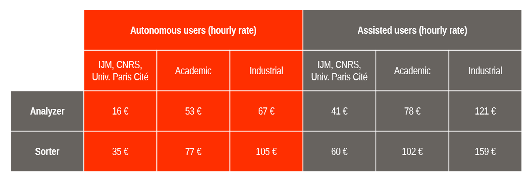
ImagoSeine flow cytometry core facility user fees
ImagoSeine light microscopy user fees
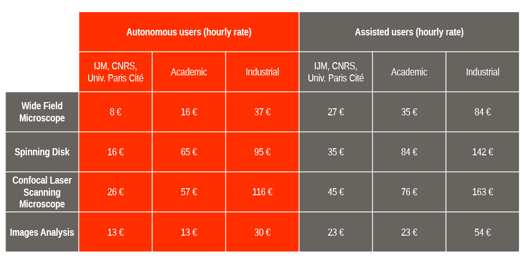
For time laps acquisitions, the price is divided by 2 between 8pm and 9am and during the weekend.
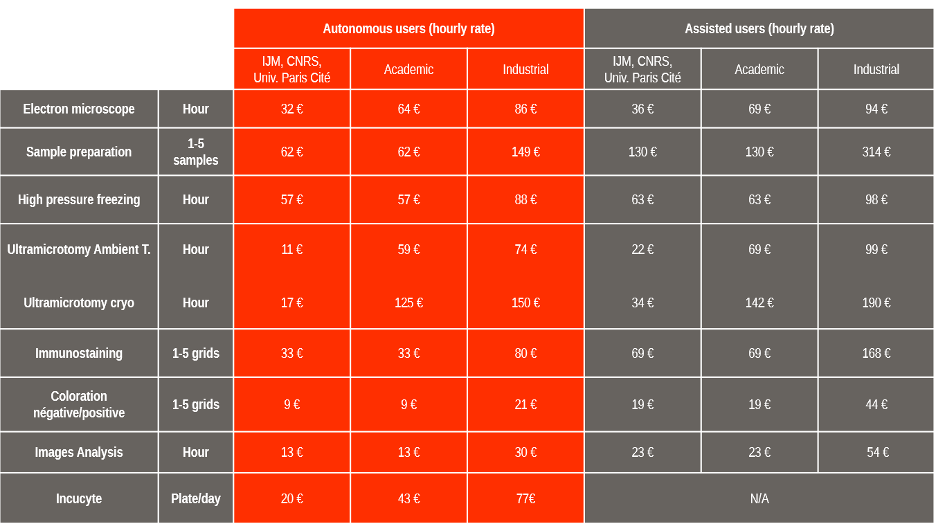
ImagoSeine electronic microscopy user fees
Janvier 2025 : Nouveau confocal à balayage Stellaris et module de Super-Résolution 3D STED
Confocal à balayage laser Leica Microsystems, équipé pour le vivant ainsi que d’un laser blanc permettant de choisir l’excitation de marqueurs fluorescents jusqu’à 790 nm. Le module STED permet une résolution inférieure à 100nm dans les trois dimensions ou jusqu’à 30nm en XY. Cet équipement a été acquis avec le soutien de France BioImaging et UPCité.
November 2024: New cytometer at ImagoSeine: the Attune CytPix (ThermoFisher).
This flow cytometer, equipped with acoustic focusing technology, enables the analysis of rare events. Its high-speed camera captures brightfield images of cells, linking morphological parameters with data obtained from conventional flow cytometry. This equipment was acquired with the support of GIS IBiSA
July 2024: Opening of a full-field Leica DMI8 in room RH19B.
The full-frame features an incubator and AFC autofocus for observation of live specimens. Numerous lenses are available.
October 2022: New LSM980 Airyscan2 scanning confocal and two-photon module available as part RH26B.
Latest generation Zeiss confocal, equipped for live experiments, allowing simultaneous spectral separation and FRAP experiments. The Airyscan 4Y module allows a 50% increase in resolution compared to a conventional confocal and an increase in acquisition speed. The two-photon microscopy module allows for more in-depth observation of samples.
September 2022 : New ELYRA7 Super-Resolution microscopy system in RH22B room.
Microscope allowing PALM and dSTORM experiments for localization accuracy down to 20nm. Lattice SIM experiments allowing a 3D resolution increase of two times the theoretical limit and compatible with the living. TIRF experiments to observe events at the interface between the cell membrane and its substrate.
June 2022: New FLIM and FCS module on the confocal LSM980 in room RH20B1.
The FLIM technique allows studying the fluorescence lifetime in the sample, which gives information on its biological state as well as the interaction between proteins by the FLIM-FRET technique. FCS techniques allow the study of the mobility of fluorescent molecules.
September 2021: Upgrade of the CSU X1 FRAP spinning disk and UV Photoablation in RH20B9.
The spinning disk X1 FRAP has a new laser bench (405nm, 445nm, 488nm, 561nm, 642nm) as well as the possibility to do FRAP with the 5 lasers and photoablation by pulsed UV.
The user and user’s manager undertake to report any publication using results obtainedbplatform and to comply with the following requirements:
- The platform must be systematically acknowledged using the following formula: “We acknowledge the ImagoSeine core facility of the Institut Jacques Monod, member of the France BioImaging infrastructure (ANR-10-INBS-04) and GIS-IBiSA”.
- In addition : “and the support of La ligue contre le Cancer (R03/75-79)” for the results obtained with the Accuri, Cyan, Facs Aria Fusion analyzer and “and the support of the Region Île-de-France (Sesame)” for results obtained with ImageStream or SBF-SEM Teneo VS.
Publications co-written by ImgoSeine members (2017-2024)
2913254
I7CUV6U5
1
apa
50
date
desc
16949
https://www.ijm.fr/wp-content/plugins/zotpress/
%7B%22status%22%3A%22success%22%2C%22updateneeded%22%3Afalse%2C%22instance%22%3Afalse%2C%22meta%22%3A%7B%22request_last%22%3A50%2C%22request_next%22%3A50%2C%22used_cache%22%3Atrue%7D%2C%22data%22%3A%5B%7B%22key%22%3A%22F43UBAFD%22%2C%22library%22%3A%7B%22id%22%3A2913254%7D%2C%22meta%22%3A%7B%22creatorSummary%22%3A%22Jouve%20et%20al.%22%2C%22parsedDate%22%3A%222025-10-09%22%2C%22numChildren%22%3A1%7D%2C%22bib%22%3A%22%26lt%3Bdiv%20class%3D%26quot%3Bcsl-bib-body%26quot%3B%20style%3D%26quot%3Bline-height%3A%202%3B%20padding-left%3A%201em%3B%20text-indent%3A-1em%3B%26quot%3B%26gt%3B%5Cn%20%20%26lt%3Bdiv%20class%3D%26quot%3Bcsl-entry%26quot%3B%26gt%3BJouve%2C%20C.%2C%20Ruiz-Velasco%2C%20A.%2C%20Donnarumma%2C%20E.%2C%20Le%20Borgne%2C%20R.%2C%20Momken%2C%20I.%2C%20Pereira%2C%20C.%2C%20Seguret%2C%20M.%2C%20Vermersch%2C%20E.%2C%20Vimont%2C%20E.%2C%20Nemazanyy%2C%20I.%2C%20Bertrand%2C%20L.%2C%20Wai%2C%20T.%2C%20Verbavatz%2C%20J.-M.%2C%20Mericksay%2C%20M.%2C%20%26amp%3B%20Hulot%2C%20J.-S.%20%282025%29.%20Myocardial%20disarray%20drives%20metabolic%20inefficiency%20in%20human%20cardiomyocytes.%20%26lt%3Bi%26gt%3BCommunications%20Biology%26lt%3B%5C%2Fi%26gt%3B%2C%20%26lt%3Bi%26gt%3B8%26lt%3B%5C%2Fi%26gt%3B%281%29%2C%201445.%20%26lt%3Ba%20class%3D%26%23039%3Bzp-DOIURL%26%23039%3B%20href%3D%26%23039%3Bhttps%3A%5C%2F%5C%2Fdoi.org%5C%2F10.1038%5C%2Fs42003-025-08842-x%26%23039%3B%26gt%3Bhttps%3A%5C%2F%5C%2Fdoi.org%5C%2F10.1038%5C%2Fs42003-025-08842-x%26lt%3B%5C%2Fa%26gt%3B%26lt%3B%5C%2Fdiv%26gt%3B%5Cn%26lt%3B%5C%2Fdiv%26gt%3B%22%2C%22data%22%3A%7B%22itemType%22%3A%22journalArticle%22%2C%22title%22%3A%22Myocardial%20disarray%20drives%20metabolic%20inefficiency%20in%20human%20cardiomyocytes%22%2C%22creators%22%3A%5B%7B%22creatorType%22%3A%22author%22%2C%22firstName%22%3A%22Charl%5Cu00e8ne%22%2C%22lastName%22%3A%22Jouve%22%7D%2C%7B%22creatorType%22%3A%22author%22%2C%22firstName%22%3A%22Andrea%22%2C%22lastName%22%3A%22Ruiz-Velasco%22%7D%2C%7B%22creatorType%22%3A%22author%22%2C%22firstName%22%3A%22Erminia%22%2C%22lastName%22%3A%22Donnarumma%22%7D%2C%7B%22creatorType%22%3A%22author%22%2C%22firstName%22%3A%22R%5Cu00e9mi%22%2C%22lastName%22%3A%22Le%20Borgne%22%7D%2C%7B%22creatorType%22%3A%22author%22%2C%22firstName%22%3A%22Iman%22%2C%22lastName%22%3A%22Momken%22%7D%2C%7B%22creatorType%22%3A%22author%22%2C%22firstName%22%3A%22C%5Cu00e9line%22%2C%22lastName%22%3A%22Pereira%22%7D%2C%7B%22creatorType%22%3A%22author%22%2C%22firstName%22%3A%22Magali%22%2C%22lastName%22%3A%22Seguret%22%7D%2C%7B%22creatorType%22%3A%22author%22%2C%22firstName%22%3A%22Eva%22%2C%22lastName%22%3A%22Vermersch%22%7D%2C%7B%22creatorType%22%3A%22author%22%2C%22firstName%22%3A%22Elodie%22%2C%22lastName%22%3A%22Vimont%22%7D%2C%7B%22creatorType%22%3A%22author%22%2C%22firstName%22%3A%22Ivan%22%2C%22lastName%22%3A%22Nemazanyy%22%7D%2C%7B%22creatorType%22%3A%22author%22%2C%22firstName%22%3A%22Luc%22%2C%22lastName%22%3A%22Bertrand%22%7D%2C%7B%22creatorType%22%3A%22author%22%2C%22firstName%22%3A%22Timothy%22%2C%22lastName%22%3A%22Wai%22%7D%2C%7B%22creatorType%22%3A%22author%22%2C%22firstName%22%3A%22Jean-Marc%22%2C%22lastName%22%3A%22Verbavatz%22%7D%2C%7B%22creatorType%22%3A%22author%22%2C%22firstName%22%3A%22Mathias%22%2C%22lastName%22%3A%22Mericksay%22%7D%2C%7B%22creatorType%22%3A%22author%22%2C%22firstName%22%3A%22Jean-S%5Cu00e9bastien%22%2C%22lastName%22%3A%22Hulot%22%7D%5D%2C%22abstractNote%22%3A%22Adult%20cardiomyocytes%20are%20embedded%20within%20a%20highly%20organized%20myocardial%20microenvironment%20that%20imposes%20critical%20geometric%20cues%20essential%20for%20the%20alignment%20and%20distribution%20of%20organelles%20and%20the%20shaping%20of%20their%20unique%2C%20rectangular%20cellular%20morphology.%20Despite%20the%20association%20of%20cardiomyocyte%20disarray%20with%20human%20heart%20disease%2C%20the%20functional%20consequences%20of%20this%20cellular%20disorganization%20remain%20poorly%20understood.%20Here%2C%20we%20leveraged%20micropatterned%20substrates%20to%20promote%20structural%20alignment%20in%20human%20induced%20pluripotent%20stem%20cell-derived%20cardiomyocytes%20%28hiPSC-CMs%29%2C%20contrasting%20the%20effects%20of%20mechanical%20alignment%20on%20mitochondrial%20form%20and%20function%20with%20hiPSC-CMs%20cultured%20under%20standard%20unconstrained%20conditions.%20Cardiomyocytes%20cultured%20under%20unconstrained%20conditions%20exhibited%20misaligned%20sarcomeres%20and%20a%20perinuclear%20mitochondrial%20distribution%20while%20micropatterned%20hiPSC-CMs%20developed%20linear%20myofibrils%20and%20reconfigured%20sarcomere%20and%20mitochondrial%20organization%2C%20which%20increased%20mitochondrial%20respiration%20without%20augmenting%20mitochondrial%20mass.%20Notably%2C%20micropatterned%20hiPSC-CMs%20exhibited%20an%20increased%20number%20of%20mitochondria-associated%20membranes%2C%20as%20determined%20by%20proximity%20ligation%20assays%20and%20transmission%20electron%20microscopy%2C%20suggesting%20enhanced%20interactions%20between%20the%20sarcoplasmic%20reticulum%20and%20mitochondria.%20Together%2C%20these%20findings%20demonstrate%20that%20mitochondrial-sarcoplasmic%20architecture%20and%20geometry%20are%20critical%20spatial%20features%20that%20ensure%20bioenergetic%20efficiency%20of%20cardiomyocytes.%20This%20work%20underscores%20the%20importance%20of%20cellular%20organization%20in%20cardiomyocyte%20metabolism%20and%20function%2C%20providing%20insights%20into%20the%20pathophysiology%20of%20cardiac%20diseases%20marked%20by%20cellular%20disarray.%22%2C%22date%22%3A%222025-10-09%22%2C%22section%22%3A%22%22%2C%22partNumber%22%3A%22%22%2C%22partTitle%22%3A%22%22%2C%22DOI%22%3A%2210.1038%5C%2Fs42003-025-08842-x%22%2C%22citationKey%22%3A%22%22%2C%22url%22%3A%22https%3A%5C%2F%5C%2Fwww.nature.com%5C%2Farticles%5C%2Fs42003-025-08842-x%22%2C%22PMID%22%3A%22%22%2C%22PMCID%22%3A%22%22%2C%22ISSN%22%3A%222399-3642%22%2C%22language%22%3A%22en%22%2C%22collections%22%3A%5B%22I7CUV6U5%22%5D%2C%22dateModified%22%3A%222025-10-13T08%3A58%3A36Z%22%7D%7D%2C%7B%22key%22%3A%22BQELGKWL%22%2C%22library%22%3A%7B%22id%22%3A2913254%7D%2C%22meta%22%3A%7B%22creatorSummary%22%3A%22El%20Mossadeq%20et%20al.%22%2C%22parsedDate%22%3A%222024-12-26%22%2C%22numChildren%22%3A1%7D%2C%22bib%22%3A%22%26lt%3Bdiv%20class%3D%26quot%3Bcsl-bib-body%26quot%3B%20style%3D%26quot%3Bline-height%3A%202%3B%20padding-left%3A%201em%3B%20text-indent%3A-1em%3B%26quot%3B%26gt%3B%5Cn%20%20%26lt%3Bdiv%20class%3D%26quot%3Bcsl-entry%26quot%3B%26gt%3BEl%20Mossadeq%2C%20L.%2C%20Bellutti%2C%20L.%2C%20Le%20Borgne%2C%20R.%2C%20Canman%2C%20J.%20C.%2C%20Pintard%2C%20L.%2C%20Verbavatz%2C%20J.-M.%2C%20Askjaer%2C%20P.%2C%20%26amp%3B%20Dumont%2C%20J.%20%282024%29.%20An%20interkinetic%20envelope%20surrounds%20chromosomes%20between%20meiosis%20I%20and%20II%20in%20C.%20elegans%20oocytes.%20%26lt%3Bi%26gt%3BJournal%20of%20Cell%20Biology%26lt%3B%5C%2Fi%26gt%3B%2C%20%26lt%3Bi%26gt%3B224%26lt%3B%5C%2Fi%26gt%3B%283%29%2C%20e202403125.%20%26lt%3Ba%20class%3D%26%23039%3Bzp-ItemURL%26%23039%3B%20href%3D%26%23039%3Bhttps%3A%5C%2F%5C%2Fdoi.org%5C%2F10.1083%5C%2Fjcb.202403125%26%23039%3B%26gt%3Bhttps%3A%5C%2F%5C%2Fdoi.org%5C%2F10.1083%5C%2Fjcb.202403125%26lt%3B%5C%2Fa%26gt%3B%26lt%3B%5C%2Fdiv%26gt%3B%5Cn%26lt%3B%5C%2Fdiv%26gt%3B%22%2C%22data%22%3A%7B%22itemType%22%3A%22journalArticle%22%2C%22title%22%3A%22An%20interkinetic%20envelope%20surrounds%20chromosomes%20between%20meiosis%20I%20and%20II%20in%20C.%20elegans%20oocytes%22%2C%22creators%22%3A%5B%7B%22creatorType%22%3A%22author%22%2C%22firstName%22%3A%22Layla%22%2C%22lastName%22%3A%22El%20Mossadeq%22%7D%2C%7B%22creatorType%22%3A%22author%22%2C%22firstName%22%3A%22Laura%22%2C%22lastName%22%3A%22Bellutti%22%7D%2C%7B%22creatorType%22%3A%22author%22%2C%22firstName%22%3A%22R%5Cu00e9mi%22%2C%22lastName%22%3A%22Le%20Borgne%22%7D%2C%7B%22creatorType%22%3A%22author%22%2C%22firstName%22%3A%22Julie%20C.%22%2C%22lastName%22%3A%22Canman%22%7D%2C%7B%22creatorType%22%3A%22author%22%2C%22firstName%22%3A%22Lionel%22%2C%22lastName%22%3A%22Pintard%22%7D%2C%7B%22creatorType%22%3A%22author%22%2C%22firstName%22%3A%22Jean-Marc%22%2C%22lastName%22%3A%22Verbavatz%22%7D%2C%7B%22creatorType%22%3A%22author%22%2C%22firstName%22%3A%22Peter%22%2C%22lastName%22%3A%22Askjaer%22%7D%2C%7B%22creatorType%22%3A%22author%22%2C%22firstName%22%3A%22Julien%22%2C%22lastName%22%3A%22Dumont%22%7D%5D%2C%22abstractNote%22%3A%22At%20the%20end%20of%20cell%20division%2C%20the%20nuclear%20envelope%20reassembles%20around%20the%20decondensing%20chromosomes.%20Female%20meiosis%20culminates%20in%20two%20consecutive%20cell%20divisions%20of%20the%20oocyte%2C%20meiosis%20I%20and%20II%2C%20which%20are%20separated%20by%20a%20brief%20transition%20phase%20known%20as%20interkinesis.%20Due%20to%20the%20absence%20of%20chromosome%20decondensation%20and%20the%20suppression%20of%20genome%20replication%20during%20interkinesis%2C%20it%20has%20been%20widely%20assumed%20that%20the%20nuclear%20envelope%20does%20not%20reassemble%20between%20meiosis%20I%20and%20II.%20By%20analyzing%20interkinesis%20in%20C.%20elegans%20oocytes%2C%20we%20instead%20show%20that%20an%20atypical%20structure%20made%20of%20two%20lipid%20bilayers%2C%20which%20we%20termed%20the%20interkinetic%20envelope%2C%20surrounds%20the%20surface%20of%20the%20segregating%20chromosomes.%20The%20interkinetic%20envelope%20shares%20common%20features%20with%20the%20nuclear%20envelope%20but%20also%20exhibits%20specific%20characteristics%20that%20distinguish%20it%2C%20including%20its%20lack%20of%20continuity%20with%20the%20endoplasmic%20reticulum%2C%20unique%20protein%20composition%2C%20assembly%20mechanism%2C%20and%20function%20in%20chromosome%20segregation.%20These%20distinct%20attributes%20collectively%20define%20the%20interkinetic%20envelope%20as%20a%20unique%20and%20specialized%20structure%20that%20has%20been%20previously%20overlooked.%22%2C%22date%22%3A%222024-12-26%22%2C%22section%22%3A%22%22%2C%22partNumber%22%3A%22%22%2C%22partTitle%22%3A%22%22%2C%22DOI%22%3A%2210.1083%5C%2Fjcb.202403125%22%2C%22citationKey%22%3A%22%22%2C%22url%22%3A%22https%3A%5C%2F%5C%2Fdoi.org%5C%2F10.1083%5C%2Fjcb.202403125%22%2C%22PMID%22%3A%22%22%2C%22PMCID%22%3A%22%22%2C%22ISSN%22%3A%220021-9525%22%2C%22language%22%3A%22%22%2C%22collections%22%3A%5B%227DC6IIUA%22%2C%22GWSKYQWE%22%2C%22I7CUV6U5%22%2C%22MH4QXJHX%22%5D%2C%22dateModified%22%3A%222025-01-02T10%3A15%3A31Z%22%7D%7D%2C%7B%22key%22%3A%22J3KTL54J%22%2C%22library%22%3A%7B%22id%22%3A2913254%7D%2C%22meta%22%3A%7B%22creatorSummary%22%3A%22Szabov%5Cu00e1%20et%20al.%22%2C%22parsedDate%22%3A%222023-06-01%22%2C%22numChildren%22%3A2%7D%2C%22bib%22%3A%22%26lt%3Bdiv%20class%3D%26quot%3Bcsl-bib-body%26quot%3B%20style%3D%26quot%3Bline-height%3A%202%3B%20padding-left%3A%201em%3B%20text-indent%3A-1em%3B%26quot%3B%26gt%3B%5Cn%20%20%26lt%3Bdiv%20class%3D%26quot%3Bcsl-entry%26quot%3B%26gt%3BSzabov%26%23xE1%3B%2C%20J.%2C%20Mravec%2C%20F.%2C%20Mokhtari%2C%20M.%2C%20Le%20Borgne%2C%20R.%2C%20Kalina%2C%20M.%2C%20%26amp%3B%20Berret%2C%20J.-F.%20%282023%29.%20N%2CN%2CN-Trimethyl%20chitosan%20as%20a%20permeation%20enhancer%20for%20inhalation%20drug%20delivery%3A%20Interaction%20with%20a%20model%20pulmonary%20surfactant.%20%26lt%3Bi%26gt%3BInternational%20Journal%20of%20Biological%20Macromolecules%26lt%3B%5C%2Fi%26gt%3B%2C%20%26lt%3Bi%26gt%3B239%26lt%3B%5C%2Fi%26gt%3B%2C%20124235.%20%26lt%3Ba%20class%3D%26%23039%3Bzp-DOIURL%26%23039%3B%20href%3D%26%23039%3Bhttps%3A%5C%2F%5C%2Fdoi.org%5C%2F10.1016%5C%2Fj.ijbiomac.2023.124235%26%23039%3B%26gt%3Bhttps%3A%5C%2F%5C%2Fdoi.org%5C%2F10.1016%5C%2Fj.ijbiomac.2023.124235%26lt%3B%5C%2Fa%26gt%3B%26lt%3B%5C%2Fdiv%26gt%3B%5Cn%26lt%3B%5C%2Fdiv%26gt%3B%22%2C%22data%22%3A%7B%22itemType%22%3A%22journalArticle%22%2C%22title%22%3A%22N%2CN%2CN-Trimethyl%20chitosan%20as%20a%20permeation%20enhancer%20for%20inhalation%20drug%20delivery%3A%20Interaction%20with%20a%20model%20pulmonary%20surfactant%22%2C%22creators%22%3A%5B%7B%22creatorType%22%3A%22author%22%2C%22firstName%22%3A%22Jana%22%2C%22lastName%22%3A%22Szabov%5Cu00e1%22%7D%2C%7B%22creatorType%22%3A%22author%22%2C%22firstName%22%3A%22Filip%22%2C%22lastName%22%3A%22Mravec%22%7D%2C%7B%22creatorType%22%3A%22author%22%2C%22firstName%22%3A%22Mostafa%22%2C%22lastName%22%3A%22Mokhtari%22%7D%2C%7B%22creatorType%22%3A%22author%22%2C%22firstName%22%3A%22R%5Cu00e9mi%22%2C%22lastName%22%3A%22Le%20Borgne%22%7D%2C%7B%22creatorType%22%3A%22author%22%2C%22firstName%22%3A%22Michal%22%2C%22lastName%22%3A%22Kalina%22%7D%2C%7B%22creatorType%22%3A%22author%22%2C%22firstName%22%3A%22Jean-Fran%5Cu00e7ois%22%2C%22lastName%22%3A%22Berret%22%7D%5D%2C%22abstractNote%22%3A%22N%2CN%2CN-Trimethyl%20chitosan%20%28TMC%29%2C%20a%20biocompatible%20and%20biodegradable%20derivative%20of%20chitosan%2C%20is%20currently%20used%20as%20a%20permeation%20enhancer%20to%20increase%20the%20translocation%20of%20drugs%20to%20the%20bloodstream%20in%20the%20lungs.%20This%20article%20discusses%20the%20effect%20of%20TMC%20on%20a%20mimetic%20pulmonary%20surfactant%2C%20Curosurf%5Cu00ae%2C%20a%20low-viscosity%20lipid%20formulation%20administered%20to%20preterm%20infants%20with%20acute%20respiratory%20distress%20syndrome.%20Curosurf%5Cu00ae%20exhibits%20a%20strong%20interaction%20with%20TMC%2C%20resulting%20in%20the%20formation%20of%20aggregates%20at%20electrostatic%20charge%20stoichiometry.%20At%20nanoscale%2C%20Curosurf%5Cu00ae%20undergoes%20a%20profound%20reorganization%20of%20its%20lipid%20vesicles%20in%20terms%20of%20size%20and%20lamellarity.%20The%20initial%20micron-sized%20vesicles%20%28average%20size%204.8%5Cu00a0%5Cu03bcm%29%20give%20way%20to%20a%20froth-like%20network%20of%20unilamellar%20vesicles%20about%20300%5Cu00a0nm%20in%20size.%20Under%20such%20conditions%2C%20neutralization%20of%20the%20cationic%20charges%20by%20pulmonary%20surfactant%20may%20inhibit%20TMC%20permeation%20enhancer%20capacity%2C%20especially%20as%20electrostatic%20charge%20complexation%20is%20found%20at%20low%20TMC%20content.%20The%20permeation%20properties%20of%20pulmonary%20surfactant-neutralized%20TMC%20should%20then%20be%20evaluated%20for%20its%20applicability%20as%20a%20permeation%20enhancer%20for%20inhalation%20in%20the%20alveolar%20region.%22%2C%22date%22%3A%222023-06-01%22%2C%22section%22%3A%22%22%2C%22partNumber%22%3A%22%22%2C%22partTitle%22%3A%22%22%2C%22DOI%22%3A%2210.1016%5C%2Fj.ijbiomac.2023.124235%22%2C%22citationKey%22%3A%22%22%2C%22url%22%3A%22%22%2C%22PMID%22%3A%22%22%2C%22PMCID%22%3A%22%22%2C%22ISSN%22%3A%221879-0003%22%2C%22language%22%3A%22eng%22%2C%22collections%22%3A%5B%22I7CUV6U5%22%5D%2C%22dateModified%22%3A%222023-12-14T10%3A34%3A23Z%22%7D%7D%2C%7B%22key%22%3A%22LU3N8MP8%22%2C%22library%22%3A%7B%22id%22%3A2913254%7D%2C%22meta%22%3A%7B%22creatorSummary%22%3A%22Dhouib%20et%20al.%22%2C%22parsedDate%22%3A%222023-05-26%22%2C%22numChildren%22%3A1%7D%2C%22bib%22%3A%22%26lt%3Bdiv%20class%3D%26quot%3Bcsl-bib-body%26quot%3B%20style%3D%26quot%3Bline-height%3A%202%3B%20padding-left%3A%201em%3B%20text-indent%3A-1em%3B%26quot%3B%26gt%3B%5Cn%20%20%26lt%3Bdiv%20class%3D%26quot%3Bcsl-entry%26quot%3B%26gt%3BDhouib%2C%20A.%2C%20Mezghrani%2C%20B.%2C%20Finocchiaro%2C%20G.%2C%20Le%20Borgne%2C%20R.%2C%20Berthet%2C%20M.%2C%20Dayd%26%23xE9%3B-Cazals%2C%20B.%2C%20Graillot%2C%20A.%2C%20Ju%2C%20X.%2C%20%26amp%3B%20Berret%2C%20J.-F.%20%282023%29.%20Synthesis%20of%20Stable%20Cerium%20Oxide%20Nanoparticles%20Coated%20with%20Phosphonic%20Acid-Based%20Functional%20Polymers.%20%26lt%3Bi%26gt%3BLangmuir%3A%20The%20ACS%20Journal%20of%20Surfaces%20and%20Colloids%26lt%3B%5C%2Fi%26gt%3B.%20%26lt%3Ba%20class%3D%26%23039%3Bzp-DOIURL%26%23039%3B%20href%3D%26%23039%3Bhttps%3A%5C%2F%5C%2Fdoi.org%5C%2F10.1021%5C%2Facs.langmuir.3c00576%26%23039%3B%26gt%3Bhttps%3A%5C%2F%5C%2Fdoi.org%5C%2F10.1021%5C%2Facs.langmuir.3c00576%26lt%3B%5C%2Fa%26gt%3B%26lt%3B%5C%2Fdiv%26gt%3B%5Cn%26lt%3B%5C%2Fdiv%26gt%3B%22%2C%22data%22%3A%7B%22itemType%22%3A%22journalArticle%22%2C%22title%22%3A%22Synthesis%20of%20Stable%20Cerium%20Oxide%20Nanoparticles%20Coated%20with%20Phosphonic%20Acid-Based%20Functional%20Polymers%22%2C%22creators%22%3A%5B%7B%22creatorType%22%3A%22author%22%2C%22firstName%22%3A%22Ameni%22%2C%22lastName%22%3A%22Dhouib%22%7D%2C%7B%22creatorType%22%3A%22author%22%2C%22firstName%22%3A%22Braham%22%2C%22lastName%22%3A%22Mezghrani%22%7D%2C%7B%22creatorType%22%3A%22author%22%2C%22firstName%22%3A%22Giusy%22%2C%22lastName%22%3A%22Finocchiaro%22%7D%2C%7B%22creatorType%22%3A%22author%22%2C%22firstName%22%3A%22R%5Cu00e9mi%22%2C%22lastName%22%3A%22Le%20Borgne%22%7D%2C%7B%22creatorType%22%3A%22author%22%2C%22firstName%22%3A%22Math%5Cu00e9o%22%2C%22lastName%22%3A%22Berthet%22%7D%2C%7B%22creatorType%22%3A%22author%22%2C%22firstName%22%3A%22B%5Cu00e9n%5Cu00e9dicte%22%2C%22lastName%22%3A%22Dayd%5Cu00e9-Cazals%22%7D%2C%7B%22creatorType%22%3A%22author%22%2C%22firstName%22%3A%22Alain%22%2C%22lastName%22%3A%22Graillot%22%7D%2C%7B%22creatorType%22%3A%22author%22%2C%22firstName%22%3A%22Xiaohui%22%2C%22lastName%22%3A%22Ju%22%7D%2C%7B%22creatorType%22%3A%22author%22%2C%22firstName%22%3A%22Jean-Fran%5Cu00e7ois%22%2C%22lastName%22%3A%22Berret%22%7D%5D%2C%22abstractNote%22%3A%22Functional%20polymers%2C%20such%20as%20poly%28ethylene%20glycol%29%20%28PEG%29%2C%20terminated%20with%20a%20single%20phosphonic%20acid%2C%20hereafter%20PEGik-Ph%20are%20often%20applied%20to%20coat%20metal%20oxide%20surfaces%20during%20post-synthesis%20steps%20but%20are%20not%20sufficient%20to%20stabilize%20sub-10%20nm%20particles%20in%20protein-rich%20biofluids.%20The%20instability%20is%20attributed%20to%20the%20weak%20binding%20affinity%20of%20post-grafted%20phosphonic%20acid%20groups%2C%20resulting%20in%20a%20gradual%20detachment%20of%20the%20polymers%20from%20the%20surface.%20Here%2C%20we%20assess%20these%20polymers%20as%20coating%20agents%20using%20an%20alternative%20route%2C%20namely%2C%20the%20one-step%20wet-chemical%20synthesis%2C%20where%20PEGik-Ph%20is%20introduced%20with%20cerium%20precursors%20during%20the%20synthesis.%20Characterization%20of%20the%20coated%20cerium%20oxide%20nanoparticles%20%28CNPs%29%20indicates%20a%20core-shell%20structure%2C%20where%20the%20cores%20are%203%20nm%20cerium%20oxide%20and%20the%20shell%20consists%20of%20functionalized%20PEG%20polymers%20in%20a%20brush%20configuration.%20Results%20show%20that%20CNPs%20coated%20with%20PEG1k-Ph%20and%20PEG2k-Ph%20are%20of%20potential%20interest%20for%20applications%20as%20nanomedicines%20due%20to%20their%20high%20Ce%28III%29%20content%20and%20increased%20colloidal%20stability%20in%20cell%20culture%20media.%20We%20further%20demonstrate%20that%20the%20CNPs%20in%20the%20presence%20of%20hydrogen%20peroxide%20show%20an%20additional%20absorbance%20band%20in%20the%20UV-vis%20spectrum%2C%20which%20is%20attributed%20to%20Ce-O22-%20peroxo-complexes%20and%20could%20be%20used%20in%20the%20evaluation%20of%20their%20catalytic%20activity%20for%20scavenging%20reactive%20oxygen%20species.%22%2C%22date%22%3A%222023-05-26%22%2C%22section%22%3A%22%22%2C%22partNumber%22%3A%22%22%2C%22partTitle%22%3A%22%22%2C%22DOI%22%3A%2210.1021%5C%2Facs.langmuir.3c00576%22%2C%22citationKey%22%3A%22%22%2C%22url%22%3A%22%22%2C%22PMID%22%3A%22%22%2C%22PMCID%22%3A%22%22%2C%22ISSN%22%3A%221520-5827%22%2C%22language%22%3A%22eng%22%2C%22collections%22%3A%5B%22I7CUV6U5%22%5D%2C%22dateModified%22%3A%222023-12-14T10%3A34%3A23Z%22%7D%7D%2C%7B%22key%22%3A%22TKC7LLDL%22%2C%22library%22%3A%7B%22id%22%3A2913254%7D%2C%22meta%22%3A%7B%22creatorSummary%22%3A%22Ferreras%20et%20al.%22%2C%22parsedDate%22%3A%222023-02-02%22%2C%22numChildren%22%3A4%7D%2C%22bib%22%3A%22%26lt%3Bdiv%20class%3D%26quot%3Bcsl-bib-body%26quot%3B%20style%3D%26quot%3Bline-height%3A%202%3B%20padding-left%3A%201em%3B%20text-indent%3A-1em%3B%26quot%3B%26gt%3B%5Cn%20%20%26lt%3Bdiv%20class%3D%26quot%3Bcsl-entry%26quot%3B%26gt%3BFerreras%2C%20S.%2C%20Singh%2C%20N.%20P.%2C%20Le%20Borgne%2C%20R.%2C%20Bun%2C%20P.%2C%20Binz%2C%20T.%2C%20Parton%2C%20R.%20G.%2C%20Verbavatz%2C%20J.-M.%2C%20Vannier%2C%20C.%2C%20%26amp%3B%20Galli%2C%20T.%20%282023%29.%20A%20synthetic%20organelle%20approach%20to%20probe%20SNARE-mediated%20membrane%20fusion%20in%20a%20bacterial%20host.%20%26lt%3Bi%26gt%3BThe%20Journal%20of%20Biological%20Chemistry%26lt%3B%5C%2Fi%26gt%3B%2C%20102974.%20%26lt%3Ba%20class%3D%26%23039%3Bzp-DOIURL%26%23039%3B%20href%3D%26%23039%3Bhttps%3A%5C%2F%5C%2Fdoi.org%5C%2F10.1016%5C%2Fj.jbc.2023.102974%26%23039%3B%26gt%3Bhttps%3A%5C%2F%5C%2Fdoi.org%5C%2F10.1016%5C%2Fj.jbc.2023.102974%26lt%3B%5C%2Fa%26gt%3B%26lt%3B%5C%2Fdiv%26gt%3B%5Cn%26lt%3B%5C%2Fdiv%26gt%3B%22%2C%22data%22%3A%7B%22itemType%22%3A%22journalArticle%22%2C%22title%22%3A%22A%20synthetic%20organelle%20approach%20to%20probe%20SNARE-mediated%20membrane%20fusion%20in%20a%20bacterial%20host%22%2C%22creators%22%3A%5B%7B%22creatorType%22%3A%22author%22%2C%22firstName%22%3A%22Soledad%22%2C%22lastName%22%3A%22Ferreras%22%7D%2C%7B%22creatorType%22%3A%22author%22%2C%22firstName%22%3A%22Neha%20Pratap%22%2C%22lastName%22%3A%22Singh%22%7D%2C%7B%22creatorType%22%3A%22author%22%2C%22firstName%22%3A%22R%5Cu00e9mi%22%2C%22lastName%22%3A%22Le%20Borgne%22%7D%2C%7B%22creatorType%22%3A%22author%22%2C%22firstName%22%3A%22Philippe%22%2C%22lastName%22%3A%22Bun%22%7D%2C%7B%22creatorType%22%3A%22author%22%2C%22firstName%22%3A%22Thomas%22%2C%22lastName%22%3A%22Binz%22%7D%2C%7B%22creatorType%22%3A%22author%22%2C%22firstName%22%3A%22Robert%20G.%22%2C%22lastName%22%3A%22Parton%22%7D%2C%7B%22creatorType%22%3A%22author%22%2C%22firstName%22%3A%22Jean-Marc%22%2C%22lastName%22%3A%22Verbavatz%22%7D%2C%7B%22creatorType%22%3A%22author%22%2C%22firstName%22%3A%22Christian%22%2C%22lastName%22%3A%22Vannier%22%7D%2C%7B%22creatorType%22%3A%22author%22%2C%22firstName%22%3A%22Thierry%22%2C%22lastName%22%3A%22Galli%22%7D%5D%2C%22abstractNote%22%3A%22In%20vivo%20and%20in%20vitro%20assays%2C%20particularly%20reconstitution%20using%20artificial%20membranes%20have%20established%20the%20role%20of%20synaptic%20soluble%20N-Ethylmaleimide%20sensitive%20attachment%20protein%20receptors%20%28SNAREs%29%20VAMP2%2C%20Syntaxin%201%2C%20and%20SNAP-25%20in%20membrane%20fusion.%20However%2C%20using%20artificial%20membranes%20requires%20challenging%20protein%20purifications%20that%20could%20be%20avoided%20in%20a%20cell-based%20assay.%20Here%20we%20developed%20a%20synthetic%20biological%20approach%20based%20on%20the%20generation%20of%20membrane%20cisternae%20by%20the%20integral%20membrane%20protein%20Caveolin%20in%20E.coli%20and%20co-expression%20of%20SNAREs.%20Syntaxin%201%5C%2FSNAP-25%5C%2FVAMP2%20complexes%20were%20formed%20and%20regulated%20by%20SNARE%20partner%20protein%20Munc-18a%20in%20the%20presence%20of%20Caveolin.%20Additionally%2C%20Syntaxin1%5C%2FSNAP-25%5C%2FVAMP2%20synthesis%20provoked%20increased%20length%20of%20E.coli%20only%20in%20the%20presence%20of%20Caveolin.%20We%20found%20that%20cell%20elongation%20required%20SNAP-25%20and%20was%20inhibited%20by%20tetanus%20neurotoxin.%20This%20elongation%20was%20not%20a%20result%20of%20cell%20division%20arrest.%20Furthermore%2C%20electron%20and%20super-resolution%20microscopies%20showed%20that%20synaptic%20SNAREs%20and%20Caveolin%20co-expression%20led%20to%20the%20partial%20loss%20of%20the%20cisternae%2C%20suggesting%20their%20fusion%20with%20the%20plasma%20membrane.%20In%20summary%2C%20we%20propose%20that%20this%20assay%20reconstitutes%20membrane%20fusion%20in%20a%20simple%20organism%20with%20an%20easy-to-observe%20phenotype%20and%20is%20amenable%20to%20structure-function%20studies%20of%20SNAREs.%22%2C%22date%22%3A%222023-02-02%22%2C%22section%22%3A%22%22%2C%22partNumber%22%3A%22%22%2C%22partTitle%22%3A%22%22%2C%22DOI%22%3A%2210.1016%5C%2Fj.jbc.2023.102974%22%2C%22citationKey%22%3A%22%22%2C%22url%22%3A%22%22%2C%22PMID%22%3A%22%22%2C%22PMCID%22%3A%22%22%2C%22ISSN%22%3A%221083-351X%22%2C%22language%22%3A%22eng%22%2C%22collections%22%3A%5B%227DC6IIUA%22%2C%22I7CUV6U5%22%5D%2C%22dateModified%22%3A%222023-12-14T10%3A32%3A13Z%22%7D%7D%2C%7B%22key%22%3A%22PLRU6K4R%22%2C%22library%22%3A%7B%22id%22%3A2913254%7D%2C%22meta%22%3A%7B%22creatorSummary%22%3A%22Siegfried%20et%20al.%22%2C%22parsedDate%22%3A%222023-01-10%22%2C%22numChildren%22%3A2%7D%2C%22bib%22%3A%22%26lt%3Bdiv%20class%3D%26quot%3Bcsl-bib-body%26quot%3B%20style%3D%26quot%3Bline-height%3A%202%3B%20padding-left%3A%201em%3B%20text-indent%3A-1em%3B%26quot%3B%26gt%3B%5Cn%20%20%26lt%3Bdiv%20class%3D%26quot%3Bcsl-entry%26quot%3B%26gt%3BSiegfried%2C%20H.%2C%20Borgne%2C%20R.%20L.%2C%20Durieu%2C%20C.%2C%20Laplace%2C%20T.%20D.%20A.%2C%20Verraes%2C%20A.%2C%20Daunas%2C%20L.%2C%20Verbavatz%2C%20J.-M.%2C%20%26amp%3B%20Heuz%26%23xE9%3B%2C%20M.%20L.%20%282023%29.%20%26lt%3Bi%26gt%3BThe%20ER%20tether%20VAPA%20is%20required%20for%20proper%20cell%20motility%20and%20anchors%20ER-PM%20contact%20sites%20to%20focal%20adhesions%26lt%3B%5C%2Fi%26gt%3B.%20bioRxiv.%20%26lt%3Ba%20class%3D%26%23039%3Bzp-DOIURL%26%23039%3B%20href%3D%26%23039%3Bhttps%3A%5C%2F%5C%2Fdoi.org%5C%2F10.1101%5C%2F2022.10.17.512434%26%23039%3B%26gt%3Bhttps%3A%5C%2F%5C%2Fdoi.org%5C%2F10.1101%5C%2F2022.10.17.512434%26lt%3B%5C%2Fa%26gt%3B%26lt%3B%5C%2Fdiv%26gt%3B%5Cn%26lt%3B%5C%2Fdiv%26gt%3B%22%2C%22data%22%3A%7B%22itemType%22%3A%22preprint%22%2C%22title%22%3A%22The%20ER%20tether%20VAPA%20is%20required%20for%20proper%20cell%20motility%20and%20anchors%20ER-PM%20contact%20sites%20to%20focal%20adhesions%22%2C%22creators%22%3A%5B%7B%22creatorType%22%3A%22author%22%2C%22firstName%22%3A%22Hugo%22%2C%22lastName%22%3A%22Siegfried%22%7D%2C%7B%22creatorType%22%3A%22author%22%2C%22firstName%22%3A%22R%5Cu00e9mi%20Le%22%2C%22lastName%22%3A%22Borgne%22%7D%2C%7B%22creatorType%22%3A%22author%22%2C%22firstName%22%3A%22Catherine%22%2C%22lastName%22%3A%22Durieu%22%7D%2C%7B%22creatorType%22%3A%22author%22%2C%22firstName%22%3A%22Tha%5Cu00efs%20De%20Azevedo%22%2C%22lastName%22%3A%22Laplace%22%7D%2C%7B%22creatorType%22%3A%22author%22%2C%22firstName%22%3A%22Agathe%22%2C%22lastName%22%3A%22Verraes%22%7D%2C%7B%22creatorType%22%3A%22author%22%2C%22firstName%22%3A%22Lucien%22%2C%22lastName%22%3A%22Daunas%22%7D%2C%7B%22creatorType%22%3A%22author%22%2C%22firstName%22%3A%22Jean-Marc%22%2C%22lastName%22%3A%22Verbavatz%22%7D%2C%7B%22creatorType%22%3A%22author%22%2C%22firstName%22%3A%22M%5Cu00e9lina%20L.%22%2C%22lastName%22%3A%22Heuz%5Cu00e9%22%7D%5D%2C%22abstractNote%22%3A%22Cell%20motility%20processes%20highly%20depend%20on%20the%20membrane%20distribution%20of%20Phosphoinositides%20%28PInst%29%2C%20giving%20rise%20to%20cytoskeleton%20reshaping%20and%20membrane%20trafficking%20events.%20Membrane%20contact%20sites%20serve%20as%20platforms%20for%20lipid%20exchange%20and%20calcium%20fluxes%20between%20two%20organelles.%20Here%2C%20we%20show%20that%20VAPA%2C%20an%20ER%20membrane-resident%20contact%20site%20tether%2C%20plays%20a%20crucial%20role%20during%20cell%20motility.%20CaCo2%20adenocarcinoma%20epithelial%20cells%20depleted%20for%20VAPA%20exhibit%20several%20collective%20and%20individual%20motility%20defects%2C%20disorganized%20actin%20cytoskeleton%20and%20altered%20protrusive%20activity.%20During%20migration%2C%20VAPA%20is%20required%20for%20the%20maintenance%20of%20PI%284%2C5%29P2%20levels%20at%20the%20plasma%20membrane%2C%20but%20not%20for%20PI%284%29P%20homeostasis%20in%20the%20Golgi%20and%20endosomal%20compartments.%20Importantly%2C%20we%20show%20that%20VAPA%20regulates%20the%20dynamics%20of%20focal%20adhesions%20%28FA%29%20through%20its%20MSP%20domain%2C%20and%20is%20essential%20to%20stabilize%20and%20anchor%20ventral%20ER-PM%20contact%20sites%20to%20FA%2C%20thus%20mediating%20microtubule-dependent%20FA%20disassembly.%20To%20conclude%2C%20our%20results%20reveal%20unprecedented%20functions%20for%20VAPA-mediated%20membrane%20contact%20sites%20during%20cell%20motility%20and%20provides%20a%20dynamic%20picture%20of%20ER-PM%20contact%20sites%20connection%20with%20FA%20mediated%20by%20VAPA.%22%2C%22genre%22%3A%22%22%2C%22repository%22%3A%22bioRxiv%22%2C%22archiveID%22%3A%22%22%2C%22date%22%3A%222023-01-10%22%2C%22DOI%22%3A%2210.1101%5C%2F2022.10.17.512434%22%2C%22citationKey%22%3A%22%22%2C%22url%22%3A%22https%3A%5C%2F%5C%2Fwww.biorxiv.org%5C%2Fcontent%5C%2F10.1101%5C%2F2022.10.17.512434v2%22%2C%22language%22%3A%22en%22%2C%22collections%22%3A%5B%2288QZGLGT%22%2C%22I7CUV6U5%22%5D%2C%22dateModified%22%3A%222023-12-14T10%3A32%3A13Z%22%7D%7D%2C%7B%22key%22%3A%228NN4SDG8%22%2C%22library%22%3A%7B%22id%22%3A2913254%7D%2C%22meta%22%3A%7B%22creatorSummary%22%3A%22Sonam%20et%20al.%22%2C%22parsedDate%22%3A%222023-01%22%2C%22numChildren%22%3A2%7D%2C%22bib%22%3A%22%26lt%3Bdiv%20class%3D%26quot%3Bcsl-bib-body%26quot%3B%20style%3D%26quot%3Bline-height%3A%202%3B%20padding-left%3A%201em%3B%20text-indent%3A-1em%3B%26quot%3B%26gt%3B%5Cn%20%20%26lt%3Bdiv%20class%3D%26quot%3Bcsl-entry%26quot%3B%26gt%3BSonam%2C%20S.%2C%20Balasubramaniam%2C%20L.%2C%20Lin%2C%20S.-Z.%2C%20Ivan%2C%20Y.%20M.%20Y.%2C%20Jaum%26%23xE0%3B%2C%20I.%20P.%2C%20Jebane%2C%20C.%2C%20Karnat%2C%20M.%2C%20Toyama%2C%20Y.%2C%20Marcq%2C%20P.%2C%20Prost%2C%20J.%2C%20M%26%23xE8%3Bge%2C%20R.-M.%2C%20Rupprecht%2C%20J.-F.%2C%20%26amp%3B%20Ladoux%2C%20B.%20%282023%29.%20Mechanical%20stress%20driven%20by%20rigidity%20sensing%20governs%20epithelial%20stability.%20%26lt%3Bi%26gt%3BNature%20Physics%26lt%3B%5C%2Fi%26gt%3B%2C%20%26lt%3Bi%26gt%3B19%26lt%3B%5C%2Fi%26gt%3B%2C%20132%26%23×2013%3B141.%20%26lt%3Ba%20class%3D%26%23039%3Bzp-DOIURL%26%23039%3B%20href%3D%26%23039%3Bhttps%3A%5C%2F%5C%2Fdoi.org%5C%2F10.1038%5C%2Fs41567-022-01826-2%26%23039%3B%26gt%3Bhttps%3A%5C%2F%5C%2Fdoi.org%5C%2F10.1038%5C%2Fs41567-022-01826-2%26lt%3B%5C%2Fa%26gt%3B%26lt%3B%5C%2Fdiv%26gt%3B%5Cn%26lt%3B%5C%2Fdiv%26gt%3B%22%2C%22data%22%3A%7B%22itemType%22%3A%22journalArticle%22%2C%22title%22%3A%22Mechanical%20stress%20driven%20by%20rigidity%20sensing%20governs%20epithelial%20stability%22%2C%22creators%22%3A%5B%7B%22creatorType%22%3A%22author%22%2C%22firstName%22%3A%22Surabhi%22%2C%22lastName%22%3A%22Sonam%22%7D%2C%7B%22creatorType%22%3A%22author%22%2C%22firstName%22%3A%22Lakshmi%22%2C%22lastName%22%3A%22Balasubramaniam%22%7D%2C%7B%22creatorType%22%3A%22author%22%2C%22firstName%22%3A%22Shao-Zhen%22%2C%22lastName%22%3A%22Lin%22%7D%2C%7B%22creatorType%22%3A%22author%22%2C%22firstName%22%3A%22Ying%20Ming%20Yow%22%2C%22lastName%22%3A%22Ivan%22%7D%2C%7B%22creatorType%22%3A%22author%22%2C%22firstName%22%3A%22Irina%20Pi%22%2C%22lastName%22%3A%22Jaum%5Cu00e0%22%7D%2C%7B%22creatorType%22%3A%22author%22%2C%22firstName%22%3A%22Cecile%22%2C%22lastName%22%3A%22Jebane%22%7D%2C%7B%22creatorType%22%3A%22author%22%2C%22firstName%22%3A%22Marc%22%2C%22lastName%22%3A%22Karnat%22%7D%2C%7B%22creatorType%22%3A%22author%22%2C%22firstName%22%3A%22Yusuke%22%2C%22lastName%22%3A%22Toyama%22%7D%2C%7B%22creatorType%22%3A%22author%22%2C%22firstName%22%3A%22Philippe%22%2C%22lastName%22%3A%22Marcq%22%7D%2C%7B%22creatorType%22%3A%22author%22%2C%22firstName%22%3A%22Jacques%22%2C%22lastName%22%3A%22Prost%22%7D%2C%7B%22creatorType%22%3A%22author%22%2C%22firstName%22%3A%22Ren%5Cu00e9-Marc%22%2C%22lastName%22%3A%22M%5Cu00e8ge%22%7D%2C%7B%22creatorType%22%3A%22author%22%2C%22firstName%22%3A%22Jean-Fran%5Cu00e7ois%22%2C%22lastName%22%3A%22Rupprecht%22%7D%2C%7B%22creatorType%22%3A%22author%22%2C%22firstName%22%3A%22Beno%5Cu00eet%22%2C%22lastName%22%3A%22Ladoux%22%7D%5D%2C%22abstractNote%22%3A%22Epithelia%20act%20as%20a%20barrier%20against%20environmental%20stress%20and%20abrasion%20and%20in%20vivo%20they%20are%20continuously%20exposed%20to%20environments%20of%20various%20mechanical%20properties.%20The%20impact%20of%20this%20environment%20on%20epithelial%20integrity%20remains%20elusive.%20By%20culturing%20epithelial%20cells%20on%202D%20hydrogels%2C%20we%20observe%20a%20loss%20of%20epithelial%20monolayer%20integrity%20through%20spontaneous%20hole%20formation%20when%20grown%20on%20soft%20substrates.%20Substrate%20stiffness%20triggers%20an%20unanticipated%20mechanical%20switch%20of%20epithelial%20monolayers%20from%20tensile%20on%20soft%20to%20compressive%20on%20stiff%20substrates.%20Through%20active%20nematic%20modelling%2C%20we%20find%20that%20spontaneous%20half-integer%20defect%20formation%20underpinning%20large%20isotropic%20stress%20fluctuations%20initiate%20hole%20opening%20events.%20Our%20data%20show%20that%20monolayer%20rupture%20due%20to%20high%20tensile%20stress%20is%20promoted%20by%20the%20weakening%20of%20cell-cell%20junctions%20that%20could%20be%20induced%20by%20cell%20division%20events%20or%20local%20cellular%20stretching.%20Our%20results%20show%20that%20substrate%20stiffness%20provides%20feedback%20on%20monolayer%20mechanical%20state%20and%20that%20topological%20defects%20can%20trigger%20stochastic%20mechanical%20failure%2C%20with%20potential%20application%20towards%20a%20mechanistic%20understanding%20of%20compromised%20epithelial%20integrity%20during%20immune%20response%20and%20morphogenesis.%22%2C%22date%22%3A%222023-01%22%2C%22section%22%3A%22%22%2C%22partNumber%22%3A%22%22%2C%22partTitle%22%3A%22%22%2C%22DOI%22%3A%2210.1038%5C%2Fs41567-022-01826-2%22%2C%22citationKey%22%3A%22%22%2C%22url%22%3A%22%22%2C%22PMID%22%3A%22%22%2C%22PMCID%22%3A%22%22%2C%22ISSN%22%3A%221745-2473%22%2C%22language%22%3A%22eng%22%2C%22collections%22%3A%5B%22I7CUV6U5%22%5D%2C%22dateModified%22%3A%222023-12-14T10%3A34%3A23Z%22%7D%7D%2C%7B%22key%22%3A%229RZRX7F7%22%2C%22library%22%3A%7B%22id%22%3A2913254%7D%2C%22meta%22%3A%7B%22creatorSummary%22%3A%22Chevalier%20et%20al.%22%2C%22parsedDate%22%3A%222023-01%22%2C%22numChildren%22%3A3%7D%2C%22bib%22%3A%22%26lt%3Bdiv%20class%3D%26quot%3Bcsl-bib-body%26quot%3B%20style%3D%26quot%3Bline-height%3A%202%3B%20padding-left%3A%201em%3B%20text-indent%3A-1em%3B%26quot%3B%26gt%3B%5Cn%20%20%26lt%3Bdiv%20class%3D%26quot%3Bcsl-entry%26quot%3B%26gt%3BChevalier%2C%20L.%2C%20Pinar%2C%20M.%2C%20Le%20Borgne%2C%20R.%2C%20Durieu%2C%20C.%2C%20Pe%26%23xF1%3Balva%2C%20M.%20A.%2C%20Boudaoud%2C%20A.%2C%20%26amp%3B%20Minc%2C%20N.%20%282023%29.%20Cell%20wall%20dynamics%20stabilize%20tip%20growth%20in%20a%20filamentous%20fungus.%20%26lt%3Bi%26gt%3BPLoS%20Biology%26lt%3B%5C%2Fi%26gt%3B%2C%20%26lt%3Bi%26gt%3B21%26lt%3B%5C%2Fi%26gt%3B%281%29%2C%20e3001981.%20%26lt%3Ba%20class%3D%26%23039%3Bzp-DOIURL%26%23039%3B%20href%3D%26%23039%3Bhttps%3A%5C%2F%5C%2Fdoi.org%5C%2F10.1371%5C%2Fjournal.pbio.3001981%26%23039%3B%26gt%3Bhttps%3A%5C%2F%5C%2Fdoi.org%5C%2F10.1371%5C%2Fjournal.pbio.3001981%26lt%3B%5C%2Fa%26gt%3B%26lt%3B%5C%2Fdiv%26gt%3B%5Cn%26lt%3B%5C%2Fdiv%26gt%3B%22%2C%22data%22%3A%7B%22itemType%22%3A%22journalArticle%22%2C%22title%22%3A%22Cell%20wall%20dynamics%20stabilize%20tip%20growth%20in%20a%20filamentous%20fungus%22%2C%22creators%22%3A%5B%7B%22creatorType%22%3A%22author%22%2C%22firstName%22%3A%22Louis%22%2C%22lastName%22%3A%22Chevalier%22%7D%2C%7B%22creatorType%22%3A%22author%22%2C%22firstName%22%3A%22Mario%22%2C%22lastName%22%3A%22Pinar%22%7D%2C%7B%22creatorType%22%3A%22author%22%2C%22firstName%22%3A%22R%5Cu00e9mi%22%2C%22lastName%22%3A%22Le%20Borgne%22%7D%2C%7B%22creatorType%22%3A%22author%22%2C%22firstName%22%3A%22Catherine%22%2C%22lastName%22%3A%22Durieu%22%7D%2C%7B%22creatorType%22%3A%22author%22%2C%22firstName%22%3A%22Miguel%20A.%22%2C%22lastName%22%3A%22Pe%5Cu00f1alva%22%7D%2C%7B%22creatorType%22%3A%22author%22%2C%22firstName%22%3A%22Arezki%22%2C%22lastName%22%3A%22Boudaoud%22%7D%2C%7B%22creatorType%22%3A%22author%22%2C%22firstName%22%3A%22Nicolas%22%2C%22lastName%22%3A%22Minc%22%7D%5D%2C%22abstractNote%22%3A%22Hyphal%20tip%20growth%20allows%20filamentous%20fungi%20to%20colonize%20space%2C%20reproduce%2C%20or%20infect.%20It%20features%20remarkable%20morphogenetic%20plasticity%20including%20unusually%20fast%20elongation%20rates%2C%20tip%20turning%2C%20branching%2C%20or%20bulging.%20These%20shape%20changes%20are%20all%20driven%20from%20the%20expansion%20of%20a%20protective%20cell%20wall%20%28CW%29%20secreted%20from%20apical%20pools%20of%20exocytic%20vesicles.%20How%20CW%20secretion%2C%20remodeling%2C%20and%20deformation%20are%20modulated%20in%20concert%20to%20support%20rapid%20tip%20growth%20and%20morphogenesis%20while%20ensuring%20surface%20integrity%20remains%20poorly%20understood.%20We%20implemented%20subresolution%20imaging%20to%20map%20the%20dynamics%20of%20CW%20thickness%20and%20secretory%20vesicles%20in%20Aspergillus%20nidulans.%20We%20found%20that%20tip%20growth%20is%20associated%20with%20balanced%20rates%20of%20CW%20secretion%20and%20expansion%2C%20which%20limit%20temporal%20fluctuations%20in%20CW%20thickness%2C%20elongation%20speed%2C%20and%20vesicle%20amount%2C%20to%20less%20than%2010%25%20to%2020%25.%20Affecting%20this%20balance%20through%20modulations%20of%20growth%20or%20trafficking%20yield%20to%20near-immediate%20changes%20in%20CW%20thickness%2C%20mechanics%2C%20and%20shape.%20We%20developed%20a%20model%20with%20mechanical%20feedback%20that%20accounts%20for%20steady%20states%20of%20hyphal%20growth%20as%20well%20as%20rapid%20adaptation%20of%20CW%20mechanics%20and%20vesicle%20recruitment%20to%20different%20perturbations.%20These%20data%20provide%20unprecedented%20details%20on%20how%20CW%20dynamics%20emerges%20from%20material%20secretion%20and%20expansion%2C%20to%20stabilize%20fungal%20tip%20growth%20as%20well%20as%20promote%20its%20morphogenetic%20plasticity.%22%2C%22date%22%3A%222023-01%22%2C%22section%22%3A%22%22%2C%22partNumber%22%3A%22%22%2C%22partTitle%22%3A%22%22%2C%22DOI%22%3A%2210.1371%5C%2Fjournal.pbio.3001981%22%2C%22citationKey%22%3A%22%22%2C%22url%22%3A%22%22%2C%22PMID%22%3A%22%22%2C%22PMCID%22%3A%22%22%2C%22ISSN%22%3A%221545-7885%22%2C%22language%22%3A%22eng%22%2C%22collections%22%3A%5B%22I7CUV6U5%22%5D%2C%22dateModified%22%3A%222023-12-14T10%3A34%3A23Z%22%7D%7D%2C%7B%22key%22%3A%22QELEV68E%22%2C%22library%22%3A%7B%22id%22%3A2913254%7D%2C%22meta%22%3A%7B%22creatorSummary%22%3A%22Gon%5Cu00e7alves%20Antunes%20et%20al.%22%2C%22parsedDate%22%3A%222022-09-09%22%2C%22numChildren%22%3A3%7D%2C%22bib%22%3A%22%26lt%3Bdiv%20class%3D%26quot%3Bcsl-bib-body%26quot%3B%20style%3D%26quot%3Bline-height%3A%202%3B%20padding-left%3A%201em%3B%20text-indent%3A-1em%3B%26quot%3B%26gt%3B%5Cn%20%20%26lt%3Bdiv%20class%3D%26quot%3Bcsl-entry%26quot%3B%26gt%3BGon%26%23xE7%3Balves%20Antunes%2C%20M.%2C%20Sanial%2C%20M.%2C%20Contremoulins%2C%20V.%2C%20Carvalho%2C%20S.%2C%20Plessis%2C%20A.%2C%20%26amp%3B%20Becam%2C%20I.%20%282022%29.%20High%20hedgehog%20signaling%20is%20transduced%20by%20a%20multikinase-dependent%20switch%20controlling%20the%20apico-basal%20distribution%20of%20the%20GPCR%20smoothened.%20%26lt%3Bi%26gt%3BeLife%26lt%3B%5C%2Fi%26gt%3B%2C%20%26lt%3Bi%26gt%3B11%26lt%3B%5C%2Fi%26gt%3B%2C%20e79843.%20%26lt%3Ba%20class%3D%26%23039%3Bzp-DOIURL%26%23039%3B%20href%3D%26%23039%3Bhttps%3A%5C%2F%5C%2Fdoi.org%5C%2F10.7554%5C%2FeLife.79843%26%23039%3B%26gt%3Bhttps%3A%5C%2F%5C%2Fdoi.org%5C%2F10.7554%5C%2FeLife.79843%26lt%3B%5C%2Fa%26gt%3B%26lt%3B%5C%2Fdiv%26gt%3B%5Cn%26lt%3B%5C%2Fdiv%26gt%3B%22%2C%22data%22%3A%7B%22itemType%22%3A%22journalArticle%22%2C%22title%22%3A%22High%20hedgehog%20signaling%20is%20transduced%20by%20a%20multikinase-dependent%20switch%20controlling%20the%20apico-basal%20distribution%20of%20the%20GPCR%20smoothened%22%2C%22creators%22%3A%5B%7B%22creatorType%22%3A%22author%22%2C%22firstName%22%3A%22Marina%22%2C%22lastName%22%3A%22Gon%5Cu00e7alves%20Antunes%22%7D%2C%7B%22creatorType%22%3A%22author%22%2C%22firstName%22%3A%22Matthieu%22%2C%22lastName%22%3A%22Sanial%22%7D%2C%7B%22creatorType%22%3A%22author%22%2C%22firstName%22%3A%22Vincent%22%2C%22lastName%22%3A%22Contremoulins%22%7D%2C%7B%22creatorType%22%3A%22author%22%2C%22firstName%22%3A%22Sandra%22%2C%22lastName%22%3A%22Carvalho%22%7D%2C%7B%22creatorType%22%3A%22author%22%2C%22firstName%22%3A%22Anne%22%2C%22lastName%22%3A%22Plessis%22%7D%2C%7B%22creatorType%22%3A%22author%22%2C%22firstName%22%3A%22Isabelle%22%2C%22lastName%22%3A%22Becam%22%7D%5D%2C%22abstractNote%22%3A%22The%20oncogenic%20G-protein-coupled%20receptor%20%28GPCR%29%20Smoothened%20%28SMO%29%20is%20a%20key%20transducer%20of%20the%20hedgehog%20%28HH%29%20morphogen%2C%20which%20plays%20an%20essential%20role%20in%20the%20patterning%20of%20epithelial%20structures.%20Here%2C%20we%20examine%20how%20HH%20controls%20SMO%20subcellular%20localization%20and%20activity%20in%20a%20polarized%20epithelium%20using%20the%20Drosophila%20wing%20imaginal%20disc%20as%20a%20model.%20We%20provide%20evidence%20that%20HH%20promotes%20the%20stabilization%20of%20SMO%20by%20switching%20its%20fate%20after%20endocytosis%20toward%20recycling.%20This%20effect%20involves%20the%20sequential%20and%20additive%20action%20of%20protein%20kinase%20A%2C%20casein%20kinase%20I%2C%20and%20the%20Fused%20%28FU%29%20kinase.%20Moreover%2C%20in%20the%20presence%20of%20very%20high%20levels%20of%20HH%2C%20the%20second%20effect%20of%20FU%20leads%20to%20the%20local%20enrichment%20of%20SMO%20in%20the%20most%20basal%20domain%20of%20the%20cell%20membrane.%20Together%2C%20these%20results%20link%20the%20morphogenetic%20effects%20of%20HH%20to%20the%20apico-basal%20distribution%20of%20SMO%20and%20provide%20a%20novel%20mechanism%20for%20the%20regulation%20of%20a%20GPCR.%22%2C%22date%22%3A%222022-09-09%22%2C%22section%22%3A%22%22%2C%22partNumber%22%3A%22%22%2C%22partTitle%22%3A%22%22%2C%22DOI%22%3A%2210.7554%5C%2FeLife.79843%22%2C%22citationKey%22%3A%22%22%2C%22url%22%3A%22%22%2C%22PMID%22%3A%22%22%2C%22PMCID%22%3A%22%22%2C%22ISSN%22%3A%222050-084X%22%2C%22language%22%3A%22eng%22%2C%22collections%22%3A%5B%22I7CUV6U5%22%5D%2C%22dateModified%22%3A%222023-12-14T10%3A34%3A23Z%22%7D%7D%2C%7B%22key%22%3A%22WTDRIMLD%22%2C%22library%22%3A%7B%22id%22%3A2913254%7D%2C%22meta%22%3A%7B%22creatorSummary%22%3A%22Zaaboub%20et%20al.%22%2C%22parsedDate%22%3A%222022-08-23%22%2C%22numChildren%22%3A2%7D%2C%22bib%22%3A%22%26lt%3Bdiv%20class%3D%26quot%3Bcsl-bib-body%26quot%3B%20style%3D%26quot%3Bline-height%3A%202%3B%20padding-left%3A%201em%3B%20text-indent%3A-1em%3B%26quot%3B%26gt%3B%5Cn%20%20%26lt%3Bdiv%20class%3D%26quot%3Bcsl-entry%26quot%3B%26gt%3BZaaboub%2C%20R.%2C%20Vimeux%2C%20L.%2C%20Contremoulins%2C%20V.%2C%20Cymbalista%2C%20F.%2C%20L%26%23xE9%3Bvy%2C%20V.%2C%20Donnadieu%2C%20E.%2C%20Varin-Blank%2C%20N.%2C%20Martin%2C%20A.%2C%20%26amp%3B%20Dondi%2C%20E.%20%282022%29.%20Nurselike%20cells%20sequester%20B%20cells%20in%20disorganized%20lymph%20nodes%20in%20chronic%20lymphocytic%20leukemia%20via%20alternative%20production%20of%20CCL21.%20%26lt%3Bi%26gt%3BBlood%20Advances%26lt%3B%5C%2Fi%26gt%3B%2C%20%26lt%3Bi%26gt%3B6%26lt%3B%5C%2Fi%26gt%3B%2816%29%2C%204691%26%23×2013%3B4704.%20%26lt%3Ba%20class%3D%26%23039%3Bzp-DOIURL%26%23039%3B%20href%3D%26%23039%3Bhttps%3A%5C%2F%5C%2Fdoi.org%5C%2F10.1182%5C%2Fbloodadvances.2021006169%26%23039%3B%26gt%3Bhttps%3A%5C%2F%5C%2Fdoi.org%5C%2F10.1182%5C%2Fbloodadvances.2021006169%26lt%3B%5C%2Fa%26gt%3B%26lt%3B%5C%2Fdiv%26gt%3B%5Cn%26lt%3B%5C%2Fdiv%26gt%3B%22%2C%22data%22%3A%7B%22itemType%22%3A%22journalArticle%22%2C%22title%22%3A%22Nurselike%20cells%20sequester%20B%20cells%20in%20disorganized%20lymph%20nodes%20in%20chronic%20lymphocytic%20leukemia%20via%20alternative%20production%20of%20CCL21%22%2C%22creators%22%3A%5B%7B%22creatorType%22%3A%22author%22%2C%22firstName%22%3A%22Rim%22%2C%22lastName%22%3A%22Zaaboub%22%7D%2C%7B%22creatorType%22%3A%22author%22%2C%22firstName%22%3A%22Lene%22%2C%22lastName%22%3A%22Vimeux%22%7D%2C%7B%22creatorType%22%3A%22author%22%2C%22firstName%22%3A%22Vincent%22%2C%22lastName%22%3A%22Contremoulins%22%7D%2C%7B%22creatorType%22%3A%22author%22%2C%22firstName%22%3A%22Florence%22%2C%22lastName%22%3A%22Cymbalista%22%7D%2C%7B%22creatorType%22%3A%22author%22%2C%22firstName%22%3A%22Vincent%22%2C%22lastName%22%3A%22L%5Cu00e9vy%22%7D%2C%7B%22creatorType%22%3A%22author%22%2C%22firstName%22%3A%22Emmanuel%22%2C%22lastName%22%3A%22Donnadieu%22%7D%2C%7B%22creatorType%22%3A%22author%22%2C%22firstName%22%3A%22Nadine%22%2C%22lastName%22%3A%22Varin-Blank%22%7D%2C%7B%22creatorType%22%3A%22author%22%2C%22firstName%22%3A%22Antoine%22%2C%22lastName%22%3A%22Martin%22%7D%2C%7B%22creatorType%22%3A%22author%22%2C%22firstName%22%3A%22Elisabetta%22%2C%22lastName%22%3A%22Dondi%22%7D%5D%2C%22abstractNote%22%3A%22Tumor%20microenvironment%20exerts%20a%20critical%20role%20in%20sustaining%20homing%2C%20retention%2C%20and%20survival%20of%20chronic%20lymphocytic%20leukemia%20%28CLL%29%20cells%20in%20secondary%20lymphoid%20organs.%20Such%20conditions%20foster%20immune%20surveillance%20escape%20and%20resistance%20to%20therapies.%20The%20physiological%20microenvironment%20is%20rendered%20tumor%20permissive%20by%20an%20interplay%20of%20chemokines%2C%20chemokine%20receptors%2C%20and%20adhesion%20molecules%20as%20well%20as%20by%20direct%20interactions%20between%20malignant%20lymphocytes%20and%20stromal%20cells%2C%20T%20cells%2C%20and%20specialized%20macrophages%20referred%20to%20as%20nurselike%20cells%20%28NLCs%29.%20To%20characterize%20this%20complex%20interplay%2C%20we%20investigated%20the%20altered%20architecture%20on%20CLL%20lymph%20nodes%20biopsies%20and%20observed%20a%20dramatic%20loss%20of%20tissue%20subcompartments%20and%20stromal%20cell%20networks%20as%20compared%20with%20nonmalignant%20lymph%20nodes.%20A%20supplemental%20high%20density%20of%20CD68%2B%20cells%20expressing%20the%20homeostatic%20chemokine%20CCL21%20was%20randomly%20distributed.%20Using%20an%20imaging%20flow%20cytometry%20approach%2C%20CCL21%20mRNA%20and%20the%20corresponding%20protein%20were%20observed%20in%20single%20CD68%2B%20NLCs%20differentiated%20in%20vitro%20from%20CLL%20peripheral%20blood%20mononuclear%20cells.%20The%20chemokine%20was%20sequestered%20at%20the%20NLC%20membrane%2C%20helping%20capture%20of%20CCR7-high-expressing%20CLL%20B%20cells.%20Inhibiting%20the%20CCL21%5C%2FCCR7%20interaction%20by%20blocking%20antibodies%20or%20using%20therapeutic%20ibrutinib%20altered%20the%20adhesion%20of%20leukemic%20cells.%20Our%20results%20indicate%20NLCs%20as%20providers%20of%20an%20alternative%20source%20of%20CCL21%2C%20taking%20over%20the%20physiological%20task%20of%20follicular%20reticular%20cells%2C%20whose%20network%20is%20deeply%20altered%20in%20CLL%20lymph%20nodes.%20By%20retaining%20malignant%20B%20cells%2C%20CCL21%20provides%20a%20protective%20environment%20for%20their%20niching%20and%20survival%2C%20thus%20allowing%20tumor%20evasion%20and%20resistance%20to%20treatment.%20These%20findings%20argue%20for%20a%20specific%20targeting%20or%20reeducation%20of%20NLCs%20as%20a%20new%20immunotherapy%20strategy%20for%20this%20disease.%22%2C%22date%22%3A%222022-08-23%22%2C%22section%22%3A%22%22%2C%22partNumber%22%3A%22%22%2C%22partTitle%22%3A%22%22%2C%22DOI%22%3A%2210.1182%5C%2Fbloodadvances.2021006169%22%2C%22citationKey%22%3A%22%22%2C%22url%22%3A%22%22%2C%22PMID%22%3A%22%22%2C%22PMCID%22%3A%22%22%2C%22ISSN%22%3A%222473-9537%22%2C%22language%22%3A%22eng%22%2C%22collections%22%3A%5B%22I7CUV6U5%22%5D%2C%22dateModified%22%3A%222023-12-14T10%3A34%3A23Z%22%7D%7D%2C%7B%22key%22%3A%22BMQJ54DN%22%2C%22library%22%3A%7B%22id%22%3A2913254%7D%2C%22meta%22%3A%7B%22creatorSummary%22%3A%22Lachat%20et%20al.%22%2C%22parsedDate%22%3A%222022-06-30%22%2C%22numChildren%22%3A4%7D%2C%22bib%22%3A%22%26lt%3Bdiv%20class%3D%26quot%3Bcsl-bib-body%26quot%3B%20style%3D%26quot%3Bline-height%3A%202%3B%20padding-left%3A%201em%3B%20text-indent%3A-1em%3B%26quot%3B%26gt%3B%5Cn%20%20%26lt%3Bdiv%20class%3D%26quot%3Bcsl-entry%26quot%3B%26gt%3BLachat%2C%20J.%2C%20Pascault%2C%20A.%2C%20Thibaut%2C%20D.%2C%20Le%20Borgne%2C%20R.%2C%20Verbavatz%2C%20J.-M.%2C%20%26amp%3B%20Weiner%2C%20A.%20%282022%29.%20Trans-cellular%20tunnels%20induced%20by%20the%20fungal%20pathogen%20Candida%20albicans%20facilitate%20invasion%20through%20successive%20epithelial%20cells%20without%20host%20damage.%20%26lt%3Bi%26gt%3BNature%20Communications%26lt%3B%5C%2Fi%26gt%3B%2C%20%26lt%3Bi%26gt%3B13%26lt%3B%5C%2Fi%26gt%3B%281%29%2C%203781.%20%26lt%3Ba%20class%3D%26%23039%3Bzp-DOIURL%26%23039%3B%20href%3D%26%23039%3Bhttps%3A%5C%2F%5C%2Fdoi.org%5C%2F10.1038%5C%2Fs41467-022-31237-z%26%23039%3B%26gt%3Bhttps%3A%5C%2F%5C%2Fdoi.org%5C%2F10.1038%5C%2Fs41467-022-31237-z%26lt%3B%5C%2Fa%26gt%3B%26lt%3B%5C%2Fdiv%26gt%3B%5Cn%26lt%3B%5C%2Fdiv%26gt%3B%22%2C%22data%22%3A%7B%22itemType%22%3A%22journalArticle%22%2C%22title%22%3A%22Trans-cellular%20tunnels%20induced%20by%20the%20fungal%20pathogen%20Candida%20albicans%20facilitate%20invasion%20through%20successive%20epithelial%20cells%20without%20host%20damage%22%2C%22creators%22%3A%5B%7B%22creatorType%22%3A%22author%22%2C%22firstName%22%3A%22Joy%22%2C%22lastName%22%3A%22Lachat%22%7D%2C%7B%22creatorType%22%3A%22author%22%2C%22firstName%22%3A%22Alice%22%2C%22lastName%22%3A%22Pascault%22%7D%2C%7B%22creatorType%22%3A%22author%22%2C%22firstName%22%3A%22Delphine%22%2C%22lastName%22%3A%22Thibaut%22%7D%2C%7B%22creatorType%22%3A%22author%22%2C%22firstName%22%3A%22R%5Cu00e9mi%22%2C%22lastName%22%3A%22Le%20Borgne%22%7D%2C%7B%22creatorType%22%3A%22author%22%2C%22firstName%22%3A%22Jean-Marc%22%2C%22lastName%22%3A%22Verbavatz%22%7D%2C%7B%22creatorType%22%3A%22author%22%2C%22firstName%22%3A%22Allon%22%2C%22lastName%22%3A%22Weiner%22%7D%5D%2C%22abstractNote%22%3A%22The%20opportunistic%20fungal%20pathogen%20Candida%20albicans%20is%20normally%20commensal%2C%20residing%20in%20the%20mucosa%20of%20most%20healthy%20individuals.%20In%20susceptible%20hosts%2C%20its%20filamentous%20hyphal%20form%20can%20invade%20epithelial%20layers%20leading%20to%20superficial%20or%20severe%20systemic%20infection.%20Although%20invasion%20is%20mainly%20intracellular%2C%20it%20causes%20no%20apparent%20damage%20to%20host%20cells%20at%20early%20stages%20of%20infection.%20Here%2C%20we%20investigate%20C.%20albicans%20invasion%20in%20vitro%20using%20live-cell%20imaging%20and%20the%20damage-sensitive%20reporter%20galectin-3.%20Quantitative%20single%20cell%20analysis%20shows%20that%20invasion%20can%20result%20in%20host%20membrane%20breaching%20at%20different%20stages%20and%20host%20cell%20death%2C%20or%20in%20traversal%20of%20host%20cells%20without%20membrane%20breaching.%20Membrane%20labelling%20and%20three-dimensional%20%26%23039%3Bvolume%26%23039%3B%20electron%20microscopy%20reveal%20that%20hyphae%20can%20traverse%20several%20host%20cells%20within%20trans-cellular%20tunnels%20that%20are%20progressively%20remodelled%20and%20may%20undergo%20%26%23039%3Binflations%26%23039%3B%20linked%20to%20host%20glycogen%20stores.%20Thus%2C%20C.%20albicans%20early%20invasion%20of%20epithelial%20tissues%20can%20lead%20to%20either%20host%20membrane%20breaching%20or%20trans-cellular%20tunnelling.%22%2C%22date%22%3A%222022-06-30%22%2C%22section%22%3A%22%22%2C%22partNumber%22%3A%22%22%2C%22partTitle%22%3A%22%22%2C%22DOI%22%3A%2210.1038%5C%2Fs41467-022-31237-z%22%2C%22citationKey%22%3A%22%22%2C%22url%22%3A%22%22%2C%22PMID%22%3A%22%22%2C%22PMCID%22%3A%22%22%2C%22ISSN%22%3A%222041-1723%22%2C%22language%22%3A%22eng%22%2C%22collections%22%3A%5B%227DC6IIUA%22%2C%22I7CUV6U5%22%5D%2C%22dateModified%22%3A%222023-12-14T10%3A32%3A13Z%22%7D%7D%2C%7B%22key%22%3A%226QFCFER6%22%2C%22library%22%3A%7B%22id%22%3A2913254%7D%2C%22meta%22%3A%7B%22creatorSummary%22%3A%22Fernandes%20et%20al.%22%2C%22parsedDate%22%3A%222022-06%22%2C%22numChildren%22%3A3%7D%2C%22bib%22%3A%22%26lt%3Bdiv%20class%3D%26quot%3Bcsl-bib-body%26quot%3B%20style%3D%26quot%3Bline-height%3A%202%3B%20padding-left%3A%201em%3B%20text-indent%3A-1em%3B%26quot%3B%26gt%3B%5Cn%20%20%26lt%3Bdiv%20class%3D%26quot%3Bcsl-entry%26quot%3B%26gt%3BFernandes%2C%20P.%2C%20Loubens%2C%20M.%2C%20Le%20Borgne%2C%20R.%2C%20Marinach%2C%20C.%2C%20Ardin%2C%20B.%2C%20Briquet%2C%20S.%2C%20Vincensini%2C%20L.%2C%20Hamada%2C%20S.%2C%20Hoareau-Coudert%2C%20B.%2C%20Verbavatz%2C%20J.-M.%2C%20Weiner%2C%20A.%2C%20%26amp%3B%20Silvie%2C%20O.%20%282022%29.%20The%20AMA1-RON%20complex%20drives%20Plasmodium%20sporozoite%20invasion%20in%20the%20mosquito%20and%20mammalian%20hosts.%20%26lt%3Bi%26gt%3BPLoS%20Pathogens%26lt%3B%5C%2Fi%26gt%3B%2C%20%26lt%3Bi%26gt%3B18%26lt%3B%5C%2Fi%26gt%3B%286%29%2C%20e1010643.%20%26lt%3Ba%20class%3D%26%23039%3Bzp-DOIURL%26%23039%3B%20href%3D%26%23039%3Bhttps%3A%5C%2F%5C%2Fdoi.org%5C%2F10.1371%5C%2Fjournal.ppat.1010643%26%23039%3B%26gt%3Bhttps%3A%5C%2F%5C%2Fdoi.org%5C%2F10.1371%5C%2Fjournal.ppat.1010643%26lt%3B%5C%2Fa%26gt%3B%26lt%3B%5C%2Fdiv%26gt%3B%5Cn%26lt%3B%5C%2Fdiv%26gt%3B%22%2C%22data%22%3A%7B%22itemType%22%3A%22journalArticle%22%2C%22title%22%3A%22The%20AMA1-RON%20complex%20drives%20Plasmodium%20sporozoite%20invasion%20in%20the%20mosquito%20and%20mammalian%20hosts%22%2C%22creators%22%3A%5B%7B%22creatorType%22%3A%22author%22%2C%22firstName%22%3A%22Priyanka%22%2C%22lastName%22%3A%22Fernandes%22%7D%2C%7B%22creatorType%22%3A%22author%22%2C%22firstName%22%3A%22Manon%22%2C%22lastName%22%3A%22Loubens%22%7D%2C%7B%22creatorType%22%3A%22author%22%2C%22firstName%22%3A%22R%5Cu00e9mi%22%2C%22lastName%22%3A%22Le%20Borgne%22%7D%2C%7B%22creatorType%22%3A%22author%22%2C%22firstName%22%3A%22Carine%22%2C%22lastName%22%3A%22Marinach%22%7D%2C%7B%22creatorType%22%3A%22author%22%2C%22firstName%22%3A%22B%5Cu00e9atrice%22%2C%22lastName%22%3A%22Ardin%22%7D%2C%7B%22creatorType%22%3A%22author%22%2C%22firstName%22%3A%22Sylvie%22%2C%22lastName%22%3A%22Briquet%22%7D%2C%7B%22creatorType%22%3A%22author%22%2C%22firstName%22%3A%22Laetitia%22%2C%22lastName%22%3A%22Vincensini%22%7D%2C%7B%22creatorType%22%3A%22author%22%2C%22firstName%22%3A%22Soumia%22%2C%22lastName%22%3A%22Hamada%22%7D%2C%7B%22creatorType%22%3A%22author%22%2C%22firstName%22%3A%22B%5Cu00e9n%5Cu00e9dicte%22%2C%22lastName%22%3A%22Hoareau-Coudert%22%7D%2C%7B%22creatorType%22%3A%22author%22%2C%22firstName%22%3A%22Jean-Marc%22%2C%22lastName%22%3A%22Verbavatz%22%7D%2C%7B%22creatorType%22%3A%22author%22%2C%22firstName%22%3A%22Allon%22%2C%22lastName%22%3A%22Weiner%22%7D%2C%7B%22creatorType%22%3A%22author%22%2C%22firstName%22%3A%22Olivier%22%2C%22lastName%22%3A%22Silvie%22%7D%5D%2C%22abstractNote%22%3A%22Plasmodium%20sporozoites%20that%20are%20transmitted%20by%20blood-feeding%20female%20Anopheles%20mosquitoes%20invade%20hepatocytes%20for%20an%20initial%20round%20of%20intracellular%20replication%2C%20leading%20to%20the%20release%20of%20merozoites%20that%20invade%20and%20multiply%20within%20red%20blood%20cells.%20Sporozoites%20and%20merozoites%20share%20a%20number%20of%20proteins%20that%20are%20expressed%20by%20both%20stages%2C%20including%20the%20Apical%20Membrane%20Antigen%201%20%28AMA1%29%20and%20the%20Rhoptry%20Neck%20Proteins%20%28RONs%29.%20Although%20AMA1%20and%20RONs%20are%20essential%20for%20merozoite%20invasion%20of%20erythrocytes%20during%20asexual%20blood%20stage%20replication%20of%20the%20parasite%2C%20their%20function%20in%20sporozoites%20was%20still%20unclear.%20Here%20we%20show%20that%20AMA1%20interacts%20with%20RONs%20in%20mature%20sporozoites.%20By%20using%20DiCre-mediated%20conditional%20gene%20deletion%20in%20P.%20berghei%2C%20we%20demonstrate%20that%20loss%20of%20AMA1%2C%20RON2%20or%20RON4%20in%20sporozoites%20impairs%20colonization%20of%20the%20mosquito%20salivary%20glands%20and%20invasion%20of%20mammalian%20hepatocytes%2C%20without%20affecting%20transcellular%20parasite%20migration.%20Three-dimensional%20electron%20microscopy%20data%20showed%20that%20sporozoites%20enter%20salivary%20gland%20cells%20through%20a%20ring-like%20structure%20and%20by%20forming%20a%20transient%20vacuole.%20The%20absence%20of%20a%20functional%20AMA1-RON%20complex%20led%20to%20an%20altered%20morphology%20of%20the%20entry%20junction%2C%20associated%20with%20epithelial%20cell%20damage.%20Our%20data%20establish%20that%20AMA1%20and%20RONs%20facilitate%20host%20cell%20invasion%20across%20Plasmodium%20invasive%20stages%2C%20and%20suggest%20that%20sporozoites%20use%20the%20AMA1-RON%20complex%20to%20efficiently%20and%20safely%20enter%20the%20mosquito%20salivary%20glands%20to%20ensure%20successful%20parasite%20transmission.%20These%20results%20open%20up%20the%20possibility%20of%20targeting%20the%20AMA1-RON%20complex%20for%20transmission-blocking%20antimalarial%20strategies.%22%2C%22date%22%3A%222022-06%22%2C%22section%22%3A%22%22%2C%22partNumber%22%3A%22%22%2C%22partTitle%22%3A%22%22%2C%22DOI%22%3A%2210.1371%5C%2Fjournal.ppat.1010643%22%2C%22citationKey%22%3A%22%22%2C%22url%22%3A%22%22%2C%22PMID%22%3A%22%22%2C%22PMCID%22%3A%22%22%2C%22ISSN%22%3A%221553-7374%22%2C%22language%22%3A%22eng%22%2C%22collections%22%3A%5B%227DC6IIUA%22%2C%22I7CUV6U5%22%5D%2C%22dateModified%22%3A%222023-12-14T10%3A32%3A13Z%22%7D%7D%2C%7B%22key%22%3A%22TMWHN2UP%22%2C%22library%22%3A%7B%22id%22%3A2913254%7D%2C%22meta%22%3A%7B%22creatorSummary%22%3A%22Gaudin%20et%20al.%22%2C%22parsedDate%22%3A%222022-03-23%22%2C%22numChildren%22%3A3%7D%2C%22bib%22%3A%22%26lt%3Bdiv%20class%3D%26quot%3Bcsl-bib-body%26quot%3B%20style%3D%26quot%3Bline-height%3A%202%3B%20padding-left%3A%201em%3B%20text-indent%3A-1em%3B%26quot%3B%26gt%3B%5Cn%20%20%26lt%3Bdiv%20class%3D%26quot%3Bcsl-entry%26quot%3B%26gt%3BGaudin%2C%20N.%2C%20Martin%20Gil%2C%20P.%2C%20Boumendjel%2C%20M.%2C%20Ershov%2C%20D.%2C%20Pioche-Durieu%2C%20C.%2C%20Bouix%2C%20M.%2C%20Delobelle%2C%20Q.%2C%20Maniscalco%2C%20L.%2C%20Phan%2C%20T.%20B.%20N.%2C%20Heyer%2C%20V.%2C%20Reina-San-Martin%2C%20B.%2C%20%26amp%3B%20Azimzadeh%2C%20J.%20%282022%29.%20Evolutionary%20conservation%20of%20centriole%20rotational%20asymmetry%20in%20the%20human%20centrosome.%20%26lt%3Bi%26gt%3BeLife%26lt%3B%5C%2Fi%26gt%3B%2C%20%26lt%3Bi%26gt%3B11%26lt%3B%5C%2Fi%26gt%3B%2C%20e72382.%20%26lt%3Ba%20class%3D%26%23039%3Bzp-DOIURL%26%23039%3B%20href%3D%26%23039%3Bhttps%3A%5C%2F%5C%2Fdoi.org%5C%2F10.7554%5C%2FeLife.72382%26%23039%3B%26gt%3Bhttps%3A%5C%2F%5C%2Fdoi.org%5C%2F10.7554%5C%2FeLife.72382%26lt%3B%5C%2Fa%26gt%3B%26lt%3B%5C%2Fdiv%26gt%3B%5Cn%26lt%3B%5C%2Fdiv%26gt%3B%22%2C%22data%22%3A%7B%22itemType%22%3A%22journalArticle%22%2C%22title%22%3A%22Evolutionary%20conservation%20of%20centriole%20rotational%20asymmetry%20in%20the%20human%20centrosome%22%2C%22creators%22%3A%5B%7B%22creatorType%22%3A%22author%22%2C%22firstName%22%3A%22No%5Cu00e9mie%22%2C%22lastName%22%3A%22Gaudin%22%7D%2C%7B%22creatorType%22%3A%22author%22%2C%22firstName%22%3A%22Paula%22%2C%22lastName%22%3A%22Martin%20Gil%22%7D%2C%7B%22creatorType%22%3A%22author%22%2C%22firstName%22%3A%22Meriem%22%2C%22lastName%22%3A%22Boumendjel%22%7D%2C%7B%22creatorType%22%3A%22author%22%2C%22firstName%22%3A%22Dmitry%22%2C%22lastName%22%3A%22Ershov%22%7D%2C%7B%22creatorType%22%3A%22author%22%2C%22firstName%22%3A%22Catherine%22%2C%22lastName%22%3A%22Pioche-Durieu%22%7D%2C%7B%22creatorType%22%3A%22author%22%2C%22firstName%22%3A%22Manon%22%2C%22lastName%22%3A%22Bouix%22%7D%2C%7B%22creatorType%22%3A%22author%22%2C%22firstName%22%3A%22Quentin%22%2C%22lastName%22%3A%22Delobelle%22%7D%2C%7B%22creatorType%22%3A%22author%22%2C%22firstName%22%3A%22Lucia%22%2C%22lastName%22%3A%22Maniscalco%22%7D%2C%7B%22creatorType%22%3A%22author%22%2C%22firstName%22%3A%22Than%20Bich%20Ngan%22%2C%22lastName%22%3A%22Phan%22%7D%2C%7B%22creatorType%22%3A%22author%22%2C%22firstName%22%3A%22Vincent%22%2C%22lastName%22%3A%22Heyer%22%7D%2C%7B%22creatorType%22%3A%22author%22%2C%22firstName%22%3A%22Bernardo%22%2C%22lastName%22%3A%22Reina-San-Martin%22%7D%2C%7B%22creatorType%22%3A%22author%22%2C%22firstName%22%3A%22Juliette%22%2C%22lastName%22%3A%22Azimzadeh%22%7D%5D%2C%22abstractNote%22%3A%22Centrioles%20are%20formed%20by%20microtubule%20triplets%20in%20a%20ninefold%20symmetric%20arrangement.%20In%20flagellated%20protists%20and%20animal%20multiciliated%20cells%2C%20accessory%20structures%20tethered%20to%20specific%20triplets%20render%20the%20centrioles%20rotationally%20asymmetric%2C%20a%20property%20that%20is%20key%20to%20cytoskeletal%20and%20cellular%20organization%20in%20these%20contexts.%20In%20contrast%2C%20centrioles%20within%20the%20centrosome%20of%20animal%20cells%20display%20no%20conspicuous%20rotational%20asymmetry.%20Here%2C%20we%20uncover%20rotationally%20asymmetric%20molecular%20features%20in%20human%20centrioles.%20Using%20ultrastructure%20expansion%20microscopy%2C%20we%20show%20that%20LRRCC1%2C%20the%20ortholog%20of%20a%20protein%20originally%20characterized%20in%20flagellate%20green%20algae%2C%20associates%20preferentially%20to%20two%20consecutive%20triplets%20in%20the%20distal%20lumen%20of%20human%20centrioles.%20LRRCC1%20partially%20co-localizes%20and%20affects%20the%20recruitment%20of%20another%20distal%20component%2C%20C2CD3%2C%20which%20also%20has%20an%20asymmetric%20localization%20pattern%20in%20the%20centriole%20lumen.%20Together%2C%20LRRCC1%20and%20C2CD3%20delineate%20a%20structure%20reminiscent%20of%20a%20filamentous%20density%20observed%20by%20electron%20microscopy%20in%20flagellates%2C%20termed%20the%20%26%23039%3Bacorn.%26%23039%3B%20Functionally%2C%20the%20depletion%20of%20LRRCC1%20in%20human%20cells%20induced%20defects%20in%20centriole%20structure%2C%20ciliary%20assembly%2C%20and%20ciliary%20signaling%2C%20supporting%20that%20LRRCC1%20cooperates%20with%20C2CD3%20to%20organizing%20the%20distal%20region%20of%20centrioles.%20Since%20a%20mutation%20in%20the%20LRRCC1%20gene%20has%20been%20identified%20in%20Joubert%20syndrome%20patients%2C%20this%20finding%20is%20relevant%20in%20the%20context%20of%20human%20ciliopathies.%20Taken%20together%2C%20our%20results%20demonstrate%20that%20rotational%20asymmetry%20is%20an%20ancient%20property%20of%20centrioles%20that%20is%20broadly%20conserved%20in%20human%20cells.%20Our%20work%20also%20reveals%20that%20asymmetrically%20localized%20proteins%20are%20key%20for%20primary%20ciliogenesis%20and%20ciliary%20signaling%20in%20human%20cells.%22%2C%22date%22%3A%222022-03-23%22%2C%22section%22%3A%22%22%2C%22partNumber%22%3A%22%22%2C%22partTitle%22%3A%22%22%2C%22DOI%22%3A%2210.7554%5C%2FeLife.72382%22%2C%22citationKey%22%3A%22%22%2C%22url%22%3A%22%22%2C%22PMID%22%3A%22%22%2C%22PMCID%22%3A%22%22%2C%22ISSN%22%3A%222050-084X%22%2C%22language%22%3A%22eng%22%2C%22collections%22%3A%5B%22I7CUV6U5%22%5D%2C%22dateModified%22%3A%222023-12-14T10%3A34%3A23Z%22%7D%7D%2C%7B%22key%22%3A%22P93Z33G6%22%2C%22library%22%3A%7B%22id%22%3A2913254%7D%2C%22meta%22%3A%7B%22creatorSummary%22%3A%22Sonam%20et%20al.%22%2C%22parsedDate%22%3A%222022-03-12%22%2C%22numChildren%22%3A3%7D%2C%22bib%22%3A%22%26lt%3Bdiv%20class%3D%26quot%3Bcsl-bib-body%26quot%3B%20style%3D%26quot%3Bline-height%3A%202%3B%20padding-left%3A%201em%3B%20text-indent%3A-1em%3B%26quot%3B%26gt%3B%5Cn%20%20%26lt%3Bdiv%20class%3D%26quot%3Bcsl-entry%26quot%3B%26gt%3BSonam%2C%20S.%2C%20Balasubramaniam%2C%20L.%2C%20Lin%2C%20S.-Z.%2C%20Ivan%2C%20Y.%20M.%20Y.%2C%20Jaum%26%23xE0%3B%2C%20I.%20P.%2C%20Jebane%2C%20C.%2C%20Karnat%2C%20M.%2C%20Toyama%2C%20Y.%2C%20Marcq%2C%20P.%2C%20Prost%2C%20J.%2C%20M%26%23xE8%3Bge%2C%20R.-M.%2C%20Rupprecht%2C%20J.-F.%2C%20%26amp%3B%20Ladoux%2C%20B.%20%282022%29.%20%26lt%3Bi%26gt%3BMechanical%20stress%20driven%20by%20rigidity%20sensing%20governs%20epithelial%20stability%26lt%3B%5C%2Fi%26gt%3B.%20bioRxiv.%20%26lt%3Ba%20class%3D%26%23039%3Bzp-DOIURL%26%23039%3B%20href%3D%26%23039%3Bhttps%3A%5C%2F%5C%2Fdoi.org%5C%2F10.1101%5C%2F2022.03.10.483785%26%23039%3B%26gt%3Bhttps%3A%5C%2F%5C%2Fdoi.org%5C%2F10.1101%5C%2F2022.03.10.483785%26lt%3B%5C%2Fa%26gt%3B%26lt%3B%5C%2Fdiv%26gt%3B%5Cn%26lt%3B%5C%2Fdiv%26gt%3B%22%2C%22data%22%3A%7B%22itemType%22%3A%22preprint%22%2C%22title%22%3A%22Mechanical%20stress%20driven%20by%20rigidity%20sensing%20governs%20epithelial%20stability%22%2C%22creators%22%3A%5B%7B%22creatorType%22%3A%22author%22%2C%22firstName%22%3A%22Surabhi%22%2C%22lastName%22%3A%22Sonam%22%7D%2C%7B%22creatorType%22%3A%22author%22%2C%22firstName%22%3A%22Lakshmi%22%2C%22lastName%22%3A%22Balasubramaniam%22%7D%2C%7B%22creatorType%22%3A%22author%22%2C%22firstName%22%3A%22Shao-Zhen%22%2C%22lastName%22%3A%22Lin%22%7D%2C%7B%22creatorType%22%3A%22author%22%2C%22firstName%22%3A%22Ying%20Ming%20Yow%22%2C%22lastName%22%3A%22Ivan%22%7D%2C%7B%22creatorType%22%3A%22author%22%2C%22firstName%22%3A%22Irina%20Pi%22%2C%22lastName%22%3A%22Jaum%5Cu00e0%22%7D%2C%7B%22creatorType%22%3A%22author%22%2C%22firstName%22%3A%22Cecile%22%2C%22lastName%22%3A%22Jebane%22%7D%2C%7B%22creatorType%22%3A%22author%22%2C%22firstName%22%3A%22Marc%22%2C%22lastName%22%3A%22Karnat%22%7D%2C%7B%22creatorType%22%3A%22author%22%2C%22firstName%22%3A%22Yusuke%22%2C%22lastName%22%3A%22Toyama%22%7D%2C%7B%22creatorType%22%3A%22author%22%2C%22firstName%22%3A%22Philippe%22%2C%22lastName%22%3A%22Marcq%22%7D%2C%7B%22creatorType%22%3A%22author%22%2C%22firstName%22%3A%22Jacques%22%2C%22lastName%22%3A%22Prost%22%7D%2C%7B%22creatorType%22%3A%22author%22%2C%22firstName%22%3A%22Ren%5Cu00e9-Marc%22%2C%22lastName%22%3A%22M%5Cu00e8ge%22%7D%2C%7B%22creatorType%22%3A%22author%22%2C%22firstName%22%3A%22Jean-Fran%5Cu00e7ois%22%2C%22lastName%22%3A%22Rupprecht%22%7D%2C%7B%22creatorType%22%3A%22author%22%2C%22firstName%22%3A%22Beno%5Cu00eet%22%2C%22lastName%22%3A%22Ladoux%22%7D%5D%2C%22abstractNote%22%3A%22Epithelia%20act%20as%20a%20barrier%20against%20environmental%20stress%20and%20abrasion%20and%20in%20vivo%20they%20are%20continuously%20exposed%20to%20environments%20of%20various%20mechanical%20properties.%20The%20impact%20of%20this%20environment%20on%20epithelial%20integrity%20remains%20elusive.%20By%20culturing%20epithelial%20cells%20on%202D%20hydrogels%2C%20we%20observe%20a%20loss%20of%20epithelial%20monolayer%20integrity%20through%20spontaneous%20hole%20formation%20when%20grown%20on%20soft%20substrates.%20Substrate%20stiffness%20triggers%20an%20unanticipated%20mechanical%20switch%20of%20epithelial%20monolayers%20from%20tensile%20on%20soft%20to%20compressive%20on%20stiff%20substrates.%20Through%20active%20nematic%20modelling%2C%20we%20find%20unique%20patterns%20of%20cell%20shape%20texture%20called%20nematic%20topological%20defects%20that%20underpin%20large%20isotropic%20stress%20fluctuations%20at%20certain%20locations%20thereby%20triggering%20mechanical%20failure%20of%20the%20monolayer%20and%20hole%20opening.%20Our%20results%20show%20that%20substrate%20stiffness%20provides%20feedback%20on%20monolayer%20mechanical%20state%20and%20that%20topological%20defects%20can%20trigger%20stochastic%20mechanical%20failure%2C%20with%20potential%20application%20towards%20a%20mechanistic%20understanding%20of%20compromised%20epithelial%20integrity%20in%20bacterial%20infection%2C%20tumor%20progression%20and%20morphogenesis.%22%2C%22genre%22%3A%22%22%2C%22repository%22%3A%22bioRxiv%22%2C%22archiveID%22%3A%22%22%2C%22date%22%3A%222022-03-12%22%2C%22DOI%22%3A%2210.1101%5C%2F2022.03.10.483785%22%2C%22citationKey%22%3A%22%22%2C%22url%22%3A%22https%3A%5C%2F%5C%2Fwww.biorxiv.org%5C%2Fcontent%5C%2F10.1101%5C%2F2022.03.10.483785v1%22%2C%22language%22%3A%22en%22%2C%22collections%22%3A%5B%22I7CUV6U5%22%5D%2C%22dateModified%22%3A%222023-12-14T10%3A34%3A23Z%22%7D%7D%2C%7B%22key%22%3A%22CBA5FL6Q%22%2C%22library%22%3A%7B%22id%22%3A2913254%7D%2C%22meta%22%3A%7B%22creatorSummary%22%3A%22Deshayes%20et%20al.%22%2C%22parsedDate%22%3A%222022-03-07%22%2C%22numChildren%22%3A3%7D%2C%22bib%22%3A%22%26lt%3Bdiv%20class%3D%26quot%3Bcsl-bib-body%26quot%3B%20style%3D%26quot%3Bline-height%3A%202%3B%20padding-left%3A%201em%3B%20text-indent%3A-1em%3B%26quot%3B%26gt%3B%5Cn%20%20%26lt%3Bdiv%20class%3D%26quot%3Bcsl-entry%26quot%3B%26gt%3BDeshayes%2C%20F.%2C%20Fradet%2C%20M.%2C%20Kaminski%2C%20S.%2C%20Viguier%2C%20M.%2C%20Frippiat%2C%20J.-P.%2C%20%26amp%3B%20Ghislin%2C%20S.%20%282022%29.%20Link%20between%20the%20EZH2%20noncanonical%20pathway%20and%20microtubule%20organization%20center%20polarization%20during%20early%20T%20lymphopoiesis.%20%26lt%3Bi%26gt%3BScientific%20Reports%26lt%3B%5C%2Fi%26gt%3B%2C%20%26lt%3Bi%26gt%3B12%26lt%3B%5C%2Fi%26gt%3B%281%29%2C%203655.%20%26lt%3Ba%20class%3D%26%23039%3Bzp-DOIURL%26%23039%3B%20href%3D%26%23039%3Bhttps%3A%5C%2F%5C%2Fdoi.org%5C%2F10.1038%5C%2Fs41598-022-07684-5%26%23039%3B%26gt%3Bhttps%3A%5C%2F%5C%2Fdoi.org%5C%2F10.1038%5C%2Fs41598-022-07684-5%26lt%3B%5C%2Fa%26gt%3B%26lt%3B%5C%2Fdiv%26gt%3B%5Cn%26lt%3B%5C%2Fdiv%26gt%3B%22%2C%22data%22%3A%7B%22itemType%22%3A%22journalArticle%22%2C%22title%22%3A%22Link%20between%20the%20EZH2%20noncanonical%20pathway%20and%20microtubule%20organization%20center%20polarization%20during%20early%20T%20lymphopoiesis%22%2C%22creators%22%3A%5B%7B%22creatorType%22%3A%22author%22%2C%22firstName%22%3A%22Frederique%22%2C%22lastName%22%3A%22Deshayes%22%7D%2C%7B%22creatorType%22%3A%22author%22%2C%22firstName%22%3A%22Magali%22%2C%22lastName%22%3A%22Fradet%22%7D%2C%7B%22creatorType%22%3A%22author%22%2C%22firstName%22%3A%22Sandra%22%2C%22lastName%22%3A%22Kaminski%22%7D%2C%7B%22creatorType%22%3A%22author%22%2C%22firstName%22%3A%22Mireille%22%2C%22lastName%22%3A%22Viguier%22%7D%2C%7B%22creatorType%22%3A%22author%22%2C%22firstName%22%3A%22Jean-Pol%22%2C%22lastName%22%3A%22Frippiat%22%7D%2C%7B%22creatorType%22%3A%22author%22%2C%22firstName%22%3A%22Stephanie%22%2C%22lastName%22%3A%22Ghislin%22%7D%5D%2C%22abstractNote%22%3A%22EZH2%20plays%20an%20essential%20role%20at%20the%20%5Cu03b2-selection%20checkpoint%20of%20T%20lymphopoiesis%20by%20regulating%20histone%20H3%20lysine%2027%20trimethylation%20%28H3K27me3%29%20via%20its%20canonical%20mode%20of%20action.%20Increasing%20data%20suggest%20that%20EZH2%20could%20also%20regulate%20other%20cellular%20functions%2C%20such%20as%20cytoskeletal%20reorganization%2C%20via%20its%20noncanonical%20pathway.%20Consequently%2C%20we%20investigated%20whether%20the%20EZH2%20noncanonical%20pathway%20could%20be%20involved%20in%20early%20T-cell%20maturation%2C%20which%20requires%20cell%20polarization.%20We%20observed%20that%20EZH2%20localization%20is%20tightly%20regulated%20during%20the%20early%20stages%20of%20T-cell%20development%20and%20that%20EZH2%20relocalizes%20in%20the%20nucleus%20of%20double-negative%20thymocytes%20enduring%20TCR%5Cu03b2%20recombination%20and%20%5Cu03b2-selection%20processes.%20Furthermore%2C%20we%20observed%20that%20EZH2%20and%20EED%2C%20but%20not%20Suz12%2C%20colocalize%20with%20the%20microtubule%20organization%20center%20%28MTOC%29%2C%20which%20might%20prevent%20its%20inappropriate%20polarization%20in%20double%20negative%20cells.%20In%20accordance%20with%20these%20results%2C%20we%20evidenced%20the%20existence%20of%20direct%20or%20indirect%20interaction%20between%20EED%20and%20%5Cu03b1-tubulin.%20Taken%20together%2C%20these%20results%20suggest%20that%20the%20EZH2%20noncanonical%20pathway%2C%20in%20association%20with%20EED%2C%20is%20involved%20in%20the%20early%20stages%20of%20T-cell%20maturation.%22%2C%22date%22%3A%222022-03-07%22%2C%22section%22%3A%22%22%2C%22partNumber%22%3A%22%22%2C%22partTitle%22%3A%22%22%2C%22DOI%22%3A%2210.1038%5C%2Fs41598-022-07684-5%22%2C%22citationKey%22%3A%22%22%2C%22url%22%3A%22%22%2C%22PMID%22%3A%22%22%2C%22PMCID%22%3A%22%22%2C%22ISSN%22%3A%222045-2322%22%2C%22language%22%3A%22eng%22%2C%22collections%22%3A%5B%22I7CUV6U5%22%5D%2C%22dateModified%22%3A%222023-12-14T10%3A34%3A23Z%22%7D%7D%2C%7B%22key%22%3A%22Y48SZLNP%22%2C%22library%22%3A%7B%22id%22%3A2913254%7D%2C%22meta%22%3A%7B%22creatorSummary%22%3A%22Xi%20et%20al.%22%2C%22parsedDate%22%3A%222022-03%22%2C%22numChildren%22%3A3%7D%2C%22bib%22%3A%22%26lt%3Bdiv%20class%3D%26quot%3Bcsl-bib-body%26quot%3B%20style%3D%26quot%3Bline-height%3A%202%3B%20padding-left%3A%201em%3B%20text-indent%3A-1em%3B%26quot%3B%26gt%3B%5Cn%20%20%26lt%3Bdiv%20class%3D%26quot%3Bcsl-entry%26quot%3B%26gt%3BXi%2C%20W.%2C%20Saleh%2C%20J.%2C%20Yamada%2C%20A.%2C%20Tomba%2C%20C.%2C%20Mercier%2C%20B.%2C%20Janel%2C%20S.%2C%20Dang%2C%20T.%2C%20Soleilhac%2C%20M.%2C%20Djemat%2C%20A.%2C%20Wu%2C%20H.%2C%20Romagnolo%2C%20B.%2C%20Lafont%2C%20F.%2C%20M%26%23xE8%3Bge%2C%20R.-M.%2C%20Chen%2C%20Y.%2C%20%26amp%3B%20Delacour%2C%20D.%20%282022%29.%20Modulation%20of%20designer%20biomimetic%20matrices%20for%20optimized%20differentiated%20intestinal%20epithelial%20cultures.%20%26lt%3Bi%26gt%3BBiomaterials%26lt%3B%5C%2Fi%26gt%3B%2C%20%26lt%3Bi%26gt%3B282%26lt%3B%5C%2Fi%26gt%3B%2C%20121380.%20%26lt%3Ba%20class%3D%26%23039%3Bzp-DOIURL%26%23039%3B%20href%3D%26%23039%3Bhttps%3A%5C%2F%5C%2Fdoi.org%5C%2F10.1016%5C%2Fj.biomaterials.2022.121380%26%23039%3B%26gt%3Bhttps%3A%5C%2F%5C%2Fdoi.org%5C%2F10.1016%5C%2Fj.biomaterials.2022.121380%26lt%3B%5C%2Fa%26gt%3B%26lt%3B%5C%2Fdiv%26gt%3B%5Cn%26lt%3B%5C%2Fdiv%26gt%3B%22%2C%22data%22%3A%7B%22itemType%22%3A%22journalArticle%22%2C%22title%22%3A%22Modulation%20of%20designer%20biomimetic%20matrices%20for%20optimized%20differentiated%20intestinal%20epithelial%20cultures%22%2C%22creators%22%3A%5B%7B%22creatorType%22%3A%22author%22%2C%22firstName%22%3A%22Wang%22%2C%22lastName%22%3A%22Xi%22%7D%2C%7B%22creatorType%22%3A%22author%22%2C%22firstName%22%3A%22Jad%22%2C%22lastName%22%3A%22Saleh%22%7D%2C%7B%22creatorType%22%3A%22author%22%2C%22firstName%22%3A%22Ayako%22%2C%22lastName%22%3A%22Yamada%22%7D%2C%7B%22creatorType%22%3A%22author%22%2C%22firstName%22%3A%22Caterina%22%2C%22lastName%22%3A%22Tomba%22%7D%2C%7B%22creatorType%22%3A%22author%22%2C%22firstName%22%3A%22Barbara%22%2C%22lastName%22%3A%22Mercier%22%7D%2C%7B%22creatorType%22%3A%22author%22%2C%22firstName%22%3A%22S%5Cu00e9bastien%22%2C%22lastName%22%3A%22Janel%22%7D%2C%7B%22creatorType%22%3A%22author%22%2C%22firstName%22%3A%22Tien%22%2C%22lastName%22%3A%22Dang%22%7D%2C%7B%22creatorType%22%3A%22author%22%2C%22firstName%22%3A%22Matis%22%2C%22lastName%22%3A%22Soleilhac%22%7D%2C%7B%22creatorType%22%3A%22author%22%2C%22firstName%22%3A%22Aur%5Cu00e9lie%22%2C%22lastName%22%3A%22Djemat%22%7D%2C%7B%22creatorType%22%3A%22author%22%2C%22firstName%22%3A%22Huiqiong%22%2C%22lastName%22%3A%22Wu%22%7D%2C%7B%22creatorType%22%3A%22author%22%2C%22firstName%22%3A%22B%5Cu00e9atrice%22%2C%22lastName%22%3A%22Romagnolo%22%7D%2C%7B%22creatorType%22%3A%22author%22%2C%22firstName%22%3A%22Frank%22%2C%22lastName%22%3A%22Lafont%22%7D%2C%7B%22creatorType%22%3A%22author%22%2C%22firstName%22%3A%22Ren%5Cu00e9-Marc%22%2C%22lastName%22%3A%22M%5Cu00e8ge%22%7D%2C%7B%22creatorType%22%3A%22author%22%2C%22firstName%22%3A%22Yong%22%2C%22lastName%22%3A%22Chen%22%7D%2C%7B%22creatorType%22%3A%22author%22%2C%22firstName%22%3A%22Delphine%22%2C%22lastName%22%3A%22Delacour%22%7D%5D%2C%22abstractNote%22%3A%22The%20field%20of%20intestinal%20biology%20is%20thirstily%20searching%20for%20different%20culture%20methods%20that%20complement%20the%20limitations%20of%20organoids%2C%20particularly%20the%20lack%20of%20a%20differentiated%20intestinal%20compartment.%20While%20being%20recognized%20as%20an%20important%20milestone%20for%20basic%20and%20translational%20biological%20studies%2C%20many%20primary%20cultures%20of%20intestinal%20epithelium%20%28IE%29%20rely%20on%20empirical%20trials%20using%20hydrogels%20of%20various%20stiffness%2C%20whose%20mechanical%20impact%20on%20epithelial%20organization%20remains%20vague%20until%20now.%20Here%2C%20we%20report%20the%20development%20of%20hydrogel%20scaffolds%20with%20a%20range%20of%20elasticities%20and%20their%20influence%20on%20IE%20expansion%2C%20organization%2C%20and%20differentiation.%20On%20stiff%20substrates%20%28%26gt%3B5%5Cu00a0kPa%29%2C%20mouse%20IE%20cells%20adopt%20a%20flat%20cell%20shape%20and%20detach%20in%20the%20short-term.%20In%20contrast%2C%20on%20soft%20substrates%20%2880-500%5Cu00a0Pa%29%2C%20they%20sustain%20for%20a%20long-term%2C%20pack%20into%20high%20density%2C%20develop%20columnar%20shape%20with%20improved%20apical-basal%20polarity%20and%20differentiation%20marker%20expression%2C%20a%20phenotype%20reminiscent%20of%20features%20in%20vivo%20mouse%20IE.%20We%20then%20developed%20a%20soft%20gel%20molding%20process%20to%20produce%203D%20Matrigel%20scaffolds%20of%20close-to-nature%20stiffness%2C%20which%20support%20and%20maintain%20a%20culture%20of%20mouse%20IE%20into%20crypt-villus%20architecture.%20Thus%2C%20the%20present%20work%20is%20up-to-date%20informative%20for%20the%20design%20of%20biomaterials%20for%20ex%20vivo%20intestinal%20models%2C%20offering%20self-renewal%20in%20vitro%20culture%20that%20emulates%20the%20mouse%20IE.%22%2C%22date%22%3A%222022-03%22%2C%22section%22%3A%22%22%2C%22partNumber%22%3A%22%22%2C%22partTitle%22%3A%22%22%2C%22DOI%22%3A%2210.1016%5C%2Fj.biomaterials.2022.121380%22%2C%22citationKey%22%3A%22%22%2C%22url%22%3A%22%22%2C%22PMID%22%3A%22%22%2C%22PMCID%22%3A%22%22%2C%22ISSN%22%3A%221878-5905%22%2C%22language%22%3A%22eng%22%2C%22collections%22%3A%5B%22I7CUV6U5%22%5D%2C%22dateModified%22%3A%222023-12-14T10%3A34%3A23Z%22%7D%7D%2C%7B%22key%22%3A%223DYY5CAP%22%2C%22library%22%3A%7B%22id%22%3A2913254%7D%2C%22meta%22%3A%7B%22creatorSummary%22%3A%22Xie%20et%20al.%22%2C%22parsedDate%22%3A%222022-02-22%22%2C%22numChildren%22%3A5%7D%2C%22bib%22%3A%22%26lt%3Bdiv%20class%3D%26quot%3Bcsl-bib-body%26quot%3B%20style%3D%26quot%3Bline-height%3A%202%3B%20padding-left%3A%201em%3B%20text-indent%3A-1em%3B%26quot%3B%26gt%3B%5Cn%20%20%26lt%3Bdiv%20class%3D%26quot%3Bcsl-entry%26quot%3B%26gt%3BXie%2C%20J.%2C%20Najafi%2C%20J.%2C%20Le%20Borgne%2C%20R.%2C%20Verbavatz%2C%20J.-M.%2C%20Durieu%2C%20C.%2C%20Sall%26%23xE9%3B%2C%20J.%2C%20%26amp%3B%20Minc%2C%20N.%20%282022%29.%20Contribution%20of%20cytoplasm%20viscoelastic%20properties%20to%20mitotic%20spindle%20positioning.%20%26lt%3Bi%26gt%3BProceedings%20of%20the%20National%20Academy%20of%20Sciences%20of%20the%20United%20States%20of%20America%26lt%3B%5C%2Fi%26gt%3B%2C%20%26lt%3Bi%26gt%3B119%26lt%3B%5C%2Fi%26gt%3B%288%29%2C%20e2115593119.%20%26lt%3Ba%20class%3D%26%23039%3Bzp-DOIURL%26%23039%3B%20href%3D%26%23039%3Bhttps%3A%5C%2F%5C%2Fdoi.org%5C%2F10.1073%5C%2Fpnas.2115593119%26%23039%3B%26gt%3Bhttps%3A%5C%2F%5C%2Fdoi.org%5C%2F10.1073%5C%2Fpnas.2115593119%26lt%3B%5C%2Fa%26gt%3B%26lt%3B%5C%2Fdiv%26gt%3B%5Cn%26lt%3B%5C%2Fdiv%26gt%3B%22%2C%22data%22%3A%7B%22itemType%22%3A%22journalArticle%22%2C%22title%22%3A%22Contribution%20of%20cytoplasm%20viscoelastic%20properties%20to%20mitotic%20spindle%20positioning%22%2C%22creators%22%3A%5B%7B%22creatorType%22%3A%22author%22%2C%22firstName%22%3A%22Jing%22%2C%22lastName%22%3A%22Xie%22%7D%2C%7B%22creatorType%22%3A%22author%22%2C%22firstName%22%3A%22Javad%22%2C%22lastName%22%3A%22Najafi%22%7D%2C%7B%22creatorType%22%3A%22author%22%2C%22firstName%22%3A%22R%5Cu00e9mi%22%2C%22lastName%22%3A%22Le%20Borgne%22%7D%2C%7B%22creatorType%22%3A%22author%22%2C%22firstName%22%3A%22Jean-Marc%22%2C%22lastName%22%3A%22Verbavatz%22%7D%2C%7B%22creatorType%22%3A%22author%22%2C%22firstName%22%3A%22Catherine%22%2C%22lastName%22%3A%22Durieu%22%7D%2C%7B%22creatorType%22%3A%22author%22%2C%22firstName%22%3A%22Jeremy%22%2C%22lastName%22%3A%22Sall%5Cu00e9%22%7D%2C%7B%22creatorType%22%3A%22author%22%2C%22firstName%22%3A%22Nicolas%22%2C%22lastName%22%3A%22Minc%22%7D%5D%2C%22abstractNote%22%3A%22Cells%20are%20filled%20with%20macromolecules%20and%20polymer%20networks%20that%20set%20scale-dependent%20viscous%20and%20elastic%20properties%20to%20the%20cytoplasm.%20Although%20the%20role%20of%20these%20parameters%20in%20molecular%20diffusion%2C%20reaction%20kinetics%2C%20and%20cellular%20biochemistry%20is%20being%20increasingly%20recognized%2C%20their%20contributions%20to%20the%20motion%20and%20positioning%20of%20larger%20organelles%2C%20such%20as%20mitotic%20spindles%20for%20cell%20division%2C%20remain%20unknown.%20Here%2C%20using%20magnetic%20tweezers%20to%20displace%20and%20rotate%20mitotic%20spindles%20in%20living%20embryos%2C%20we%20uncovered%20that%20the%20cytoplasm%20can%20impart%20viscoelastic%20reactive%20forces%20that%20move%20spindles%2C%20or%20passive%20objects%20with%20similar%20size%2C%20back%20to%20their%20original%20positions.%20These%20forces%20are%20independent%20of%20cytoskeletal%20force%20generators%20yet%20reach%20hundreds%20of%20piconewtons%20and%20scale%20with%20cytoplasm%20crowding.%20Spindle%20motion%20shears%20and%20fluidizes%20the%20cytoplasm%2C%20dissipating%20elastic%20energy%20and%20limiting%20spindle%20recoils%20with%20functional%20implications%20for%20asymmetric%20and%20oriented%20divisions.%20These%20findings%20suggest%20that%20bulk%20cytoplasm%20material%20properties%20may%20constitute%20important%20control%20elements%20for%20the%20regulation%20of%20division%20positioning%20and%20cellular%20organization.%22%2C%22date%22%3A%222022-02-22%22%2C%22section%22%3A%22%22%2C%22partNumber%22%3A%22%22%2C%22partTitle%22%3A%22%22%2C%22DOI%22%3A%2210.1073%5C%2Fpnas.2115593119%22%2C%22citationKey%22%3A%22%22%2C%22url%22%3A%22%22%2C%22PMID%22%3A%22%22%2C%22PMCID%22%3A%22%22%2C%22ISSN%22%3A%221091-6490%22%2C%22language%22%3A%22eng%22%2C%22collections%22%3A%5B%227DC6IIUA%22%2C%22I7CUV6U5%22%5D%2C%22dateModified%22%3A%222023-12-14T10%3A32%3A13Z%22%7D%7D%2C%7B%22key%22%3A%22QSXXNJ6L%22%2C%22library%22%3A%7B%22id%22%3A2913254%7D%2C%22meta%22%3A%7B%22creatorSummary%22%3A%22Dussouchaud%20et%20al.%22%2C%22parsedDate%22%3A%222022%22%2C%22numChildren%22%3A5%7D%2C%22bib%22%3A%22%26lt%3Bdiv%20class%3D%26quot%3Bcsl-bib-body%26quot%3B%20style%3D%26quot%3Bline-height%3A%202%3B%20padding-left%3A%201em%3B%20text-indent%3A-1em%3B%26quot%3B%26gt%3B%5Cn%20%20%26lt%3Bdiv%20class%3D%26quot%3Bcsl-entry%26quot%3B%26gt%3BDussouchaud%2C%20A.%2C%20Jacob%2C%20J.%2C%20Secq%2C%20C.%2C%20Verbavatz%2C%20J.-M.%2C%20Moras%2C%20M.%2C%20Larghero%2C%20J.%2C%20Fader%2C%20C.%20M.%2C%20Ostuni%2C%20M.%20A.%2C%20%26amp%3B%20Lefevre%2C%20S.%20D.%20%282022%29.%20Transmission%20Electron%20Microscopy%20to%20Follow%20Ultrastructural%20Modifications%20of%20Erythroblasts%20Upon%20ex%20vivo%20Human%20Erythropoiesis.%20%26lt%3Bi%26gt%3BFrontiers%20in%20Physiology%26lt%3B%5C%2Fi%26gt%3B%2C%20%26lt%3Bi%26gt%3B12%26lt%3B%5C%2Fi%26gt%3B%2C%20791691.%20%26lt%3Ba%20class%3D%26%23039%3Bzp-DOIURL%26%23039%3B%20href%3D%26%23039%3Bhttps%3A%5C%2F%5C%2Fdoi.org%5C%2F10.3389%5C%2Ffphys.2021.791691%26%23039%3B%26gt%3Bhttps%3A%5C%2F%5C%2Fdoi.org%5C%2F10.3389%5C%2Ffphys.2021.791691%26lt%3B%5C%2Fa%26gt%3B%26lt%3B%5C%2Fdiv%26gt%3B%5Cn%26lt%3B%5C%2Fdiv%26gt%3B%22%2C%22data%22%3A%7B%22itemType%22%3A%22journalArticle%22%2C%22title%22%3A%22Transmission%20Electron%20Microscopy%20to%20Follow%20Ultrastructural%20Modifications%20of%20Erythroblasts%20Upon%20ex%20vivo%20Human%20Erythropoiesis%22%2C%22creators%22%3A%5B%7B%22creatorType%22%3A%22author%22%2C%22firstName%22%3A%22Alice%22%2C%22lastName%22%3A%22Dussouchaud%22%7D%2C%7B%22creatorType%22%3A%22author%22%2C%22firstName%22%3A%22Julieta%22%2C%22lastName%22%3A%22Jacob%22%7D%2C%7B%22creatorType%22%3A%22author%22%2C%22firstName%22%3A%22Charles%22%2C%22lastName%22%3A%22Secq%22%7D%2C%7B%22creatorType%22%3A%22author%22%2C%22firstName%22%3A%22Jean-Marc%22%2C%22lastName%22%3A%22Verbavatz%22%7D%2C%7B%22creatorType%22%3A%22author%22%2C%22firstName%22%3A%22Martina%22%2C%22lastName%22%3A%22Moras%22%7D%2C%7B%22creatorType%22%3A%22author%22%2C%22firstName%22%3A%22J%5Cu00e9r%5Cu00f4me%22%2C%22lastName%22%3A%22Larghero%22%7D%2C%7B%22creatorType%22%3A%22author%22%2C%22firstName%22%3A%22Claudio%20M.%22%2C%22lastName%22%3A%22Fader%22%7D%2C%7B%22creatorType%22%3A%22author%22%2C%22firstName%22%3A%22Mariano%20A.%22%2C%22lastName%22%3A%22Ostuni%22%7D%2C%7B%22creatorType%22%3A%22author%22%2C%22firstName%22%3A%22Sophie%20D.%22%2C%22lastName%22%3A%22Lefevre%22%7D%5D%2C%22abstractNote%22%3A%22Throughout%20mammal%20erythroid%20differentiation%2C%20erythroblasts%20undergo%20enucleation%20and%20organelle%20clearance%20becoming%20mature%20red%20blood%20cell.%20Organelles%20are%20cleared%20by%20autophagic%20pathways%20non-specifically%20targeting%20organelles%20and%20cytosolic%20content%20or%20by%20specific%20mitophagy%20targeting%20mitochondria.%20Mitochondrial%20functions%20are%20essential%20to%20coordinate%20metabolism%20reprogramming%2C%20cell%20death%2C%20and%20differentiation%20balance%2C%20and%20also%20synthesis%20of%20heme%2C%20the%20prosthetic%20group%20needed%20in%20hemoglobin%20assembly.%20In%20mammals%2C%20mitochondria%20subcellular%20localization%20and%20mitochondria%20interaction%20with%20other%20structures%20as%20endoplasmic%20reticulum%20and%20nucleus%20might%20be%20of%20importance%20for%20the%20removal%20of%20the%20nucleus%2C%20that%20is%2C%20the%20enucleation.%20Here%2C%20we%20aim%20to%20characterize%20by%20electron%20microscopy%20the%20changes%20in%20ultrastructure%20of%20cells%20over%20successive%20stages%20of%20human%20erythroblast%20differentiation.%20We%20focus%20on%20mitochondria%20to%20gain%20insights%20into%20intracellular%20localization%2C%20ultrastructure%2C%20and%20contact%20with%20other%20organelles.%20We%20found%20that%20mitochondria%20are%20progressively%20cleared%20with%20a%20significant%20switch%20between%20PolyE%20and%20OrthoE%20stages%2C%20acquiring%20a%20rounded%20shape%20and%20losing%20contact%20sites%20with%20both%20ER%20%28MAM%29%20and%20nucleus%20%28NAM%29.%20We%20studied%20intracellular%20vesicle%20trafficking%20and%20found%20that%20endosomes%20and%20MVBs%2C%20known%20to%20be%20involved%20in%20iron%20traffic%20and%20heme%20synthesis%2C%20are%20increased%20during%20BasoE%20to%20PolyE%20transition%3B%20autophagic%20structures%20such%20as%20autophagosomes%20increase%20from%20ProE%20to%20OrthoE%20stages.%20Finally%2C%20consistent%20with%20metabolic%20switch%2C%20glycogen%20accumulation%20was%20observed%20in%20OrthoE%20stage.%22%2C%22date%22%3A%222022%22%2C%22section%22%3A%22%22%2C%22partNumber%22%3A%22%22%2C%22partTitle%22%3A%22%22%2C%22DOI%22%3A%2210.3389%5C%2Ffphys.2021.791691%22%2C%22citationKey%22%3A%22%22%2C%22url%22%3A%22%22%2C%22PMID%22%3A%22%22%2C%22PMCID%22%3A%22%22%2C%22ISSN%22%3A%221664-042X%22%2C%22language%22%3A%22eng%22%2C%22collections%22%3A%5B%227DC6IIUA%22%2C%22I7CUV6U5%22%5D%2C%22dateModified%22%3A%222023-12-14T10%3A32%3A13Z%22%7D%7D%2C%7B%22key%22%3A%222SJILF2M%22%2C%22library%22%3A%7B%22id%22%3A2913254%7D%2C%22meta%22%3A%7B%22creatorSummary%22%3A%22Nedara%20et%20al.%22%2C%22parsedDate%22%3A%222022%22%2C%22numChildren%22%3A3%7D%2C%22bib%22%3A%22%26lt%3Bdiv%20class%3D%26quot%3Bcsl-bib-body%26quot%3B%20style%3D%26quot%3Bline-height%3A%202%3B%20padding-left%3A%201em%3B%20text-indent%3A-1em%3B%26quot%3B%26gt%3B%5Cn%20%20%26lt%3Bdiv%20class%3D%26quot%3Bcsl-entry%26quot%3B%26gt%3BNedara%2C%20K.%2C%20Reinhardt%2C%20C.%2C%20Lebraud%2C%20E.%2C%20Arena%2C%20G.%2C%20Gracia%2C%20C.%2C%20Buard%2C%20V.%2C%20Pioche-Durieu%2C%20C.%2C%20Castelli%2C%20F.%2C%20Colsch%2C%20B.%2C%20B%26%23xE9%3Bnit%2C%20P.%2C%20Rustin%2C%20P.%2C%20Albaud%2C%20B.%2C%20Gestraud%2C%20P.%2C%20Baulande%2C%20S.%2C%20Servant%2C%20N.%2C%20Deutsch%2C%20E.%2C%20Verbavatz%2C%20J.-M.%2C%20Brenner%2C%20C.%2C%20Milliat%2C%20F.%2C%20%26amp%3B%20Modjtahedi%2C%20N.%20%282022%29.%20Relevance%20of%20the%20TRIAP1%5C%2Fp53%20axis%20in%20colon%20cancer%20cell%20proliferation%20and%20adaptation%20to%20glutamine%20deprivation.%20%26lt%3Bi%26gt%3BFrontiers%20in%20Oncology%26lt%3B%5C%2Fi%26gt%3B%2C%20%26lt%3Bi%26gt%3B12%26lt%3B%5C%2Fi%26gt%3B%2C%20958155.%20%26lt%3Ba%20class%3D%26%23039%3Bzp-DOIURL%26%23039%3B%20href%3D%26%23039%3Bhttps%3A%5C%2F%5C%2Fdoi.org%5C%2F10.3389%5C%2Ffonc.2022.958155%26%23039%3B%26gt%3Bhttps%3A%5C%2F%5C%2Fdoi.org%5C%2F10.3389%5C%2Ffonc.2022.958155%26lt%3B%5C%2Fa%26gt%3B%26lt%3B%5C%2Fdiv%26gt%3B%5Cn%26lt%3B%5C%2Fdiv%26gt%3B%22%2C%22data%22%3A%7B%22itemType%22%3A%22journalArticle%22%2C%22title%22%3A%22Relevance%20of%20the%20TRIAP1%5C%2Fp53%20axis%20in%20colon%20cancer%20cell%20proliferation%20and%20adaptation%20to%20glutamine%20deprivation%22%2C%22creators%22%3A%5B%7B%22creatorType%22%3A%22author%22%2C%22firstName%22%3A%22Kenza%22%2C%22lastName%22%3A%22Nedara%22%7D%2C%7B%22creatorType%22%3A%22author%22%2C%22firstName%22%3A%22Camille%22%2C%22lastName%22%3A%22Reinhardt%22%7D%2C%7B%22creatorType%22%3A%22author%22%2C%22firstName%22%3A%22Emilie%22%2C%22lastName%22%3A%22Lebraud%22%7D%2C%7B%22creatorType%22%3A%22author%22%2C%22firstName%22%3A%22Giuseppe%22%2C%22lastName%22%3A%22Arena%22%7D%2C%7B%22creatorType%22%3A%22author%22%2C%22firstName%22%3A%22C%5Cu00e9line%22%2C%22lastName%22%3A%22Gracia%22%7D%2C%7B%22creatorType%22%3A%22author%22%2C%22firstName%22%3A%22Val%5Cu00e9rie%22%2C%22lastName%22%3A%22Buard%22%7D%2C%7B%22creatorType%22%3A%22author%22%2C%22firstName%22%3A%22Catherine%22%2C%22lastName%22%3A%22Pioche-Durieu%22%7D%2C%7B%22creatorType%22%3A%22author%22%2C%22firstName%22%3A%22Florence%22%2C%22lastName%22%3A%22Castelli%22%7D%2C%7B%22creatorType%22%3A%22author%22%2C%22firstName%22%3A%22Benoit%22%2C%22lastName%22%3A%22Colsch%22%7D%2C%7B%22creatorType%22%3A%22author%22%2C%22firstName%22%3A%22Paule%22%2C%22lastName%22%3A%22B%5Cu00e9nit%22%7D%2C%7B%22creatorType%22%3A%22author%22%2C%22firstName%22%3A%22Pierre%22%2C%22lastName%22%3A%22Rustin%22%7D%2C%7B%22creatorType%22%3A%22author%22%2C%22firstName%22%3A%22Benoit%22%2C%22lastName%22%3A%22Albaud%22%7D%2C%7B%22creatorType%22%3A%22author%22%2C%22firstName%22%3A%22Pierre%22%2C%22lastName%22%3A%22Gestraud%22%7D%2C%7B%22creatorType%22%3A%22author%22%2C%22firstName%22%3A%22Sylvain%22%2C%22lastName%22%3A%22Baulande%22%7D%2C%7B%22creatorType%22%3A%22author%22%2C%22firstName%22%3A%22Nicolas%22%2C%22lastName%22%3A%22Servant%22%7D%2C%7B%22creatorType%22%3A%22author%22%2C%22firstName%22%3A%22Eric%22%2C%22lastName%22%3A%22Deutsch%22%7D%2C%7B%22creatorType%22%3A%22author%22%2C%22firstName%22%3A%22Jean-Marc%22%2C%22lastName%22%3A%22Verbavatz%22%7D%2C%7B%22creatorType%22%3A%22author%22%2C%22firstName%22%3A%22Catherine%22%2C%22lastName%22%3A%22Brenner%22%7D%2C%7B%22creatorType%22%3A%22author%22%2C%22firstName%22%3A%22Fabien%22%2C%22lastName%22%3A%22Milliat%22%7D%2C%7B%22creatorType%22%3A%22author%22%2C%22firstName%22%3A%22Nazanine%22%2C%22lastName%22%3A%22Modjtahedi%22%7D%5D%2C%22abstractNote%22%3A%22Human%20TRIAP1%20%28TP53-regulated%20inhibitor%20of%20apoptosis%201%3B%20also%20known%20as%20p53CSV%20for%20p53-inducible%20cell%20survival%20factor%29%20is%20the%20homolog%20of%20yeast%20Mdm35%2C%20a%20well-known%20chaperone%20that%20interacts%20with%20the%20Ups%5C%2FPRELI%20family%20proteins%20and%20participates%20in%20the%20intramitochondrial%20transfer%20of%20lipids%20for%20the%20synthesis%20of%20cardiolipin%20%28CL%29%20and%20phosphatidylethanolamine.%20Although%20recent%20reports%20indicate%20that%20TRIAP1%20is%20a%20prosurvival%20factor%20abnormally%20overexpressed%20in%20various%20types%20of%20cancer%2C%20knowledge%20about%20its%20molecular%20and%20metabolic%20function%20in%20human%20cells%20is%20still%20elusive.%20It%20is%20therefore%20critical%20to%20understand%20the%20metabolic%20and%20proliferative%20advantages%20that%20TRIAP1%20expression%20provides%20to%20cancer%20cells.%20Here%2C%20in%20a%20colorectal%20cancer%20cell%20model%2C%20we%20report%20that%20the%20expression%20of%20TRIAP1%20supports%20cancer%20cell%20proliferation%20and%20tumorigenesis.%20Depletion%20of%20TRIAP1%20perturbed%20the%20mitochondrial%20ultrastructure%2C%20without%20a%20major%20impact%20on%20CL%20levels%20and%20mitochondrial%20activity.%20TRIAP1%20depletion%20caused%20extramitochondrial%20perturbations%20resulting%20in%20changes%20in%20the%20endoplasmic%20reticulum-dependent%20lipid%20homeostasis%20and%20induction%20of%20a%20p53-mediated%20stress%20response.%20Furthermore%2C%20we%20observed%20that%20TRIAP1%20depletion%20conferred%20a%20robust%20p53-mediated%20resistance%20to%20the%20metabolic%20stress%20caused%20by%20glutamine%20deprivation.%20These%20findings%20highlight%20the%20importance%20of%20TRIAP1%20in%20tumorigenesis%20and%20indicate%20that%20the%20loss%20of%20TRIAP1%20has%20extramitochondrial%20consequences%20that%20could%20impact%20on%20the%20metabolic%20plasticity%20of%20cancer%20cells%20and%20their%20response%20to%20conditions%20of%20nutrient%20deprivation.%22%2C%22date%22%3A%222022%22%2C%22section%22%3A%22%22%2C%22partNumber%22%3A%22%22%2C%22partTitle%22%3A%22%22%2C%22DOI%22%3A%2210.3389%5C%2Ffonc.2022.958155%22%2C%22citationKey%22%3A%22%22%2C%22url%22%3A%22%22%2C%22PMID%22%3A%22%22%2C%22PMCID%22%3A%22%22%2C%22ISSN%22%3A%222234-943X%22%2C%22language%22%3A%22eng%22%2C%22collections%22%3A%5B%227DC6IIUA%22%2C%22I7CUV6U5%22%5D%2C%22dateModified%22%3A%222023-12-14T10%3A32%3A13Z%22%7D%7D%2C%7B%22key%22%3A%228DELLCKU%22%2C%22library%22%3A%7B%22id%22%3A2913254%7D%2C%22meta%22%3A%7B%22creatorSummary%22%3A%22Sonam%20et%20al.%22%2C%22parsedDate%22%3A%222021-11%22%2C%22numChildren%22%3A3%7D%2C%22bib%22%3A%22%26lt%3Bdiv%20class%3D%26quot%3Bcsl-bib-body%26quot%3B%20style%3D%26quot%3Bline-height%3A%202%3B%20padding-left%3A%201em%3B%20text-indent%3A-1em%3B%26quot%3B%26gt%3B%5Cn%20%20%26lt%3Bdiv%20class%3D%26quot%3Bcsl-entry%26quot%3B%26gt%3BSonam%2C%20S.%2C%20Vigouroux%2C%20C.%2C%20J%26%23xE9%3Bgou%2C%20A.%2C%20Romet-Lemonne%2C%20G.%2C%20Le%20Clainche%2C%20C.%2C%20Ladoux%2C%20B.%2C%20%26amp%3B%20M%26%23xE8%3Bge%2C%20R.%20M.%20%282021%29.%20Direct%20measurement%20of%20near-nano-Newton%20forces%20developed%20by%20self-organizing%20actomyosin%20fibers%20bound%20%26%23x3B1%3B-catenin.%20%26lt%3Bi%26gt%3BBiology%20of%20the%20Cell%26lt%3B%5C%2Fi%26gt%3B%2C%20%26lt%3Bi%26gt%3B113%26lt%3B%5C%2Fi%26gt%3B%2811%29%2C%20441%26%23×2013%3B449.%20%26lt%3Ba%20class%3D%26%23039%3Bzp-DOIURL%26%23039%3B%20href%3D%26%23039%3Bhttps%3A%5C%2F%5C%2Fdoi.org%5C%2F10.1111%5C%2Fboc.202100014%26%23039%3B%26gt%3Bhttps%3A%5C%2F%5C%2Fdoi.org%5C%2F10.1111%5C%2Fboc.202100014%26lt%3B%5C%2Fa%26gt%3B%26lt%3B%5C%2Fdiv%26gt%3B%5Cn%26lt%3B%5C%2Fdiv%26gt%3B%22%2C%22data%22%3A%7B%22itemType%22%3A%22journalArticle%22%2C%22title%22%3A%22Direct%20measurement%20of%20near-nano-Newton%20forces%20developed%20by%20self-organizing%20actomyosin%20fibers%20bound%20%5Cu03b1-catenin%22%2C%22creators%22%3A%5B%7B%22creatorType%22%3A%22author%22%2C%22firstName%22%3A%22Surabhi%22%2C%22lastName%22%3A%22Sonam%22%7D%2C%7B%22creatorType%22%3A%22author%22%2C%22firstName%22%3A%22Cl%5Cu00e9mence%22%2C%22lastName%22%3A%22Vigouroux%22%7D%2C%7B%22creatorType%22%3A%22author%22%2C%22firstName%22%3A%22Antoine%22%2C%22lastName%22%3A%22J%5Cu00e9gou%22%7D%2C%7B%22creatorType%22%3A%22author%22%2C%22firstName%22%3A%22Guillaume%22%2C%22lastName%22%3A%22Romet-Lemonne%22%7D%2C%7B%22creatorType%22%3A%22author%22%2C%22firstName%22%3A%22Christophe%22%2C%22lastName%22%3A%22Le%20Clainche%22%7D%2C%7B%22creatorType%22%3A%22author%22%2C%22firstName%22%3A%22Benoit%22%2C%22lastName%22%3A%22Ladoux%22%7D%2C%7B%22creatorType%22%3A%22author%22%2C%22firstName%22%3A%22Ren%5Cu00e9%20Marc%22%2C%22lastName%22%3A%22M%5Cu00e8ge%22%7D%5D%2C%22abstractNote%22%3A%22BACKGROUND%20INFORMATION%3A%20Actin%20cytoskeleton%20contractility%20plays%20a%20critical%20role%20in%20morphogenetic%20processes%20by%20generating%20forces%20that%20are%20then%20transmitted%20to%20cell-cell%20and%20cell-ECM%20adhesion%20complexes.%20In%20turn%2C%20mechanical%20properties%20of%20the%20environment%20are%20sensed%20and%20transmitted%20to%20the%20cytoskeleton%20at%20cell%20adhesion%20sites%2C%20influencing%20cellular%20processes%20such%20as%20cell%20migration%2C%20differentiation%20and%20survival.%20Anchoring%20of%20the%20actomyosin%20cytoskeleton%20to%20adhesion%20sites%20is%20mediated%20by%20adaptor%20proteins%20such%20as%20talin%20or%20%5Cu03b1-catenin%20that%20link%20F-actin%20to%20transmembrane%20cell%20adhesion%20receptors%2C%20thereby%20allowing%20mechanical%20coupling%20between%20the%20intracellular%20and%20extracellular%20compartments.%20Thus%2C%20a%20key%20issue%20is%20to%20be%20able%20to%20measure%20the%20forces%20generated%20by%20actomyosin%20and%20transmitted%20to%20the%20adhesion%20complexes.%20Approaches%20developed%20in%20cells%20and%20those%20probing%20single%20molecule%20mechanical%20properties%20of%20%5Cu03b1-catenin%20molecules%20allowed%20to%20identify%20%5Cu03b1-catenin%2C%20an%20F-actin%20binding%20protein%20which%20binds%20to%20the%20cadherin%20complexes%20as%20a%20major%20player%20in%20cadherin-based%20mechanotransduction.%20However%2C%20it%20is%20still%20very%20difficult%20to%20bridge%20intercellular%20forces%20measured%20at%20cellular%20levels%20and%20those%20measured%20at%20the%20single-molecule%20level.%5CnRESULTS%3A%20Here%2C%20we%20applied%20an%20intermediate%20approach%20allowing%20reconstruction%20of%20the%20actomyosin-%5Cu03b1-catenin%20complex%20in%20acellular%20conditions%20to%20probe%20directly%20the%20transmitted%20forces.%20For%20this%2C%20we%20combined%20micropatterning%20of%20purified%20%5Cu03b1-catenin%20and%20spontaneous%20actomyosin%20network%20assembly%20in%20the%20presence%20of%20G-actin%20and%20Myosin%20II%20with%20microforce%20sensor%20arrays%20used%20so%20far%20to%20measure%20cell-generated%20forces.%5CnCONCLUSIONS%3A%20Using%20this%20method%2C%20we%20show%20that%20self-organizing%20actomyosin%20bundles%20bound%20to%20micrometric%20%5Cu03b1-catenin%20patches%20can%20apply%20near-nano-Newton%20forces.%5CnSIGNIFICANCE%3A%20Our%20results%20pave%20the%20way%20for%20future%20studies%20on%20molecular%5C%2Fcellular%20mechanotransduction%20and%20mechanosensing.%22%2C%22date%22%3A%222021-11%22%2C%22section%22%3A%22%22%2C%22partNumber%22%3A%22%22%2C%22partTitle%22%3A%22%22%2C%22DOI%22%3A%2210.1111%5C%2Fboc.202100014%22%2C%22citationKey%22%3A%22%22%2C%22url%22%3A%22%22%2C%22PMID%22%3A%22%22%2C%22PMCID%22%3A%22%22%2C%22ISSN%22%3A%221768-322X%22%2C%22language%22%3A%22eng%22%2C%22collections%22%3A%5B%22I7CUV6U5%22%5D%2C%22dateModified%22%3A%222023-12-14T10%3A34%3A23Z%22%7D%7D%2C%7B%22key%22%3A%22A38QNUK2%22%2C%22library%22%3A%7B%22id%22%3A2913254%7D%2C%22meta%22%3A%7B%22creatorSummary%22%3A%22Balasubramaniam%20et%20al.%22%2C%22parsedDate%22%3A%222021-08%22%2C%22numChildren%22%3A3%7D%2C%22bib%22%3A%22%26lt%3Bdiv%20class%3D%26quot%3Bcsl-bib-body%26quot%3B%20style%3D%26quot%3Bline-height%3A%202%3B%20padding-left%3A%201em%3B%20text-indent%3A-1em%3B%26quot%3B%26gt%3B%5Cn%20%20%26lt%3Bdiv%20class%3D%26quot%3Bcsl-entry%26quot%3B%26gt%3BBalasubramaniam%2C%20L.%2C%20Doostmohammadi%2C%20A.%2C%20Saw%2C%20T.%20B.%2C%20Narayana%2C%20G.%20H.%20N.%20S.%2C%20Mueller%2C%20R.%2C%20Dang%2C%20T.%2C%20Thomas%2C%20M.%2C%20Gupta%2C%20S.%2C%20Sonam%2C%20S.%2C%20Yap%2C%20A.%20S.%2C%20Toyama%2C%20Y.%2C%20M%26%23xE8%3Bge%2C%20R.-M.%2C%20Yeomans%2C%20J.%20M.%2C%20%26amp%3B%20Ladoux%2C%20B.%20%282021%29.%20Investigating%20the%20nature%20of%20active%20forces%20in%20tissues%20reveals%20how%20contractile%20cells%20can%20form%20extensile%20monolayers.%20%26lt%3Bi%26gt%3BNature%20Materials%26lt%3B%5C%2Fi%26gt%3B%2C%20%26lt%3Bi%26gt%3B20%26lt%3B%5C%2Fi%26gt%3B%288%29%2C%201156%26%23×2013%3B1166.%20%26lt%3Ba%20class%3D%26%23039%3Bzp-DOIURL%26%23039%3B%20href%3D%26%23039%3Bhttps%3A%5C%2F%5C%2Fdoi.org%5C%2F10.1038%5C%2Fs41563-021-00919-2%26%23039%3B%26gt%3Bhttps%3A%5C%2F%5C%2Fdoi.org%5C%2F10.1038%5C%2Fs41563-021-00919-2%26lt%3B%5C%2Fa%26gt%3B%26lt%3B%5C%2Fdiv%26gt%3B%5Cn%26lt%3B%5C%2Fdiv%26gt%3B%22%2C%22data%22%3A%7B%22itemType%22%3A%22journalArticle%22%2C%22title%22%3A%22Investigating%20the%20nature%20of%20active%20forces%20in%20tissues%20reveals%20how%20contractile%20cells%20can%20form%20extensile%20monolayers%22%2C%22creators%22%3A%5B%7B%22creatorType%22%3A%22author%22%2C%22firstName%22%3A%22Lakshmi%22%2C%22lastName%22%3A%22Balasubramaniam%22%7D%2C%7B%22creatorType%22%3A%22author%22%2C%22firstName%22%3A%22Amin%22%2C%22lastName%22%3A%22Doostmohammadi%22%7D%2C%7B%22creatorType%22%3A%22author%22%2C%22firstName%22%3A%22Thuan%20Beng%22%2C%22lastName%22%3A%22Saw%22%7D%2C%7B%22creatorType%22%3A%22author%22%2C%22firstName%22%3A%22Gautham%20Hari%20Narayana%20Sankara%22%2C%22lastName%22%3A%22Narayana%22%7D%2C%7B%22creatorType%22%3A%22author%22%2C%22firstName%22%3A%22Romain%22%2C%22lastName%22%3A%22Mueller%22%7D%2C%7B%22creatorType%22%3A%22author%22%2C%22firstName%22%3A%22Tien%22%2C%22lastName%22%3A%22Dang%22%7D%2C%7B%22creatorType%22%3A%22author%22%2C%22firstName%22%3A%22Minnah%22%2C%22lastName%22%3A%22Thomas%22%7D%2C%7B%22creatorType%22%3A%22author%22%2C%22firstName%22%3A%22Shafali%22%2C%22lastName%22%3A%22Gupta%22%7D%2C%7B%22creatorType%22%3A%22author%22%2C%22firstName%22%3A%22Surabhi%22%2C%22lastName%22%3A%22Sonam%22%7D%2C%7B%22creatorType%22%3A%22author%22%2C%22firstName%22%3A%22Alpha%20S.%22%2C%22lastName%22%3A%22Yap%22%7D%2C%7B%22creatorType%22%3A%22author%22%2C%22firstName%22%3A%22Yusuke%22%2C%22lastName%22%3A%22Toyama%22%7D%2C%7B%22creatorType%22%3A%22author%22%2C%22firstName%22%3A%22Ren%5Cu00e9-Marc%22%2C%22lastName%22%3A%22M%5Cu00e8ge%22%7D%2C%7B%22creatorType%22%3A%22author%22%2C%22firstName%22%3A%22Julia%20M.%22%2C%22lastName%22%3A%22Yeomans%22%7D%2C%7B%22creatorType%22%3A%22author%22%2C%22firstName%22%3A%22Beno%5Cu00eet%22%2C%22lastName%22%3A%22Ladoux%22%7D%5D%2C%22abstractNote%22%3A%22Actomyosin%20machinery%20endows%20cells%20with%20contractility%20at%20a%20single-cell%20level.%20However%2C%20within%20a%20monolayer%2C%20cells%20can%20be%20contractile%20or%20extensile%20based%20on%20the%20direction%20of%20pushing%20or%20pulling%20forces%20exerted%20by%20their%20neighbours%20or%20on%20the%20substrate.%20It%20has%20been%20shown%20that%20a%20monolayer%20of%20fibroblasts%20behaves%20as%20a%20contractile%20system%20while%20epithelial%20or%20neural%20progentior%20monolayers%20behave%20as%20an%20extensile%20system.%20Through%20a%20combination%20of%20cell%20culture%20experiments%20and%20in%20silico%20modelling%2C%20we%20reveal%20the%20mechanism%20behind%20this%20switch%20in%20extensile%20to%20contractile%20as%20the%20weakening%20of%20intercellular%20contacts.%20This%20switch%20promotes%20the%20build-up%20of%20tension%20at%20the%20cell-substrate%20interface%20through%20an%20increase%20in%20actin%20stress%20fibres%20and%20traction%20forces.%20This%20is%20accompanied%20by%20mechanotransductive%20changes%20in%20vinculin%20and%20YAP%20activation.%20We%20further%20show%20that%20contractile%20and%20extensile%20differences%20in%20cell%20activity%20sort%20cells%20in%20mixtures%2C%20uncovering%20a%20generic%20mechanism%20for%20pattern%20formation%20during%20cell%20competition%2C%20and%20morphogenesis.%22%2C%22date%22%3A%222021-08%22%2C%22section%22%3A%22%22%2C%22partNumber%22%3A%22%22%2C%22partTitle%22%3A%22%22%2C%22DOI%22%3A%2210.1038%5C%2Fs41563-021-00919-2%22%2C%22citationKey%22%3A%22%22%2C%22url%22%3A%22%22%2C%22PMID%22%3A%22%22%2C%22PMCID%22%3A%22%22%2C%22ISSN%22%3A%221476-1122%22%2C%22language%22%3A%22eng%22%2C%22collections%22%3A%5B%22I7CUV6U5%22%5D%2C%22dateModified%22%3A%222023-12-14T10%3A34%3A23Z%22%7D%7D%2C%7B%22key%22%3A%22LQIQ63JE%22%2C%22library%22%3A%7B%22id%22%3A2913254%7D%2C%22meta%22%3A%7B%22creatorSummary%22%3A%22d%27Alessandro%20et%20al.%22%2C%22parsedDate%22%3A%222021-07-05%22%2C%22numChildren%22%3A3%7D%2C%22bib%22%3A%22%26lt%3Bdiv%20class%3D%26quot%3Bcsl-bib-body%26quot%3B%20style%3D%26quot%3Bline-height%3A%202%3B%20padding-left%3A%201em%3B%20text-indent%3A-1em%3B%26quot%3B%26gt%3B%5Cn%20%20%26lt%3Bdiv%20class%3D%26quot%3Bcsl-entry%26quot%3B%26gt%3Bd%26%23×2019%3BAlessandro%2C%20J.%2C%20Barbier-Chebbah%2C%20A.%2C%20Cellerin%2C%20V.%2C%20Benichou%2C%20O.%2C%20M%26%23xE8%3Bge%2C%20R.%20M.%2C%20Voituriez%2C%20R.%2C%20%26amp%3B%20Ladoux%2C%20B.%20%282021%29.%20Cell%20migration%20guided%20by%20long-lived%20spatial%20memory.%20%26lt%3Bi%26gt%3BNature%20Communications%26lt%3B%5C%2Fi%26gt%3B%2C%20%26lt%3Bi%26gt%3B12%26lt%3B%5C%2Fi%26gt%3B%281%29%2C%204118.%20%26lt%3Ba%20class%3D%26%23039%3Bzp-DOIURL%26%23039%3B%20href%3D%26%23039%3Bhttps%3A%5C%2F%5C%2Fdoi.org%5C%2F10.1038%5C%2Fs41467-021-24249-8%26%23039%3B%26gt%3Bhttps%3A%5C%2F%5C%2Fdoi.org%5C%2F10.1038%5C%2Fs41467-021-24249-8%26lt%3B%5C%2Fa%26gt%3B%26lt%3B%5C%2Fdiv%26gt%3B%5Cn%26lt%3B%5C%2Fdiv%26gt%3B%22%2C%22data%22%3A%7B%22itemType%22%3A%22journalArticle%22%2C%22title%22%3A%22Cell%20migration%20guided%20by%20long-lived%20spatial%20memory%22%2C%22creators%22%3A%5B%7B%22creatorType%22%3A%22author%22%2C%22firstName%22%3A%22Joseph%22%2C%22lastName%22%3A%22d%27Alessandro%22%7D%2C%7B%22creatorType%22%3A%22author%22%2C%22firstName%22%3A%22Alex%22%2C%22lastName%22%3A%22Barbier-Chebbah%22%7D%2C%7B%22creatorType%22%3A%22author%22%2C%22firstName%22%3A%22Victor%22%2C%22lastName%22%3A%22Cellerin%22%7D%2C%7B%22creatorType%22%3A%22author%22%2C%22firstName%22%3A%22Olivier%22%2C%22lastName%22%3A%22Benichou%22%7D%2C%7B%22creatorType%22%3A%22author%22%2C%22firstName%22%3A%22Ren%5Cu00e9%20Marc%22%2C%22lastName%22%3A%22M%5Cu00e8ge%22%7D%2C%7B%22creatorType%22%3A%22author%22%2C%22firstName%22%3A%22Rapha%5Cu00ebl%22%2C%22lastName%22%3A%22Voituriez%22%7D%2C%7B%22creatorType%22%3A%22author%22%2C%22firstName%22%3A%22Beno%5Cu00eet%22%2C%22lastName%22%3A%22Ladoux%22%7D%5D%2C%22abstractNote%22%3A%22Living%20cells%20actively%20migrate%20in%20their%20environment%20to%20perform%20key%20biological%20functions-from%20unicellular%20organisms%20looking%20for%20food%20to%20single%20cells%20such%20as%20fibroblasts%2C%20leukocytes%20or%20cancer%20cells%20that%20can%20shape%2C%20patrol%20or%20invade%20tissues.%20Cell%20migration%20results%20from%20complex%20intracellular%20processes%20that%20enable%20cell%20self-propulsion%2C%20and%20has%20been%20shown%20to%20also%20integrate%20various%20chemical%20or%20physical%20extracellular%20signals.%20While%20it%20is%20established%20that%20cells%20can%20modify%20their%20environment%20by%20depositing%20biochemical%20signals%20or%20mechanically%20remodelling%20the%20extracellular%20matrix%2C%20the%20impact%20of%20such%20self-induced%20environmental%20perturbations%20on%20cell%20trajectories%20at%20various%20scales%20remains%20unexplored.%20Here%2C%20we%20show%20that%20cells%20can%20retrieve%20their%20path%3A%20by%20confining%20motile%20cells%20on%201D%20and%202D%20micropatterned%20surfaces%2C%20we%20demonstrate%20that%20they%20leave%20long-lived%20physicochemical%20footprints%20along%20their%20way%2C%20which%20determine%20their%20future%20path.%20On%20this%20basis%2C%20we%20argue%20that%20cell%20trajectories%20belong%20to%20the%20general%20class%20of%20self-interacting%20random%20walks%2C%20and%20show%20that%20self-interactions%20can%20rule%20large%20scale%20exploration%20by%20inducing%20long-lived%20ageing%2C%20subdiffusion%20and%20anomalous%20first-passage%20statistics.%20Altogether%2C%20our%20joint%20experimental%20and%20theoretical%20approach%20points%20to%20a%20generic%20coupling%20between%20motile%20cells%20and%20their%20environment%2C%20which%20endows%20cells%20with%20a%20spatial%20memory%20of%20their%20path%20and%20can%20dramatically%20change%20their%20space%20exploration.%22%2C%22date%22%3A%222021-07-05%22%2C%22section%22%3A%22%22%2C%22partNumber%22%3A%22%22%2C%22partTitle%22%3A%22%22%2C%22DOI%22%3A%2210.1038%5C%2Fs41467-021-24249-8%22%2C%22citationKey%22%3A%22%22%2C%22url%22%3A%22%22%2C%22PMID%22%3A%22%22%2C%22PMCID%22%3A%22%22%2C%22ISSN%22%3A%222041-1723%22%2C%22language%22%3A%22eng%22%2C%22collections%22%3A%5B%22I7CUV6U5%22%5D%2C%22dateModified%22%3A%222023-12-14T10%3A34%3A23Z%22%7D%7D%2C%7B%22key%22%3A%22WLWTN5KD%22%2C%22library%22%3A%7B%22id%22%3A2913254%7D%2C%22meta%22%3A%7B%22creatorSummary%22%3A%22Sellis%20et%20al.%22%2C%22parsedDate%22%3A%222021-07%22%2C%22numChildren%22%3A3%7D%2C%22bib%22%3A%22%26lt%3Bdiv%20class%3D%26quot%3Bcsl-bib-body%26quot%3B%20style%3D%26quot%3Bline-height%3A%202%3B%20padding-left%3A%201em%3B%20text-indent%3A-1em%3B%26quot%3B%26gt%3B%5Cn%20%20%26lt%3Bdiv%20class%3D%26quot%3Bcsl-entry%26quot%3B%26gt%3BSellis%2C%20D.%2C%20Gu%26%23xE9%3Brin%2C%20F.%2C%20Arnaiz%2C%20O.%2C%20Pett%2C%20W.%2C%20Lerat%2C%20E.%2C%20Boggetto%2C%20N.%2C%20Krenek%2C%20S.%2C%20Berendonk%2C%20T.%2C%20Couloux%2C%20A.%2C%20Aury%2C%20J.-M.%2C%20Labadie%2C%20K.%2C%20Malinsky%2C%20S.%2C%20Bhullar%2C%20S.%2C%20Meyer%2C%20E.%2C%20Sperling%2C%20L.%2C%20Duret%2C%20L.%2C%20%26amp%3B%20Duharcourt%2C%20S.%20%282021%29.%20Massive%20colonization%20of%20protein-coding%20exons%20by%20selfish%20genetic%20elements%20in%20Paramecium%20germline%20genomes.%20%26lt%3Bi%26gt%3BPLoS%20Biology%26lt%3B%5C%2Fi%26gt%3B%2C%20%26lt%3Bi%26gt%3B19%26lt%3B%5C%2Fi%26gt%3B%287%29%2C%20e3001309.%20%26lt%3Ba%20class%3D%26%23039%3Bzp-DOIURL%26%23039%3B%20href%3D%26%23039%3Bhttps%3A%5C%2F%5C%2Fdoi.org%5C%2F10.1371%5C%2Fjournal.pbio.3001309%26%23039%3B%26gt%3Bhttps%3A%5C%2F%5C%2Fdoi.org%5C%2F10.1371%5C%2Fjournal.pbio.3001309%26lt%3B%5C%2Fa%26gt%3B%26lt%3B%5C%2Fdiv%26gt%3B%5Cn%26lt%3B%5C%2Fdiv%26gt%3B%22%2C%22data%22%3A%7B%22itemType%22%3A%22journalArticle%22%2C%22title%22%3A%22Massive%20colonization%20of%20protein-coding%20exons%20by%20selfish%20genetic%20elements%20in%20Paramecium%20germline%20genomes%22%2C%22creators%22%3A%5B%7B%22creatorType%22%3A%22author%22%2C%22firstName%22%3A%22Diamantis%22%2C%22lastName%22%3A%22Sellis%22%7D%2C%7B%22creatorType%22%3A%22author%22%2C%22firstName%22%3A%22Fr%5Cu00e9d%5Cu00e9ric%22%2C%22lastName%22%3A%22Gu%5Cu00e9rin%22%7D%2C%7B%22creatorType%22%3A%22author%22%2C%22firstName%22%3A%22Olivier%22%2C%22lastName%22%3A%22Arnaiz%22%7D%2C%7B%22creatorType%22%3A%22author%22%2C%22firstName%22%3A%22Walker%22%2C%22lastName%22%3A%22Pett%22%7D%2C%7B%22creatorType%22%3A%22author%22%2C%22firstName%22%3A%22Emmanuelle%22%2C%22lastName%22%3A%22Lerat%22%7D%2C%7B%22creatorType%22%3A%22author%22%2C%22firstName%22%3A%22Nicole%22%2C%22lastName%22%3A%22Boggetto%22%7D%2C%7B%22creatorType%22%3A%22author%22%2C%22firstName%22%3A%22Sascha%22%2C%22lastName%22%3A%22Krenek%22%7D%2C%7B%22creatorType%22%3A%22author%22%2C%22firstName%22%3A%22Thomas%22%2C%22lastName%22%3A%22Berendonk%22%7D%2C%7B%22creatorType%22%3A%22author%22%2C%22firstName%22%3A%22Arnaud%22%2C%22lastName%22%3A%22Couloux%22%7D%2C%7B%22creatorType%22%3A%22author%22%2C%22firstName%22%3A%22Jean-Marc%22%2C%22lastName%22%3A%22Aury%22%7D%2C%7B%22creatorType%22%3A%22author%22%2C%22firstName%22%3A%22Karine%22%2C%22lastName%22%3A%22Labadie%22%7D%2C%7B%22creatorType%22%3A%22author%22%2C%22firstName%22%3A%22Sophie%22%2C%22lastName%22%3A%22Malinsky%22%7D%2C%7B%22creatorType%22%3A%22author%22%2C%22firstName%22%3A%22Simran%22%2C%22lastName%22%3A%22Bhullar%22%7D%2C%7B%22creatorType%22%3A%22author%22%2C%22firstName%22%3A%22Eric%22%2C%22lastName%22%3A%22Meyer%22%7D%2C%7B%22creatorType%22%3A%22author%22%2C%22firstName%22%3A%22Linda%22%2C%22lastName%22%3A%22Sperling%22%7D%2C%7B%22creatorType%22%3A%22author%22%2C%22firstName%22%3A%22Laurent%22%2C%22lastName%22%3A%22Duret%22%7D%2C%7B%22creatorType%22%3A%22author%22%2C%22firstName%22%3A%22Sandra%22%2C%22lastName%22%3A%22Duharcourt%22%7D%5D%2C%22abstractNote%22%3A%22Ciliates%20are%20unicellular%20eukaryotes%20with%20both%20a%20germline%20genome%20and%20a%20somatic%20genome%20in%20the%20same%20cytoplasm.%20The%20somatic%20macronucleus%20%28MAC%29%2C%20responsible%20for%20gene%20expression%2C%20is%20not%20sexually%20transmitted%20but%20develops%20from%20a%20copy%20of%20the%20germline%20micronucleus%20%28MIC%29%20at%20each%20sexual%20generation.%20In%20the%20MIC%20genome%20of%20Paramecium%20tetraurelia%2C%20genes%20are%20interrupted%20by%20tens%20of%20thousands%20of%20unique%20intervening%20sequences%20called%20internal%20eliminated%20sequences%20%28IESs%29%2C%20which%20have%20to%20be%20precisely%20excised%20during%20the%20development%20of%20the%20new%20MAC%20to%20restore%20functional%20genes.%20To%20understand%20the%20evolutionary%20origin%20of%20this%20peculiar%20genomic%20architecture%2C%20we%20sequenced%20the%20MIC%20genomes%20of%209%20Paramecium%20species%20%28from%20approximately%20100%20Mb%20in%20Paramecium%20aurelia%20species%20to%20%26gt%3B1.5%20Gb%20in%20Paramecium%20caudatum%29.%20We%20detected%20several%20waves%20of%20IES%20gains%2C%20both%20in%20ancestral%20and%20in%20more%20recent%20lineages.%20While%20the%20vast%20majority%20of%20IESs%20are%20single%20copy%20in%20present-day%20genomes%2C%20we%20identified%20several%20families%20of%20mobile%20IESs%2C%20including%20nonautonomous%20elements%20acquired%20via%20horizontal%20transfer%2C%20which%20generated%20tens%20to%20thousands%20of%20new%20copies.%20These%20observations%20provide%20the%20first%20direct%20evidence%20that%20transposable%20elements%20can%20account%20for%20the%20massive%20proliferation%20of%20IESs%20in%20Paramecium.%20The%20comparison%20of%20IESs%20of%20different%20evolutionary%20ages%20indicates%20that%2C%20over%20time%2C%20IESs%20shorten%20and%20diverge%20rapidly%20in%20sequence%20while%20they%20acquire%20features%20that%20allow%20them%20to%20be%20more%20efficiently%20excised.%20We%20nevertheless%20identified%20rare%20cases%20of%20IESs%20that%20are%20under%20strong%20purifying%20selection%20across%20the%20aurelia%20clade.%20The%20cases%20examined%20contain%20or%20overlap%20cellular%20genes%20that%20are%20inactivated%20by%20excision%20during%20development%2C%20suggesting%20conserved%20regulatory%20mechanisms.%20Similar%20to%20the%20evolution%20of%20introns%20in%20eukaryotes%2C%20the%20evolution%20of%20Paramecium%20IESs%20highlights%20the%20major%20role%20played%20by%20selfish%20genetic%20elements%20in%20shaping%20the%20complexity%20of%20genome%20architecture%20and%20gene%20expression.%22%2C%22date%22%3A%222021-07%22%2C%22section%22%3A%22%22%2C%22partNumber%22%3A%22%22%2C%22partTitle%22%3A%22%22%2C%22DOI%22%3A%2210.1371%5C%2Fjournal.pbio.3001309%22%2C%22citationKey%22%3A%22%22%2C%22url%22%3A%22%22%2C%22PMID%22%3A%22%22%2C%22PMCID%22%3A%22%22%2C%22ISSN%22%3A%221545-7885%22%2C%22language%22%3A%22eng%22%2C%22collections%22%3A%5B%22I7CUV6U5%22%5D%2C%22dateModified%22%3A%222023-12-14T10%3A34%3A23Z%22%7D%7D%2C%7B%22key%22%3A%22CDBKT6ZP%22%2C%22library%22%3A%7B%22id%22%3A2913254%7D%2C%22meta%22%3A%7B%22creatorSummary%22%3A%22Bernard%20et%20al.%22%2C%22parsedDate%22%3A%222021%22%2C%22numChildren%22%3A3%7D%2C%22bib%22%3A%22%26lt%3Bdiv%20class%3D%26quot%3Bcsl-bib-body%26quot%3B%20style%3D%26quot%3Bline-height%3A%202%3B%20padding-left%3A%201em%3B%20text-indent%3A-1em%3B%26quot%3B%26gt%3B%5Cn%20%20%26lt%3Bdiv%20class%3D%26quot%3Bcsl-entry%26quot%3B%26gt%3BBernard%2C%20F.%2C%20Jouette%2C%20J.%2C%20Durieu%2C%20C.%2C%20Le%20Borgne%2C%20R.%2C%20Guichet%2C%20A.%2C%20%26amp%3B%20Claret%2C%20S.%20%282021%29.%20GFP-Tagged%20Protein%20Detection%20by%20Electron%20Microscopy%20Using%20a%20GBP-APEX%20Tool%20in%20Drosophila.%20%26lt%3Bi%26gt%3BFrontiers%20in%20Cell%20and%20Developmental%20Biology%26lt%3B%5C%2Fi%26gt%3B%2C%20%26lt%3Bi%26gt%3B9%26lt%3B%5C%2Fi%26gt%3B%2C%20719582.%20%26lt%3Ba%20class%3D%26%23039%3Bzp-DOIURL%26%23039%3B%20href%3D%26%23039%3Bhttps%3A%5C%2F%5C%2Fdoi.org%5C%2F10.3389%5C%2Ffcell.2021.719582%26%23039%3B%26gt%3Bhttps%3A%5C%2F%5C%2Fdoi.org%5C%2F10.3389%5C%2Ffcell.2021.719582%26lt%3B%5C%2Fa%26gt%3B%26lt%3B%5C%2Fdiv%26gt%3B%5Cn%26lt%3B%5C%2Fdiv%26gt%3B%22%2C%22data%22%3A%7B%22itemType%22%3A%22journalArticle%22%2C%22title%22%3A%22GFP-Tagged%20Protein%20Detection%20by%20Electron%20Microscopy%20Using%20a%20GBP-APEX%20Tool%20in%20Drosophila%22%2C%22creators%22%3A%5B%7B%22creatorType%22%3A%22author%22%2C%22firstName%22%3A%22Fred%22%2C%22lastName%22%3A%22Bernard%22%7D%2C%7B%22creatorType%22%3A%22author%22%2C%22firstName%22%3A%22Julie%22%2C%22lastName%22%3A%22Jouette%22%7D%2C%7B%22creatorType%22%3A%22author%22%2C%22firstName%22%3A%22Catherine%22%2C%22lastName%22%3A%22Durieu%22%7D%2C%7B%22creatorType%22%3A%22author%22%2C%22firstName%22%3A%22R%5Cu00e9mi%22%2C%22lastName%22%3A%22Le%20Borgne%22%7D%2C%7B%22creatorType%22%3A%22author%22%2C%22firstName%22%3A%22Antoine%22%2C%22lastName%22%3A%22Guichet%22%7D%2C%7B%22creatorType%22%3A%22author%22%2C%22firstName%22%3A%22Sandra%22%2C%22lastName%22%3A%22Claret%22%7D%5D%2C%22abstractNote%22%3A%22In%20cell%20biology%2C%20detection%20of%20protein%20subcellular%20localizations%20is%20often%20achieved%20by%20optical%20microscopy%20techniques%20and%20more%20rarely%20by%20electron%20microscopy%20%28EM%29%20despite%20the%20greater%20resolution%20offered%20by%20EM.%20One%20of%20the%20possible%20reasons%20was%20that%20protein%20detection%20by%20EM%20required%20specific%20antibodies%20whereas%20this%20need%20could%20be%20circumvented%20by%20using%20fluorescently-tagged%20proteins%20in%20optical%20microscopy%20approaches.%20Recently%2C%20the%20description%20of%20a%20genetically%20encodable%20EM%20tag%2C%20the%20engineered%20ascorbate%20peroxidase%20%28APEX%29%2C%20whose%20activity%20can%20be%20monitored%20by%20electron-dense%20DAB%20precipitates%2C%20has%20widened%20the%20possibilities%20of%20specific%20protein%20detection%20in%20EM.%20However%2C%20this%20technique%20still%20requires%20the%20generation%20of%20new%20molecular%20constructions.%20Thus%2C%20we%20decided%20to%20develop%20a%20versatile%20method%20that%20would%20take%20advantage%20of%20the%20numerous%20GFP-tagged%20proteins%20already%20existing%20and%20create%20a%20tool%20combining%20a%20nanobody%20anti-GFP%20%28GBP%29%20with%20APEX.%20This%20GBP-APEX%20tool%20allows%20a%20simple%20and%20efficient%20detection%20of%20any%20GFP%20fusion%20proteins%20without%20the%20needs%20of%20specific%20antibodies%20nor%20the%20generation%20of%20additional%20constructions.%20We%20have%20shown%20the%20feasibility%20and%20efficiency%20of%20this%20method%20to%20detect%20various%20proteins%20in%20Drosophila%20ovarian%20follicles%20such%20as%20nuclear%20proteins%2C%20proteins%20associated%20with%20endocytic%20vesicles%2C%20plasma%20membranes%20or%20nuclear%20envelopes.%20Lastly%2C%20we%20expressed%20this%20tool%20in%20Drosophila%20with%20the%20UAS%5C%2FGAL4%20system%20that%20enables%20spatiotemporal%20control%20of%20the%20protein%20detection.%22%2C%22date%22%3A%222021%22%2C%22section%22%3A%22%22%2C%22partNumber%22%3A%22%22%2C%22partTitle%22%3A%22%22%2C%22DOI%22%3A%2210.3389%5C%2Ffcell.2021.719582%22%2C%22citationKey%22%3A%22%22%2C%22url%22%3A%22%22%2C%22PMID%22%3A%22%22%2C%22PMCID%22%3A%22%22%2C%22ISSN%22%3A%222296-634X%22%2C%22language%22%3A%22eng%22%2C%22collections%22%3A%5B%22I7CUV6U5%22%5D%2C%22dateModified%22%3A%222023-12-14T10%3A34%3A23Z%22%7D%7D%2C%7B%22key%22%3A%22CFGKKLFF%22%2C%22library%22%3A%7B%22id%22%3A2913254%7D%2C%22meta%22%3A%7B%22creatorSummary%22%3A%22Gonzalez%20Curto%20et%20al.%22%2C%22parsedDate%22%3A%222020-11%22%2C%22numChildren%22%3A2%7D%2C%22bib%22%3A%22%26lt%3Bdiv%20class%3D%26quot%3Bcsl-bib-body%26quot%3B%20style%3D%26quot%3Bline-height%3A%202%3B%20padding-left%3A%201em%3B%20text-indent%3A-1em%3B%26quot%3B%26gt%3B%5Cn%20%20%26lt%3Bdiv%20class%3D%26quot%3Bcsl-entry%26quot%3B%26gt%3BGonzalez%20Curto%2C%20G.%2C%20Der%20Vartanian%2C%20A.%2C%20Frarma%2C%20Y.%20E.-M.%2C%20Manceau%2C%20L.%2C%20Baldi%2C%20L.%2C%20Prisco%2C%20S.%2C%20Elarouci%2C%20N.%2C%20Causeret%2C%20F.%2C%20Korenkov%2C%20D.%2C%20Rigolet%2C%20M.%2C%20Aurade%2C%20F.%2C%20De%20Reynies%2C%20A.%2C%20Contremoulins%2C%20V.%2C%20Relaix%2C%20F.%2C%20Faklaris%2C%20O.%2C%20Briscoe%2C%20J.%2C%20Gilardi-Hebenstreit%2C%20P.%2C%20%26amp%3B%20Ribes%2C%20V.%20%282020%29.%20The%20PAX-FOXO1s%20trigger%20fast%20trans-differentiation%20of%20chick%20embryonic%20neural%20cells%20into%20alveolar%20rhabdomyosarcoma%20with%20tissue%20invasive%20properties%20limited%20by%20S%20phase%20entry%20inhibition.%20%26lt%3Bi%26gt%3BPLoS%20Genetics%26lt%3B%5C%2Fi%26gt%3B%2C%20%26lt%3Bi%26gt%3B16%26lt%3B%5C%2Fi%26gt%3B%2811%29%2C%20e1009164.%20%26lt%3Ba%20class%3D%26%23039%3Bzp-DOIURL%26%23039%3B%20href%3D%26%23039%3Bhttps%3A%5C%2F%5C%2Fdoi.org%5C%2F10.1371%5C%2Fjournal.pgen.1009164%26%23039%3B%26gt%3Bhttps%3A%5C%2F%5C%2Fdoi.org%5C%2F10.1371%5C%2Fjournal.pgen.1009164%26lt%3B%5C%2Fa%26gt%3B%26lt%3B%5C%2Fdiv%26gt%3B%5Cn%26lt%3B%5C%2Fdiv%26gt%3B%22%2C%22data%22%3A%7B%22itemType%22%3A%22journalArticle%22%2C%22title%22%3A%22The%20PAX-FOXO1s%20trigger%20fast%20trans-differentiation%20of%20chick%20embryonic%20neural%20cells%20into%20alveolar%20rhabdomyosarcoma%20with%20tissue%20invasive%20properties%20limited%20by%20S%20phase%20entry%20inhibition%22%2C%22creators%22%3A%5B%7B%22creatorType%22%3A%22author%22%2C%22firstName%22%3A%22Gloria%22%2C%22lastName%22%3A%22Gonzalez%20Curto%22%7D%2C%7B%22creatorType%22%3A%22author%22%2C%22firstName%22%3A%22Audrey%22%2C%22lastName%22%3A%22Der%20Vartanian%22%7D%2C%7B%22creatorType%22%3A%22author%22%2C%22firstName%22%3A%22Youcef%20El-Mokhtar%22%2C%22lastName%22%3A%22Frarma%22%7D%2C%7B%22creatorType%22%3A%22author%22%2C%22firstName%22%3A%22Line%22%2C%22lastName%22%3A%22Manceau%22%7D%2C%7B%22creatorType%22%3A%22author%22%2C%22firstName%22%3A%22Lorenzo%22%2C%22lastName%22%3A%22Baldi%22%7D%2C%7B%22creatorType%22%3A%22author%22%2C%22firstName%22%3A%22Selene%22%2C%22lastName%22%3A%22Prisco%22%7D%2C%7B%22creatorType%22%3A%22author%22%2C%22firstName%22%3A%22Nabila%22%2C%22lastName%22%3A%22Elarouci%22%7D%2C%7B%22creatorType%22%3A%22author%22%2C%22firstName%22%3A%22Fr%5Cu00e9d%5Cu00e9ric%22%2C%22lastName%22%3A%22Causeret%22%7D%2C%7B%22creatorType%22%3A%22author%22%2C%22firstName%22%3A%22Daniil%22%2C%22lastName%22%3A%22Korenkov%22%7D%2C%7B%22creatorType%22%3A%22author%22%2C%22firstName%22%3A%22Muriel%22%2C%22lastName%22%3A%22Rigolet%22%7D%2C%7B%22creatorType%22%3A%22author%22%2C%22firstName%22%3A%22Fr%5Cu00e9d%5Cu00e9ric%22%2C%22lastName%22%3A%22Aurade%22%7D%2C%7B%22creatorType%22%3A%22author%22%2C%22firstName%22%3A%22Aur%5Cu00e9lien%22%2C%22lastName%22%3A%22De%20Reynies%22%7D%2C%7B%22creatorType%22%3A%22author%22%2C%22firstName%22%3A%22Vincent%22%2C%22lastName%22%3A%22Contremoulins%22%7D%2C%7B%22creatorType%22%3A%22author%22%2C%22firstName%22%3A%22Fr%5Cu00e9d%5Cu00e9ric%22%2C%22lastName%22%3A%22Relaix%22%7D%2C%7B%22creatorType%22%3A%22author%22%2C%22firstName%22%3A%22Orestis%22%2C%22lastName%22%3A%22Faklaris%22%7D%2C%7B%22creatorType%22%3A%22author%22%2C%22firstName%22%3A%22James%22%2C%22lastName%22%3A%22Briscoe%22%7D%2C%7B%22creatorType%22%3A%22author%22%2C%22firstName%22%3A%22Pascale%22%2C%22lastName%22%3A%22Gilardi-Hebenstreit%22%7D%2C%7B%22creatorType%22%3A%22author%22%2C%22firstName%22%3A%22Vanessa%22%2C%22lastName%22%3A%22Ribes%22%7D%5D%2C%22abstractNote%22%3A%22The%20chromosome%20translocations%20generating%20PAX3-FOXO1%20and%20PAX7-FOXO1%20chimeric%20proteins%20are%20the%20primary%20hallmarks%20of%20the%20paediatric%20fusion-positive%20alveolar%20subtype%20of%20Rhabdomyosarcoma%20%28FP-RMS%29.%20Despite%20the%20ability%20of%20these%20transcription%20factors%20to%20remodel%20chromatin%20landscapes%20and%20promote%20the%20expression%20of%20tumour%20driver%20genes%2C%20they%20only%20inefficiently%20promote%20malignant%20transformation%20in%20vivo.%20The%20reason%20for%20this%20is%20unclear.%20To%20address%20this%2C%20we%20developed%20an%20in%20ovo%20model%20to%20follow%20the%20response%20of%20spinal%20cord%20progenitors%20to%20PAX-FOXO1s.%20Our%20data%20demonstrate%20that%20PAX-FOXO1s%2C%20but%20not%20wild-type%20PAX3%20or%20PAX7%2C%20trigger%20the%20trans-differentiation%20of%20neural%20cells%20into%20FP-RMS-like%20cells%20with%20myogenic%20characteristics.%20In%20parallel%2C%20PAX-FOXO1s%20remodel%20the%20neural%20pseudo-stratified%20epithelium%20into%20a%20cohesive%20mesenchyme%20capable%20of%20tissue%20invasion.%20Surprisingly%2C%20expression%20of%20PAX-FOXO1s%2C%20similar%20to%20wild-type%20PAX3%5C%2F7%2C%20reduce%20the%20levels%20of%20CDK-CYCLIN%20activity%20and%20increase%20the%20fraction%20of%20cells%20in%20G1.%20Introduction%20of%20CYCLIN%20D1%20or%20MYCN%20overcomes%20this%20PAX-FOXO1-mediated%20cell%20cycle%20inhibition%20and%20promotes%20tumour%20growth.%20Together%2C%20our%20findings%20reveal%20a%20mechanism%20that%20can%20explain%20the%20apparent%20limited%20oncogenicity%20of%20PAX-FOXO1%20fusion%20transcription%20factors.%20They%20are%20also%20consistent%20with%20certain%20clinical%20reports%20indicative%20of%20a%20neural%20origin%20of%20FP-RMS.%22%2C%22date%22%3A%222020-11%22%2C%22section%22%3A%22%22%2C%22partNumber%22%3A%22%22%2C%22partTitle%22%3A%22%22%2C%22DOI%22%3A%2210.1371%5C%2Fjournal.pgen.1009164%22%2C%22citationKey%22%3A%22%22%2C%22url%22%3A%22%22%2C%22PMID%22%3A%22%22%2C%22PMCID%22%3A%22%22%2C%22ISSN%22%3A%221553-7404%22%2C%22language%22%3A%22eng%22%2C%22collections%22%3A%5B%22I7CUV6U5%22%5D%2C%22dateModified%22%3A%222023-12-14T10%3A34%3A23Z%22%7D%7D%2C%7B%22key%22%3A%2248FA342T%22%2C%22library%22%3A%7B%22id%22%3A2913254%7D%2C%22meta%22%3A%7B%22creatorSummary%22%3A%22Velez-Aguilera%20et%20al.%22%2C%22parsedDate%22%3A%222020-10-08%22%2C%22numChildren%22%3A4%7D%2C%22bib%22%3A%22%26lt%3Bdiv%20class%3D%26quot%3Bcsl-bib-body%26quot%3B%20style%3D%26quot%3Bline-height%3A%202%3B%20padding-left%3A%201em%3B%20text-indent%3A-1em%3B%26quot%3B%26gt%3B%5Cn%20%20%26lt%3Bdiv%20class%3D%26quot%3Bcsl-entry%26quot%3B%26gt%3BVelez-Aguilera%2C%20G.%2C%20Nkombo%20Nkoula%2C%20S.%2C%20Ossareh-Nazari%2C%20B.%2C%20Link%2C%20J.%2C%20Paouneskou%2C%20D.%2C%20Van%20Hove%2C%20L.%2C%20Joly%2C%20N.%2C%20Tavernier%2C%20N.%2C%20Verbavatz%2C%20J.-M.%2C%20Jantsch%2C%20V.%2C%20%26amp%3B%20Pintard%2C%20L.%20%282020%29.%20PLK-1%20promotes%20the%20merger%20of%20the%20parental%20genome%20into%20a%20single%20nucleus%20by%20triggering%20lamina%20disassembly.%20%26lt%3Bi%26gt%3BeLife%26lt%3B%5C%2Fi%26gt%3B%2C%20%26lt%3Bi%26gt%3B9%26lt%3B%5C%2Fi%26gt%3B%2C%20e59510.%20%26lt%3Ba%20class%3D%26%23039%3Bzp-DOIURL%26%23039%3B%20href%3D%26%23039%3Bhttps%3A%5C%2F%5C%2Fdoi.org%5C%2F10.7554%5C%2FeLife.59510%26%23039%3B%26gt%3Bhttps%3A%5C%2F%5C%2Fdoi.org%5C%2F10.7554%5C%2FeLife.59510%26lt%3B%5C%2Fa%26gt%3B%26lt%3B%5C%2Fdiv%26gt%3B%5Cn%26lt%3B%5C%2Fdiv%26gt%3B%22%2C%22data%22%3A%7B%22itemType%22%3A%22journalArticle%22%2C%22title%22%3A%22PLK-1%20promotes%20the%20merger%20of%20the%20parental%20genome%20into%20a%20single%20nucleus%20by%20triggering%20lamina%20disassembly%22%2C%22creators%22%3A%5B%7B%22creatorType%22%3A%22author%22%2C%22firstName%22%3A%22Griselda%22%2C%22lastName%22%3A%22Velez-Aguilera%22%7D%2C%7B%22creatorType%22%3A%22author%22%2C%22firstName%22%3A%22Sylvia%22%2C%22lastName%22%3A%22Nkombo%20Nkoula%22%7D%2C%7B%22creatorType%22%3A%22author%22%2C%22firstName%22%3A%22Batool%22%2C%22lastName%22%3A%22Ossareh-Nazari%22%7D%2C%7B%22creatorType%22%3A%22author%22%2C%22firstName%22%3A%22Jana%22%2C%22lastName%22%3A%22Link%22%7D%2C%7B%22creatorType%22%3A%22author%22%2C%22firstName%22%3A%22Dimitra%22%2C%22lastName%22%3A%22Paouneskou%22%7D%2C%7B%22creatorType%22%3A%22author%22%2C%22firstName%22%3A%22Lucie%22%2C%22lastName%22%3A%22Van%20Hove%22%7D%2C%7B%22creatorType%22%3A%22author%22%2C%22firstName%22%3A%22Nicolas%22%2C%22lastName%22%3A%22Joly%22%7D%2C%7B%22creatorType%22%3A%22author%22%2C%22firstName%22%3A%22Nicolas%22%2C%22lastName%22%3A%22Tavernier%22%7D%2C%7B%22creatorType%22%3A%22author%22%2C%22firstName%22%3A%22Jean-Marc%22%2C%22lastName%22%3A%22Verbavatz%22%7D%2C%7B%22creatorType%22%3A%22author%22%2C%22firstName%22%3A%22Verena%22%2C%22lastName%22%3A%22Jantsch%22%7D%2C%7B%22creatorType%22%3A%22author%22%2C%22firstName%22%3A%22Lionel%22%2C%22lastName%22%3A%22Pintard%22%7D%5D%2C%22abstractNote%22%3A%22Life%20of%20sexually%20reproducing%20organisms%20starts%20with%20the%20fusion%20of%20the%20haploid%20egg%20and%20sperm%20gametes%20to%20form%20the%20genome%20of%20a%20new%20diploid%20organism.%20Using%20the%20newly%20fertilized%20Caenorhabditis%20elegans%20zygote%2C%20we%20show%20that%20the%20mitotic%20Polo-like%20kinase%20PLK-1%20phosphorylates%20the%20lamin%20LMN-1%20to%20promote%20timely%20lamina%20disassembly%20and%20subsequent%20merging%20of%20the%20parental%20genomes%20into%20a%20single%20nucleus%20after%20mitosis.%20Expression%20of%20non-phosphorylatable%20versions%20of%20LMN-1%2C%20which%20affect%20lamina%20depolymerization%20during%20mitosis%2C%20is%20sufficient%20to%20prevent%20the%20mixing%20of%20the%20parental%20chromosomes%20into%20a%20single%20nucleus%20in%20daughter%20cells.%20Finally%2C%20we%20recapitulate%20lamina%20depolymerization%20by%20PLK-1%20in%20vitro%20demonstrating%20that%20LMN-1%20is%20a%20direct%20PLK-1%20target.%20Our%20findings%20indicate%20that%20the%20timely%20removal%20of%20lamin%20is%20essential%20for%20the%20merging%20of%20parental%20chromosomes%20at%20the%20beginning%20of%20life%20in%20C.%20elegans%20and%20possibly%20also%20in%20humans%2C%20where%20a%20defect%20in%20this%20process%20might%20be%20fatal%20for%20embryo%20development.%22%2C%22date%22%3A%222020-10-08%22%2C%22section%22%3A%22%22%2C%22partNumber%22%3A%22%22%2C%22partTitle%22%3A%22%22%2C%22DOI%22%3A%2210.7554%5C%2FeLife.59510%22%2C%22citationKey%22%3A%22%22%2C%22url%22%3A%22%22%2C%22PMID%22%3A%22%22%2C%22PMCID%22%3A%22%22%2C%22ISSN%22%3A%222050-084X%22%2C%22language%22%3A%22eng%22%2C%22collections%22%3A%5B%227DC6IIUA%22%2C%22I7CUV6U5%22%5D%2C%22dateModified%22%3A%222023-12-14T10%3A32%3A13Z%22%7D%7D%2C%7B%22key%22%3A%22R7KK3TL6%22%2C%22library%22%3A%7B%22id%22%3A2913254%7D%2C%22meta%22%3A%7B%22creatorSummary%22%3A%22D%5Cu00e9jardin%20et%20al.%22%2C%22parsedDate%22%3A%222020-10-05%22%2C%22numChildren%22%3A2%7D%2C%22bib%22%3A%22%26lt%3Bdiv%20class%3D%26quot%3Bcsl-bib-body%26quot%3B%20style%3D%26quot%3Bline-height%3A%202%3B%20padding-left%3A%201em%3B%20text-indent%3A-1em%3B%26quot%3B%26gt%3B%5Cn%20%20%26lt%3Bdiv%20class%3D%26quot%3Bcsl-entry%26quot%3B%26gt%3BD%26%23xE9%3Bjardin%2C%20T.%2C%20Carollo%2C%20P.%20S.%2C%20Sipieter%2C%20F.%2C%20Davidson%2C%20P.%20M.%2C%20Seiler%2C%20C.%2C%20Cuvelier%2C%20D.%2C%20Cadot%2C%20B.%2C%20Sykes%2C%20C.%2C%20Gomes%2C%20E.%20R.%2C%20%26amp%3B%20Borghi%2C%20N.%20%282020%29.%20Nesprins%20are%20mechanotransducers%20that%20discriminate%20epithelial-mesenchymal%20transition%20programs.%20%26lt%3Bi%26gt%3BThe%20Journal%20of%20Cell%20Biology%26lt%3B%5C%2Fi%26gt%3B%2C%20%26lt%3Bi%26gt%3B219%26lt%3B%5C%2Fi%26gt%3B%2810%29%2C%20e201908036.%20%26lt%3Ba%20class%3D%26%23039%3Bzp-DOIURL%26%23039%3B%20href%3D%26%23039%3Bhttps%3A%5C%2F%5C%2Fdoi.org%5C%2F10.1083%5C%2Fjcb.201908036%26%23039%3B%26gt%3Bhttps%3A%5C%2F%5C%2Fdoi.org%5C%2F10.1083%5C%2Fjcb.201908036%26lt%3B%5C%2Fa%26gt%3B%26lt%3B%5C%2Fdiv%26gt%3B%5Cn%26lt%3B%5C%2Fdiv%26gt%3B%22%2C%22data%22%3A%7B%22itemType%22%3A%22journalArticle%22%2C%22title%22%3A%22Nesprins%20are%20mechanotransducers%20that%20discriminate%20epithelial-mesenchymal%20transition%20programs%22%2C%22creators%22%3A%5B%7B%22creatorType%22%3A%22author%22%2C%22firstName%22%3A%22Th%5Cu00e9ophile%22%2C%22lastName%22%3A%22D%5Cu00e9jardin%22%7D%2C%7B%22creatorType%22%3A%22author%22%2C%22firstName%22%3A%22Pietro%20Salvatore%22%2C%22lastName%22%3A%22Carollo%22%7D%2C%7B%22creatorType%22%3A%22author%22%2C%22firstName%22%3A%22Fran%5Cu00e7ois%22%2C%22lastName%22%3A%22Sipieter%22%7D%2C%7B%22creatorType%22%3A%22author%22%2C%22firstName%22%3A%22Patricia%20M.%22%2C%22lastName%22%3A%22Davidson%22%7D%2C%7B%22creatorType%22%3A%22author%22%2C%22firstName%22%3A%22Cynthia%22%2C%22lastName%22%3A%22Seiler%22%7D%2C%7B%22creatorType%22%3A%22author%22%2C%22firstName%22%3A%22Damien%22%2C%22lastName%22%3A%22Cuvelier%22%7D%2C%7B%22creatorType%22%3A%22author%22%2C%22firstName%22%3A%22Bruno%22%2C%22lastName%22%3A%22Cadot%22%7D%2C%7B%22creatorType%22%3A%22author%22%2C%22firstName%22%3A%22Cecile%22%2C%22lastName%22%3A%22Sykes%22%7D%2C%7B%22creatorType%22%3A%22author%22%2C%22firstName%22%3A%22Edgar%20R.%22%2C%22lastName%22%3A%22Gomes%22%7D%2C%7B%22creatorType%22%3A%22author%22%2C%22firstName%22%3A%22Nicolas%22%2C%22lastName%22%3A%22Borghi%22%7D%5D%2C%22abstractNote%22%3A%22LINC%20complexes%20are%20transmembrane%20protein%20assemblies%20that%20physically%20connect%20the%20nucleoskeleton%20and%20cytoskeleton%20through%20the%20nuclear%20envelope.%20Dysfunctions%20of%20LINC%20complexes%20are%20associated%20with%20pathologies%20such%20as%20cancer%20and%20muscular%20disorders.%20The%20mechanical%20roles%20of%20LINC%20complexes%20are%20poorly%20understood.%20To%20address%20this%2C%20we%20used%20genetically%20encoded%20FRET%20biosensors%20of%20molecular%20tension%20in%20a%20nesprin%20protein%20of%20the%20LINC%20complex%20of%20fibroblastic%20and%20epithelial%20cells%20in%20culture.%20We%20exposed%20cells%20to%20mechanical%2C%20genetic%2C%20and%20pharmacological%20perturbations%2C%20mimicking%20a%20range%20of%20physiological%20and%20pathological%20situations.%20We%20show%20that%20nesprin%20experiences%20tension%20generated%20by%20the%20cytoskeleton%20and%20acts%20as%20a%20mechanical%20sensor%20of%20cell%20packing.%20Moreover%2C%20nesprin%20discriminates%20between%20inductions%20of%20partial%20and%20complete%20epithelial-mesenchymal%20transitions.%20We%20identify%20the%20implicated%20mechanisms%2C%20which%20involve%20%5Cu03b1-catenin%20capture%20at%20the%20nuclear%20envelope%20by%20nesprin%20upon%20its%20relaxation%2C%20thereby%20regulating%20%5Cu03b2-catenin%20transcription.%20Our%20data%20thus%20implicate%20LINC%20complex%20proteins%20as%20mechanotransducers%20that%20fine-tune%20%5Cu03b2-catenin%20signaling%20in%20a%20manner%20dependent%20on%20the%20epithelial-mesenchymal%20transition%20program.%22%2C%22date%22%3A%222020-10-05%22%2C%22section%22%3A%22%22%2C%22partNumber%22%3A%22%22%2C%22partTitle%22%3A%22%22%2C%22DOI%22%3A%2210.1083%5C%2Fjcb.201908036%22%2C%22citationKey%22%3A%22%22%2C%22url%22%3A%22%22%2C%22PMID%22%3A%22%22%2C%22PMCID%22%3A%22%22%2C%22ISSN%22%3A%221540-8140%22%2C%22language%22%3A%22eng%22%2C%22collections%22%3A%5B%22I7CUV6U5%22%5D%2C%22dateModified%22%3A%222023-12-14T10%3A34%3A23Z%22%7D%7D%2C%7B%22key%22%3A%224F6V7ZZ4%22%2C%22library%22%3A%7B%22id%22%3A2913254%7D%2C%22meta%22%3A%7B%22creatorSummary%22%3A%22Raich%20et%20al.%22%2C%22parsedDate%22%3A%222020-08-27%22%2C%22numChildren%22%3A2%7D%2C%22bib%22%3A%22%26lt%3Bdiv%20class%3D%26quot%3Bcsl-bib-body%26quot%3B%20style%3D%26quot%3Bline-height%3A%202%3B%20padding-left%3A%201em%3B%20text-indent%3A-1em%3B%26quot%3B%26gt%3B%5Cn%20%20%26lt%3Bdiv%20class%3D%26quot%3Bcsl-entry%26quot%3B%26gt%3BRaich%2C%20N.%2C%20Contremoulins%2C%20V.%2C%20%26amp%3B%20Karess%2C%20R.%20E.%20%282020%29.%20Immunostaining%20of%20Whole-Mount%20Drosophila%20Testes%20for%203D%20Confocal%20Analysis%20of%20Large%20Spermatocytes.%20%26lt%3Bi%26gt%3BJournal%20of%20Visualized%20Experiments%3A%20JoVE%26lt%3B%5C%2Fi%26gt%3B%2C%20%28162%29.%20%26lt%3Ba%20class%3D%26%23039%3Bzp-DOIURL%26%23039%3B%20href%3D%26%23039%3Bhttps%3A%5C%2F%5C%2Fdoi.org%5C%2F10.3791%5C%2F61061%26%23039%3B%26gt%3Bhttps%3A%5C%2F%5C%2Fdoi.org%5C%2F10.3791%5C%2F61061%26lt%3B%5C%2Fa%26gt%3B%26lt%3B%5C%2Fdiv%26gt%3B%5Cn%26lt%3B%5C%2Fdiv%26gt%3B%22%2C%22data%22%3A%7B%22itemType%22%3A%22journalArticle%22%2C%22title%22%3A%22Immunostaining%20of%20Whole-Mount%20Drosophila%20Testes%20for%203D%20Confocal%20Analysis%20of%20Large%20Spermatocytes%22%2C%22creators%22%3A%5B%7B%22creatorType%22%3A%22author%22%2C%22firstName%22%3A%22Natacha%22%2C%22lastName%22%3A%22Raich%22%7D%2C%7B%22creatorType%22%3A%22author%22%2C%22firstName%22%3A%22Vincent%22%2C%22lastName%22%3A%22Contremoulins%22%7D%2C%7B%22creatorType%22%3A%22author%22%2C%22firstName%22%3A%22Roger%20E.%22%2C%22lastName%22%3A%22Karess%22%7D%5D%2C%22abstractNote%22%3A%22Drosophila%20testes%20are%20a%20powerful%20model%20system%20for%20studying%20biological%20processes%20including%20stem%20cell%20biology%2C%20nuclear%20architecture%2C%20meiosis%20and%20sperm%20development.%20However%2C%20immunolabeling%20of%20the%20whole%20Drosophila%20testis%20is%20often%20associated%20with%20significant%20non-uniformity%20of%20staining%20due%20to%20antibody%20penetration.%20Squashed%20preparations%20only%20partially%20overcome%20the%20problem%20since%20it%20decreases%20the%203D%20quality%20of%20the%20analyses.%20Herein%2C%20we%20describe%20a%20whole-mount%20protocol%20using%20NP40%20and%20heptane%20during%20fixation%20together%20with%20immunolabeling%20in%20liquid%20media.%20It%20preserves%20the%20volume%20suitable%20for%20confocal%20microscopy%20together%20with%20reproducible%20and%20reliable%20labeling.%20We%20show%20different%20examples%20of%203D%20reconstitution%20of%20spermatocyte%20nuclei%20from%20confocal%20sections.%20The%20intra-%20and%20inter-testes%20reproducibility%20allows%203D%20quantification%20and%20comparison%20of%20fluorescence%20between%20single%20cells%20from%20different%20genotypes.%20We%20used%20different%20components%20of%20the%20intranuclear%20MINT%20structure%20%28Mad1-containing%20Intra%20Nuclear%20Territory%29%20as%20well%20as%20two%20components%20associated%20with%20the%20nuclear%20pore%20complex%20to%20illustrate%20this%20protocol%20and%20its%20applications%20on%20the%20largest%20cells%20of%20the%20testis%2C%20the%20S4-S5%20spermatocytes.%22%2C%22date%22%3A%222020-08-27%22%2C%22section%22%3A%22%22%2C%22partNumber%22%3A%22%22%2C%22partTitle%22%3A%22%22%2C%22DOI%22%3A%2210.3791%5C%2F61061%22%2C%22citationKey%22%3A%22%22%2C%22url%22%3A%22%22%2C%22PMID%22%3A%22%22%2C%22PMCID%22%3A%22%22%2C%22ISSN%22%3A%221940-087X%22%2C%22language%22%3A%22eng%22%2C%22collections%22%3A%5B%22I7CUV6U5%22%5D%2C%22dateModified%22%3A%222023-12-14T10%3A34%3A23Z%22%7D%7D%2C%7B%22key%22%3A%22XW4PH6D4%22%2C%22library%22%3A%7B%22id%22%3A2913254%7D%2C%22meta%22%3A%7B%22creatorSummary%22%3A%22Jain%20et%20al.%22%2C%22parsedDate%22%3A%222020-07%22%2C%22numChildren%22%3A2%7D%2C%22bib%22%3A%22%26lt%3Bdiv%20class%3D%26quot%3Bcsl-bib-body%26quot%3B%20style%3D%26quot%3Bline-height%3A%202%3B%20padding-left%3A%201em%3B%20text-indent%3A-1em%3B%26quot%3B%26gt%3B%5Cn%20%20%26lt%3Bdiv%20class%3D%26quot%3Bcsl-entry%26quot%3B%26gt%3BJain%2C%20S.%2C%20Cachoux%2C%20V.%20M.%20L.%2C%20Narayana%2C%20G.%20H.%20N.%20S.%2C%20de%20Beco%2C%20S.%2C%20D%26%23×2019%3BAlessandro%2C%20J.%2C%20Cellerin%2C%20V.%2C%20Chen%2C%20T.%2C%20Heuz%26%23xE9%3B%2C%20M.%20L.%2C%20Marcq%2C%20P.%2C%20M%26%23xE8%3Bge%2C%20R.-M.%2C%20Kabla%2C%20A.%20J.%2C%20Lim%2C%20C.%20T.%2C%20%26amp%3B%20Ladoux%2C%20B.%20%282020%29.%20The%20role%20of%20single%20cell%20mechanical%20behavior%20and%20polarity%20in%20driving%20collective%20cell%20migration.%20%26lt%3Bi%26gt%3BNature%20Physics%26lt%3B%5C%2Fi%26gt%3B%2C%20%26lt%3Bi%26gt%3B16%26lt%3B%5C%2Fi%26gt%3B%287%29%2C%20802%26%23×2013%3B809.%20%26lt%3Ba%20class%3D%26%23039%3Bzp-DOIURL%26%23039%3B%20href%3D%26%23039%3Bhttps%3A%5C%2F%5C%2Fdoi.org%5C%2F10.1038%5C%2Fs41567-020-0875-z%26%23039%3B%26gt%3Bhttps%3A%5C%2F%5C%2Fdoi.org%5C%2F10.1038%5C%2Fs41567-020-0875-z%26lt%3B%5C%2Fa%26gt%3B%26lt%3B%5C%2Fdiv%26gt%3B%5Cn%26lt%3B%5C%2Fdiv%26gt%3B%22%2C%22data%22%3A%7B%22itemType%22%3A%22journalArticle%22%2C%22title%22%3A%22The%20role%20of%20single%20cell%20mechanical%20behavior%20and%20polarity%20in%20driving%20collective%20cell%20migration%22%2C%22creators%22%3A%5B%7B%22creatorType%22%3A%22author%22%2C%22firstName%22%3A%22Shreyansh%22%2C%22lastName%22%3A%22Jain%22%7D%2C%7B%22creatorType%22%3A%22author%22%2C%22firstName%22%3A%22Victoire%20M.%20L.%22%2C%22lastName%22%3A%22Cachoux%22%7D%2C%7B%22creatorType%22%3A%22author%22%2C%22firstName%22%3A%22Gautham%20H.%20N.%20S.%22%2C%22lastName%22%3A%22Narayana%22%7D%2C%7B%22creatorType%22%3A%22author%22%2C%22firstName%22%3A%22Simon%22%2C%22lastName%22%3A%22de%20Beco%22%7D%2C%7B%22creatorType%22%3A%22author%22%2C%22firstName%22%3A%22Joseph%22%2C%22lastName%22%3A%22D%27Alessandro%22%7D%2C%7B%22creatorType%22%3A%22author%22%2C%22firstName%22%3A%22Victor%22%2C%22lastName%22%3A%22Cellerin%22%7D%2C%7B%22creatorType%22%3A%22author%22%2C%22firstName%22%3A%22Tianchi%22%2C%22lastName%22%3A%22Chen%22%7D%2C%7B%22creatorType%22%3A%22author%22%2C%22firstName%22%3A%22M%5Cu00e9lina%20L.%22%2C%22lastName%22%3A%22Heuz%5Cu00e9%22%7D%2C%7B%22creatorType%22%3A%22author%22%2C%22firstName%22%3A%22Philippe%22%2C%22lastName%22%3A%22Marcq%22%7D%2C%7B%22creatorType%22%3A%22author%22%2C%22firstName%22%3A%22Ren%5Cu00e9-Marc%22%2C%22lastName%22%3A%22M%5Cu00e8ge%22%7D%2C%7B%22creatorType%22%3A%22author%22%2C%22firstName%22%3A%22Alexandre%20J.%22%2C%22lastName%22%3A%22Kabla%22%7D%2C%7B%22creatorType%22%3A%22author%22%2C%22firstName%22%3A%22Chwee%20Teck%22%2C%22lastName%22%3A%22Lim%22%7D%2C%7B%22creatorType%22%3A%22author%22%2C%22firstName%22%3A%22Benoit%22%2C%22lastName%22%3A%22Ladoux%22%7D%5D%2C%22abstractNote%22%3A%22The%20directed%20migration%20of%20cell%20collectives%20is%20essential%20in%20various%20physiological%20processes%2C%20such%20as%20epiboly%2C%20intestinal%20epithelial%20turnover%2C%20and%20convergent%20extension%20during%20morphogenesis%20as%20well%20as%20during%20pathological%20events%20like%20wound%20healing%20and%20cancer%20metastasis.%20Collective%20cell%20migration%20leads%20to%20the%20emergence%20of%20coordinated%20movements%20over%20multiple%20cells.%20Our%20current%20understanding%20emphasizes%20that%20these%20movements%20are%20mainly%20driven%20by%20large-scale%20transmission%20of%20signals%20through%20adherens%20junctions.%20In%20this%20study%2C%20we%20show%20that%20collective%20movements%20of%20epithelial%20cells%20can%20be%20triggered%20by%20polarity%20signals%20at%20the%20single%20cell%20level%20through%20the%20establishment%20of%20coordinated%20lamellipodial%20protrusions.%20We%20designed%20a%20minimalistic%20model%20system%20to%20generate%20one-dimensional%20epithelial%20trains%20confined%20in%20ring%20shaped%20patterns%20that%20recapitulate%20rotational%20movements%20observed%20in%20vitro%20in%20cellular%20monolayers%20and%20in%20vivo%20in%20genitalia%20or%20follicular%20cell%20rotation.%20Using%20our%20system%2C%20we%20demonstrated%20that%20cells%20follow%20coordinated%20rotational%20movements%20after%20the%20establishment%20of%20directed%20Rac1-dependent%20polarity%20over%20the%20entire%20monolayer.%20Our%20experimental%20and%20numerical%20approaches%20show%20that%20the%20maintenance%20of%20coordinated%20migration%20requires%20the%20acquisition%20of%20a%20front-back%20polarity%20within%20each%20single%20cell%20but%20does%20not%20require%20the%20maintenance%20of%20cell-cell%20junctions.%20Taken%20together%2C%20these%20unexpected%20findings%20demonstrate%20that%20collective%20cell%20dynamics%20in%20closed%20environments%20as%20observed%20in%20multiple%20in%20vitro%20and%20in%20vivo%20situations%20can%20arise%20from%20single%20cell%20behavior%20through%20a%20sustained%20memory%20of%20cell%20polarity.%22%2C%22date%22%3A%222020-07%22%2C%22section%22%3A%22%22%2C%22partNumber%22%3A%22%22%2C%22partTitle%22%3A%22%22%2C%22DOI%22%3A%2210.1038%5C%2Fs41567-020-0875-z%22%2C%22citationKey%22%3A%22%22%2C%22url%22%3A%22%22%2C%22PMID%22%3A%22%22%2C%22PMCID%22%3A%22%22%2C%22ISSN%22%3A%221745-2473%22%2C%22language%22%3A%22eng%22%2C%22collections%22%3A%5B%22I7CUV6U5%22%5D%2C%22dateModified%22%3A%222023-12-14T10%3A34%3A23Z%22%7D%7D%2C%7B%22key%22%3A%228QXB3ZDU%22%2C%22library%22%3A%7B%22id%22%3A2913254%7D%2C%22meta%22%3A%7B%22creatorSummary%22%3A%22Bott%5Cu00e9%20et%20al.%22%2C%22parsedDate%22%3A%222020-06-24%22%2C%22numChildren%22%3A2%7D%2C%22bib%22%3A%22%26lt%3Bdiv%20class%3D%26quot%3Bcsl-bib-body%26quot%3B%20style%3D%26quot%3Bline-height%3A%202%3B%20padding-left%3A%201em%3B%20text-indent%3A-1em%3B%26quot%3B%26gt%3B%5Cn%20%20%26lt%3Bdiv%20class%3D%26quot%3Bcsl-entry%26quot%3B%26gt%3BBott%26%23xE9%3B%2C%20A.%2C%20Lain%26%23xE9%3B%2C%20J.%2C%20Xicota%2C%20L.%2C%20Heiligenstein%2C%20X.%2C%20Fontaine%2C%20G.%2C%20Kasri%2C%20A.%2C%20Rivals%2C%20I.%2C%20Goh%2C%20P.%2C%20Faklaris%2C%20O.%2C%20Cossec%2C%20J.-C.%2C%20Morel%2C%20E.%2C%20Rebillat%2C%20A.-S.%2C%20Nizetic%2C%20D.%2C%20Raposo%2C%20G.%2C%20%26amp%3B%20Potier%2C%20M.-C.%20%282020%29.%20Ultrastructural%20and%20dynamic%20studies%20of%20the%20endosomal%20compartment%20in%20Down%20syndrome.%20%26lt%3Bi%26gt%3BActa%20Neuropathologica%20Communications%26lt%3B%5C%2Fi%26gt%3B%2C%20%26lt%3Bi%26gt%3B8%26lt%3B%5C%2Fi%26gt%3B%281%29%2C%2089.%20%26lt%3Ba%20class%3D%26%23039%3Bzp-DOIURL%26%23039%3B%20href%3D%26%23039%3Bhttps%3A%5C%2F%5C%2Fdoi.org%5C%2F10.1186%5C%2Fs40478-020-00956-z%26%23039%3B%26gt%3Bhttps%3A%5C%2F%5C%2Fdoi.org%5C%2F10.1186%5C%2Fs40478-020-00956-z%26lt%3B%5C%2Fa%26gt%3B%26lt%3B%5C%2Fdiv%26gt%3B%5Cn%26lt%3B%5C%2Fdiv%26gt%3B%22%2C%22data%22%3A%7B%22itemType%22%3A%22journalArticle%22%2C%22title%22%3A%22Ultrastructural%20and%20dynamic%20studies%20of%20the%20endosomal%20compartment%20in%20Down%20syndrome%22%2C%22creators%22%3A%5B%7B%22creatorType%22%3A%22author%22%2C%22firstName%22%3A%22Alexandra%22%2C%22lastName%22%3A%22Bott%5Cu00e9%22%7D%2C%7B%22creatorType%22%3A%22author%22%2C%22firstName%22%3A%22Jeanne%22%2C%22lastName%22%3A%22Lain%5Cu00e9%22%7D%2C%7B%22creatorType%22%3A%22author%22%2C%22firstName%22%3A%22Laura%22%2C%22lastName%22%3A%22Xicota%22%7D%2C%7B%22creatorType%22%3A%22author%22%2C%22firstName%22%3A%22Xavier%22%2C%22lastName%22%3A%22Heiligenstein%22%7D%2C%7B%22creatorType%22%3A%22author%22%2C%22firstName%22%3A%22Ga%5Cu00eblle%22%2C%22lastName%22%3A%22Fontaine%22%7D%2C%7B%22creatorType%22%3A%22author%22%2C%22firstName%22%3A%22Amal%22%2C%22lastName%22%3A%22Kasri%22%7D%2C%7B%22creatorType%22%3A%22author%22%2C%22firstName%22%3A%22Isabelle%22%2C%22lastName%22%3A%22Rivals%22%7D%2C%7B%22creatorType%22%3A%22author%22%2C%22firstName%22%3A%22Pollyanna%22%2C%22lastName%22%3A%22Goh%22%7D%2C%7B%22creatorType%22%3A%22author%22%2C%22firstName%22%3A%22Orestis%22%2C%22lastName%22%3A%22Faklaris%22%7D%2C%7B%22creatorType%22%3A%22author%22%2C%22firstName%22%3A%22Jack-Christophe%22%2C%22lastName%22%3A%22Cossec%22%7D%2C%7B%22creatorType%22%3A%22author%22%2C%22firstName%22%3A%22Etienne%22%2C%22lastName%22%3A%22Morel%22%7D%2C%7B%22creatorType%22%3A%22author%22%2C%22firstName%22%3A%22Anne-Sophie%22%2C%22lastName%22%3A%22Rebillat%22%7D%2C%7B%22creatorType%22%3A%22author%22%2C%22firstName%22%3A%22Dean%22%2C%22lastName%22%3A%22Nizetic%22%7D%2C%7B%22creatorType%22%3A%22author%22%2C%22firstName%22%3A%22Gra%5Cu00e7a%22%2C%22lastName%22%3A%22Raposo%22%7D%2C%7B%22creatorType%22%3A%22author%22%2C%22firstName%22%3A%22Marie-Claude%22%2C%22lastName%22%3A%22Potier%22%7D%5D%2C%22abstractNote%22%3A%22Enlarged%20early%20endosomes%20have%20been%20visualized%20in%20Alzheimer%26%23039%3Bs%20disease%20%28AD%29%20and%20Down%20syndrome%20%28DS%29%20using%20conventional%20confocal%20microscopy%20at%20a%20resolution%20corresponding%20to%20endosomal%20size%20%28hundreds%20of%20nm%29.%20In%20order%20to%20overtake%20the%20diffraction%20limit%2C%20we%20used%20super-resolution%20structured%20illumination%20microscopy%20%28SR-SIM%29%20and%20transmission%20electron%20microscopies%20%28TEM%29%20to%20analyze%20the%20early%20endosomal%20compartment%20in%20DS.By%20immunofluorescence%20and%20confocal%20microscopy%2C%20we%20confirmed%20that%20the%20volume%20of%20Early%20Endosome%20Antigen%201%20%28EEA1%29-positive%20puncta%20was%2013-19%25%20larger%20in%20fibroblasts%20and%20iPSC-derived%20neurons%20from%20individuals%20with%20DS%2C%20and%20in%20basal%20forebrain%20cholinergic%20neurons%20%28BFCN%29%20of%20the%20Ts65Dn%20mice%20modelling%20DS.%20However%2C%20EEA1-positive%20structures%20imaged%20by%20TEM%20or%20SR-SIM%20after%20chemical%20fixation%20had%20a%20normal%20size%20but%20appeared%20clustered.%20In%20order%20to%20disentangle%20these%20discrepancies%2C%20we%20imaged%20optimally%20preserved%20High%20Pressure%20Freezing%20%28HPF%29-vitrified%20DS%20fibroblasts%20by%20TEM%20and%20found%20that%20early%20endosomes%20were%2075%25%20denser%20but%20remained%20normal-sized.RNA%20sequencing%20of%20DS%20and%20euploid%20fibroblasts%20revealed%20a%20subgroup%20of%20differentially-expressed%20genes%20related%20to%20cargo%20sorting%20at%20multivesicular%20bodies%20%28MVBs%29.%20We%20thus%20studied%20the%20dynamics%20of%20endocytosis%2C%20recycling%20and%20MVB-dependent%20degradation%20in%20DS%20fibroblasts.%20We%20found%20no%20change%20in%20endocytosis%2C%20increased%20recycling%20and%20delayed%20degradation%2C%20suggesting%20a%20%26quot%3Btraffic%20jam%26quot%3B%20in%20the%20endosomal%20compartment.Finally%2C%20we%20show%20that%20the%20phosphoinositide%20PI%20%283%29%20P%2C%20involved%20in%20early%20endosome%20fusion%2C%20is%20decreased%20in%20DS%20fibroblasts%2C%20unveiling%20a%20new%20mechanism%20for%20endosomal%20dysfunctions%20in%20DS%20and%20a%20target%20for%20pharmacotherapy.%22%2C%22date%22%3A%222020-06-24%22%2C%22section%22%3A%22%22%2C%22partNumber%22%3A%22%22%2C%22partTitle%22%3A%22%22%2C%22DOI%22%3A%2210.1186%5C%2Fs40478-020-00956-z%22%2C%22citationKey%22%3A%22%22%2C%22url%22%3A%22%22%2C%22PMID%22%3A%22%22%2C%22PMCID%22%3A%22%22%2C%22ISSN%22%3A%222051-5960%22%2C%22language%22%3A%22eng%22%2C%22collections%22%3A%5B%22I7CUV6U5%22%5D%2C%22dateModified%22%3A%222023-12-14T10%3A34%3A23Z%22%7D%7D%2C%7B%22key%22%3A%22YMJ9TZH7%22%2C%22library%22%3A%7B%22id%22%3A2913254%7D%2C%22meta%22%3A%7B%22creatorSummary%22%3A%22Doss%20et%20al.%22%2C%22parsedDate%22%3A%222020-06-09%22%2C%22numChildren%22%3A2%7D%2C%22bib%22%3A%22%26lt%3Bdiv%20class%3D%26quot%3Bcsl-bib-body%26quot%3B%20style%3D%26quot%3Bline-height%3A%202%3B%20padding-left%3A%201em%3B%20text-indent%3A-1em%3B%26quot%3B%26gt%3B%5Cn%20%20%26lt%3Bdiv%20class%3D%26quot%3Bcsl-entry%26quot%3B%26gt%3BDoss%2C%20B.%20L.%2C%20Pan%2C%20M.%2C%20Gupta%2C%20M.%2C%20Grenci%2C%20G.%2C%20M%26%23xE8%3Bge%2C%20R.-M.%2C%20Lim%2C%20C.%20T.%2C%20Sheetz%2C%20M.%20P.%2C%20Voituriez%2C%20R.%2C%20%26amp%3B%20Ladoux%2C%20B.%20%282020%29.%20Cell%20response%20to%20substrate%20rigidity%20is%20regulated%20by%20active%20and%20passive%20cytoskeletal%20stress.%20%26lt%3Bi%26gt%3BProceedings%20of%20the%20National%20Academy%20of%20Sciences%20of%20the%20United%20States%20of%20America%26lt%3B%5C%2Fi%26gt%3B%2C%20%26lt%3Bi%26gt%3B117%26lt%3B%5C%2Fi%26gt%3B%2823%29%2C%2012817%26%23×2013%3B12825.%20%26lt%3Ba%20class%3D%26%23039%3Bzp-DOIURL%26%23039%3B%20href%3D%26%23039%3Bhttps%3A%5C%2F%5C%2Fdoi.org%5C%2F10.1073%5C%2Fpnas.1917555117%26%23039%3B%26gt%3Bhttps%3A%5C%2F%5C%2Fdoi.org%5C%2F10.1073%5C%2Fpnas.1917555117%26lt%3B%5C%2Fa%26gt%3B%26lt%3B%5C%2Fdiv%26gt%3B%5Cn%26lt%3B%5C%2Fdiv%26gt%3B%22%2C%22data%22%3A%7B%22itemType%22%3A%22journalArticle%22%2C%22title%22%3A%22Cell%20response%20to%20substrate%20rigidity%20is%20regulated%20by%20active%20and%20passive%20cytoskeletal%20stress%22%2C%22creators%22%3A%5B%7B%22creatorType%22%3A%22author%22%2C%22firstName%22%3A%22Bryant%20L.%22%2C%22lastName%22%3A%22Doss%22%7D%2C%7B%22creatorType%22%3A%22author%22%2C%22firstName%22%3A%22Meng%22%2C%22lastName%22%3A%22Pan%22%7D%2C%7B%22creatorType%22%3A%22author%22%2C%22firstName%22%3A%22Mukund%22%2C%22lastName%22%3A%22Gupta%22%7D%2C%7B%22creatorType%22%3A%22author%22%2C%22firstName%22%3A%22Gianluca%22%2C%22lastName%22%3A%22Grenci%22%7D%2C%7B%22creatorType%22%3A%22author%22%2C%22firstName%22%3A%22Ren%5Cu00e9-Marc%22%2C%22lastName%22%3A%22M%5Cu00e8ge%22%7D%2C%7B%22creatorType%22%3A%22author%22%2C%22firstName%22%3A%22Chwee%20Teck%22%2C%22lastName%22%3A%22Lim%22%7D%2C%7B%22creatorType%22%3A%22author%22%2C%22firstName%22%3A%22Michael%20P.%22%2C%22lastName%22%3A%22Sheetz%22%7D%2C%7B%22creatorType%22%3A%22author%22%2C%22firstName%22%3A%22Rapha%5Cu00ebl%22%2C%22lastName%22%3A%22Voituriez%22%7D%2C%7B%22creatorType%22%3A%22author%22%2C%22firstName%22%3A%22Beno%5Cu00eet%22%2C%22lastName%22%3A%22Ladoux%22%7D%5D%2C%22abstractNote%22%3A%22Morphogenesis%2C%20tumor%20formation%2C%20and%20wound%20healing%20are%20regulated%20by%20tissue%20rigidity.%20Focal%20adhesion%20behavior%20is%20locally%20regulated%20by%20stiffness%3B%20however%2C%20how%20cells%20globally%20adapt%2C%20detect%2C%20and%20respond%20to%20rigidity%20remains%20unknown.%20Here%2C%20we%20studied%20the%20interplay%20between%20the%20rheological%20properties%20of%20the%20cytoskeleton%20and%20matrix%20rigidity.%20We%20seeded%20fibroblasts%20onto%20flexible%20microfabricated%20pillar%20arrays%20with%20varying%20stiffness%20and%20simultaneously%20measured%20the%20cytoskeleton%20organization%2C%20traction%20forces%2C%20and%20cell-rigidity%20responses%20at%20both%20the%20adhesion%20and%20cell%20scale.%20Cells%20adopted%20a%20rigidity-dependent%20phenotype%20whereby%20the%20actin%20cytoskeleton%20polarized%20on%20stiff%20substrates%20but%20not%20on%20soft.%20We%20further%20showed%20a%20crucial%20role%20of%20active%20and%20passive%20cross-linkers%20in%20rigidity-sensing%20responses.%20By%20reducing%20myosin%20II%20activity%20or%20knocking%20down%20%5Cu03b1-actinin%2C%20we%20found%20that%20both%20promoted%20cell%20polarization%20on%20soft%20substrates%2C%20whereas%20%5Cu03b1-actinin%20overexpression%20prevented%20polarization%20on%20stiff%20substrates.%20Atomic%20force%20microscopy%20indentation%20experiments%20showed%20that%20this%20polarization%20response%20correlated%20with%20cell%20stiffness%2C%20whereby%20cell%20stiffness%20decreased%20when%20active%20or%20passive%20cross-linking%20was%20reduced%20and%20softer%20cells%20polarized%20on%20softer%20matrices.%20Theoretical%20modeling%20of%20the%20actin%20network%20as%20an%20active%20gel%20suggests%20that%20adaptation%20to%20matrix%20rigidity%20is%20controlled%20by%20internal%20mechanical%20properties%20of%20the%20cytoskeleton%20and%20puts%20forward%20a%20universal%20scaling%20between%20nematic%20order%20of%20the%20actin%20cytoskeleton%20and%20the%20substrate-to-cell%20elastic%20modulus%20ratio.%20Altogether%2C%20our%20study%20demonstrates%20the%20implication%20of%20cell-scale%20mechanosensing%20through%20the%20internal%20stress%20within%20the%20actomyosin%20cytoskeleton%20and%20its%20coupling%20with%20local%20rigidity%20sensing%20at%20focal%20adhesions%20in%20the%20regulation%20of%20cell%20shape%20changes%20and%20polarity.%22%2C%22date%22%3A%222020-06-09%22%2C%22section%22%3A%22%22%2C%22partNumber%22%3A%22%22%2C%22partTitle%22%3A%22%22%2C%22DOI%22%3A%2210.1073%5C%2Fpnas.1917555117%22%2C%22citationKey%22%3A%22%22%2C%22url%22%3A%22%22%2C%22PMID%22%3A%22%22%2C%22PMCID%22%3A%22%22%2C%22ISSN%22%3A%221091-6490%22%2C%22language%22%3A%22eng%22%2C%22collections%22%3A%5B%22I7CUV6U5%22%5D%2C%22dateModified%22%3A%222023-12-14T10%3A34%3A23Z%22%7D%7D%2C%7B%22key%22%3A%22SXSN7WTZ%22%2C%22library%22%3A%7B%22id%22%3A2913254%7D%2C%22meta%22%3A%7B%22creatorSummary%22%3A%22Bidel%20et%20al.%22%2C%22parsedDate%22%3A%222020-06%22%2C%22numChildren%22%3A2%7D%2C%22bib%22%3A%22%26lt%3Bdiv%20class%3D%26quot%3Bcsl-bib-body%26quot%3B%20style%3D%26quot%3Bline-height%3A%202%3B%20padding-left%3A%201em%3B%20text-indent%3A-1em%3B%26quot%3B%26gt%3B%5Cn%20%20%26lt%3Bdiv%20class%3D%26quot%3Bcsl-entry%26quot%3B%26gt%3BBidel%2C%20L.%20P.%20R.%2C%20Meyer%2C%20S.%2C%20Talhou%26%23xEB%3Bt%2C%20A.-C.%2C%20Baudin%2C%20X.%2C%20Daniel%2C%20C.%2C%20Cazals%2C%20G.%2C%20%26amp%3B%20Streb%2C%20P.%20%282020%29.%20Epidermal%20UVA%20screening%20capacity%20measured%20in%20situ%20as%20an%20indicator%20of%20light%20acclimation%20state%20of%20leaves%20of%20a%20very%20plastic%20alpine%20plant%20Soldanella%20alpina%20L.%20%26lt%3Bi%26gt%3BPlant%20Physiology%20and%20Biochemistry%3A%20PPB%26lt%3B%5C%2Fi%26gt%3B%2C%20%26lt%3Bi%26gt%3B151%26lt%3B%5C%2Fi%26gt%3B%2C%2010%26%23×2013%3B20.%20%26lt%3Ba%20class%3D%26%23039%3Bzp-DOIURL%26%23039%3B%20href%3D%26%23039%3Bhttps%3A%5C%2F%5C%2Fdoi.org%5C%2F10.1016%5C%2Fj.plaphy.2020.02.045%26%23039%3B%26gt%3Bhttps%3A%5C%2F%5C%2Fdoi.org%5C%2F10.1016%5C%2Fj.plaphy.2020.02.045%26lt%3B%5C%2Fa%26gt%3B%26lt%3B%5C%2Fdiv%26gt%3B%5Cn%26lt%3B%5C%2Fdiv%26gt%3B%22%2C%22data%22%3A%7B%22itemType%22%3A%22journalArticle%22%2C%22title%22%3A%22Epidermal%20UVA%20screening%20capacity%20measured%20in%20situ%20as%20an%20indicator%20of%20light%20acclimation%20state%20of%20leaves%20of%20a%20very%20plastic%20alpine%20plant%20Soldanella%20alpina%20L%22%2C%22creators%22%3A%5B%7B%22creatorType%22%3A%22author%22%2C%22firstName%22%3A%22Luc%20P.%20R.%22%2C%22lastName%22%3A%22Bidel%22%7D%2C%7B%22creatorType%22%3A%22author%22%2C%22firstName%22%3A%22Sylvie%22%2C%22lastName%22%3A%22Meyer%22%7D%2C%7B%22creatorType%22%3A%22author%22%2C%22firstName%22%3A%22Anne-Claire%22%2C%22lastName%22%3A%22Talhou%5Cu00ebt%22%7D%2C%7B%22creatorType%22%3A%22author%22%2C%22firstName%22%3A%22Xavier%22%2C%22lastName%22%3A%22Baudin%22%7D%2C%7B%22creatorType%22%3A%22author%22%2C%22firstName%22%3A%22Caroline%22%2C%22lastName%22%3A%22Daniel%22%7D%2C%7B%22creatorType%22%3A%22author%22%2C%22firstName%22%3A%22Guillaume%22%2C%22lastName%22%3A%22Cazals%22%7D%2C%7B%22creatorType%22%3A%22author%22%2C%22firstName%22%3A%22Peter%22%2C%22lastName%22%3A%22Streb%22%7D%5D%2C%22abstractNote%22%3A%22Soldanella%20alpina%20differing%20in%20leaf%20epidermal%20UV-A%20absorbance%20%28DEA375%29%2C%20as%20measured%20with%20the%20Dualex%2C%20was%20investigated%20as%20a%20model%20alpine%20plant%20for%20the%20flavonoid%20%28Flav%29%20composition%20and%20concentration%20and%20for%20anatomical%20and%20pigment%20characteristics.%20In%20sun%20leaves%2C%20twenty-three%20flavones%20were%20characterised%20by%20their%20mass%20formula%2C%20their%20maximum%20absorption%2C%20their%20glycosylation%2C%20their%20methylation%20and%20dehydroxylation%20pattern.%20The%20flavones%20belonged%20to%20four%20subfamilies%20%28tetra-hydroxy-flavones%2C%20penta-hydroxy-flavones%2C%20penta-hydroxy-methyl-flavones%20and%20tri-hydroxy-di-methoxy-flavones%29%2C%20abundant%20in%20sun%20and%20shade%20leaves.%20Their%20concentration%20was%20estimated%20by%20their%20absorption%20at%20350%5Cu00a0nm%20after%20HPLC%20separation.%20Sun%20leaves%20contained%20relatively%20higher%20contents%20of%20penta-hydroxy-methyl-flavones%20and%20shade%20leaves%20higher%20contents%20of%20tetra-hydroxy-flavones.%20The%20flavones%20were%20present%20mainly%20in%20vacuoles%2C%20all%20over%20the%20leaf.%20After%20shade-sun%20transfer%2C%20the%20content%20of%20most%20flavones%20increased%2C%20irrespective%20of%20the%20presence%20or%20absence%20of%20UV%20radiation.%20Highly%20significant%20correlations%20with%20the%20log-transformed%20DEA375%20suggest%20that%20DEA375%20can%20be%20readily%20applied%20to%20predict%20the%20flavone%20content%20of%20S.%20alpina%20leaves.%20Shade-sun%20transfer%20of%20leaves%20decreased%20the%20hydroxycinnamic%20acid%20%28HCA%29%20content%2C%20the%20mass-based%20chlorophyll%20%28Chl%29%20a%2Bb%20content%20and%20the%20Chl%5C%2FCarotenoid%20%28Car%29%20ratio%20but%20increased%20DEA375%2C%20and%20the%20Car%20content.%20Together%20with%20previously%20reported%20anatomical%20characteristics%20all%20these%20parameters%20correlated%20significantly%20with%20the%20DEA375.%20The%20Flav%20content%20is%20therefore%20correlated%20to%20most%20of%20the%20structural%20characteristics%20of%20leaf%20acclimation%20to%20light%20and%20this%20can%20be%20probed%20in%20situ%20by%20DEA375.%22%2C%22date%22%3A%222020-06%22%2C%22section%22%3A%22%22%2C%22partNumber%22%3A%22%22%2C%22partTitle%22%3A%22%22%2C%22DOI%22%3A%2210.1016%5C%2Fj.plaphy.2020.02.045%22%2C%22citationKey%22%3A%22%22%2C%22url%22%3A%22%22%2C%22PMID%22%3A%22%22%2C%22PMCID%22%3A%22%22%2C%22ISSN%22%3A%221873-2690%22%2C%22language%22%3A%22eng%22%2C%22collections%22%3A%5B%22I7CUV6U5%22%5D%2C%22dateModified%22%3A%222023-12-14T10%3A34%3A23Z%22%7D%7D%2C%7B%22key%22%3A%22AEX8LCVZ%22%2C%22library%22%3A%7B%22id%22%3A2913254%7D%2C%22meta%22%3A%7B%22creatorSummary%22%3A%22Debernardi%20et%20al.%22%2C%22parsedDate%22%3A%222020-05-11%22%2C%22numChildren%22%3A2%7D%2C%22bib%22%3A%22%26lt%3Bdiv%20class%3D%26quot%3Bcsl-bib-body%26quot%3B%20style%3D%26quot%3Bline-height%3A%202%3B%20padding-left%3A%201em%3B%20text-indent%3A-1em%3B%26quot%3B%26gt%3B%5Cn%20%20%26lt%3Bdiv%20class%3D%26quot%3Bcsl-entry%26quot%3B%26gt%3BDebernardi%2C%20J.%2C%20Pioche-Durieu%2C%20C.%2C%20Cam%2C%20E.%20L.%2C%20Wiels%2C%20J.%2C%20%26amp%3B%20Robert%2C%20A.%20%282020%29.%20Verotoxin-1-Induced%20ER%20Stress%20Triggers%20Apoptotic%20or%20Survival%20Pathways%20in%20Burkitt%20Lymphoma%20Cells.%20%26lt%3Bi%26gt%3BToxins%26lt%3B%5C%2Fi%26gt%3B%2C%20%26lt%3Bi%26gt%3B12%26lt%3B%5C%2Fi%26gt%3B%285%29%2C%20316.%20%26lt%3Ba%20class%3D%26%23039%3Bzp-DOIURL%26%23039%3B%20href%3D%26%23039%3Bhttps%3A%5C%2F%5C%2Fdoi.org%5C%2F10.3390%5C%2Ftoxins12050316%26%23039%3B%26gt%3Bhttps%3A%5C%2F%5C%2Fdoi.org%5C%2F10.3390%5C%2Ftoxins12050316%26lt%3B%5C%2Fa%26gt%3B%26lt%3B%5C%2Fdiv%26gt%3B%5Cn%26lt%3B%5C%2Fdiv%26gt%3B%22%2C%22data%22%3A%7B%22itemType%22%3A%22journalArticle%22%2C%22title%22%3A%22Verotoxin-1-Induced%20ER%20Stress%20Triggers%20Apoptotic%20or%20Survival%20Pathways%20in%20Burkitt%20Lymphoma%20Cells%22%2C%22creators%22%3A%5B%7B%22creatorType%22%3A%22author%22%2C%22firstName%22%3A%22Justine%22%2C%22lastName%22%3A%22Debernardi%22%7D%2C%7B%22creatorType%22%3A%22author%22%2C%22firstName%22%3A%22Catherine%22%2C%22lastName%22%3A%22Pioche-Durieu%22%7D%2C%7B%22creatorType%22%3A%22author%22%2C%22firstName%22%3A%22Eric%20Le%22%2C%22lastName%22%3A%22Cam%22%7D%2C%7B%22creatorType%22%3A%22author%22%2C%22firstName%22%3A%22Jo%5Cu00eblle%22%2C%22lastName%22%3A%22Wiels%22%7D%2C%7B%22creatorType%22%3A%22author%22%2C%22firstName%22%3A%22Aude%22%2C%22lastName%22%3A%22Robert%22%7D%5D%2C%22abstractNote%22%3A%22Shiga%20toxins%20%28Stxs%29%20expressed%20by%20the%20enterohaemorrhagic%20Escherichia%20coli%20and%20enteric%20Shigella%20dysenteriae%201%20pathogens%20are%20protein%20synthesis%20inhibitors.%20Stxs%20have%20been%20shown%20to%20induce%20apoptosis%20via%20the%20activation%20of%20extrinsic%20and%20intrinsic%20pathways%20in%20many%20cell%20types%20%28epithelial%2C%20endothelial%2C%20and%20B%20cells%29%20but%20the%20link%20between%20the%20protein%20synthesis%20inhibition%20and%20caspase%20activation%20is%20still%20unclear.%20Endoplasmic%20reticulum%20%28ER%29%20stress%20induced%20by%20the%20inhibition%20of%20protein%20synthesis%20may%20be%20this%20missing%20link.%20Here%2C%20we%20show%20that%20the%20treatment%20of%20Burkitt%20lymphoma%20%28BL%29%20cells%20with%20verotoxin-1%20%28VT-1%20or%20Stx1%29%20consistently%20induced%20the%20ER%20stress%20response%20by%20activation%20of%20IRE1%20and%20ATF6-two%20ER%20stress%20sensors-followed%20by%20increased%20expression%20of%20the%20transcription%20factor%20C%5C%2FREB%20homologous%20protein%20%28CHOP%29.%20However%2C%20our%20data%20suggest%20that%2C%20although%20ER%20stress%20is%20systematically%20induced%20by%20VT-1%20in%20BL%20cells%2C%20its%20role%20in%20cell%20death%20appears%20to%20be%20cell%20specific%20and%20can%20be%20the%20opposite%3A%20ER%20stress%20may%20enhance%20VT-1-induced%20apoptosis%20through%20CHOP%20or%20play%20a%20protective%20role%20through%20ER-phagy%2C%20depending%20on%20the%20cell%20line.%20Several%20engineered%20Stxs%20are%20currently%20under%20investigation%20as%20potential%20anti-cancer%20agents.%20Our%20results%20suggest%20that%20a%20better%20understanding%20of%20the%20signaling%20pathways%20induced%20by%20Stxs%20is%20needed%20before%20using%20them%20in%20the%20clinic.%22%2C%22date%22%3A%222020-05-11%22%2C%22section%22%3A%22%22%2C%22partNumber%22%3A%22%22%2C%22partTitle%22%3A%22%22%2C%22DOI%22%3A%2210.3390%5C%2Ftoxins12050316%22%2C%22citationKey%22%3A%22%22%2C%22url%22%3A%22%22%2C%22PMID%22%3A%22%22%2C%22PMCID%22%3A%22%22%2C%22ISSN%22%3A%222072-6651%22%2C%22language%22%3A%22eng%22%2C%22collections%22%3A%5B%22I7CUV6U5%22%5D%2C%22dateModified%22%3A%222023-12-14T10%3A34%3A23Z%22%7D%7D%2C%7B%22key%22%3A%2232Q23RLT%22%2C%22library%22%3A%7B%22id%22%3A2913254%7D%2C%22meta%22%3A%7B%22creatorSummary%22%3A%22Plan%20Sangnier%20et%20al.%22%2C%22parsedDate%22%3A%222020-04%22%2C%22numChildren%22%3A3%7D%2C%22bib%22%3A%22%26lt%3Bdiv%20class%3D%26quot%3Bcsl-bib-body%26quot%3B%20style%3D%26quot%3Bline-height%3A%202%3B%20padding-left%3A%201em%3B%20text-indent%3A-1em%3B%26quot%3B%26gt%3B%5Cn%20%20%26lt%3Bdiv%20class%3D%26quot%3Bcsl-entry%26quot%3B%26gt%3BPlan%20Sangnier%2C%20A.%2C%20Van%20de%20Walle%2C%20A.%2C%20Aufaure%2C%20R.%2C%20Fradet%2C%20M.%2C%20Motte%2C%20L.%2C%20Gu%26%23xE9%3Bnin%2C%20E.%2C%20Lalatonne%2C%20Y.%2C%20%26amp%3B%20Wilhelm%2C%20C.%20%282020%29.%20Endosomal%20Confinement%20of%20Gold%20Nanospheres%2C%20Nanorods%2C%20and%20Nanoraspberries%20Governs%20Their%20Photothermal%20Identity%20and%20Is%20Beneficial%20for%20Cancer%20Cell%20Therapy.%20%26lt%3Bi%26gt%3BAdvanced%20Biosystems%26lt%3B%5C%2Fi%26gt%3B%2C%20%26lt%3Bi%26gt%3B4%26lt%3B%5C%2Fi%26gt%3B%284%29%2C%20e1900284.%20%26lt%3Ba%20class%3D%26%23039%3Bzp-DOIURL%26%23039%3B%20href%3D%26%23039%3Bhttps%3A%5C%2F%5C%2Fdoi.org%5C%2F10.1002%5C%2Fadbi.201900284%26%23039%3B%26gt%3Bhttps%3A%5C%2F%5C%2Fdoi.org%5C%2F10.1002%5C%2Fadbi.201900284%26lt%3B%5C%2Fa%26gt%3B%26lt%3B%5C%2Fdiv%26gt%3B%5Cn%26lt%3B%5C%2Fdiv%26gt%3B%22%2C%22data%22%3A%7B%22itemType%22%3A%22journalArticle%22%2C%22title%22%3A%22Endosomal%20Confinement%20of%20Gold%20Nanospheres%2C%20Nanorods%2C%20and%20Nanoraspberries%20Governs%20Their%20Photothermal%20Identity%20and%20Is%20Beneficial%20for%20Cancer%20Cell%20Therapy%22%2C%22creators%22%3A%5B%7B%22creatorType%22%3A%22author%22%2C%22firstName%22%3A%22Anouchka%22%2C%22lastName%22%3A%22Plan%20Sangnier%22%7D%2C%7B%22creatorType%22%3A%22author%22%2C%22firstName%22%3A%22Aurore%22%2C%22lastName%22%3A%22Van%20de%20Walle%22%7D%2C%7B%22creatorType%22%3A%22author%22%2C%22firstName%22%3A%22Romain%22%2C%22lastName%22%3A%22Aufaure%22%7D%2C%7B%22creatorType%22%3A%22author%22%2C%22firstName%22%3A%22Magali%22%2C%22lastName%22%3A%22Fradet%22%7D%2C%7B%22creatorType%22%3A%22author%22%2C%22firstName%22%3A%22Laurence%22%2C%22lastName%22%3A%22Motte%22%7D%2C%7B%22creatorType%22%3A%22author%22%2C%22firstName%22%3A%22Erwann%22%2C%22lastName%22%3A%22Gu%5Cu00e9nin%22%7D%2C%7B%22creatorType%22%3A%22author%22%2C%22firstName%22%3A%22Yoann%22%2C%22lastName%22%3A%22Lalatonne%22%7D%2C%7B%22creatorType%22%3A%22author%22%2C%22firstName%22%3A%22Claire%22%2C%22lastName%22%3A%22Wilhelm%22%7D%5D%2C%22abstractNote%22%3A%22Gold%20nanoparticles%20can%20act%20as%20photothermal%20agents%20to%20generate%20local%20tumor%20heating%20and%20subsequent%20depletion%20upon%20laser%20exposure.%20Herein%2C%20photothermal%20heating%20of%20four%20gold%20nanoparticles%20and%20the%20resulting%20induced%20cancer%20cell%20death%20are%20systematically%20assessed%2C%20within%20extra-%20or%20intracellular%20localizations.%20Two%20state-of-the-art%20gold%20nanorods%20are%20compared%20with%20small%20nanospheres%20%28single-core%29%20and%20nanoraspberries%20%28multicore%29.%20Heat%20generation%20is%20measured%20in%20water%20dispersion%20and%20in%20cancer%20cells%2C%20using%20lasers%20at%20wavelengths%20of%20680%2C%20808%2C%20and%201064%20nm%2C%20covering%20the%20entire%20range%20used%20in%20photothermal%20therapy%2C%20defined%20as%20near%20infrared%20first%20%28NIR-I%29%20and%20second%20%28NIR-II%29%20windows%2C%20with%20NIR-II%20offering%20more%20tissue%20penetration.%20When%20dispersed%20in%20water%2C%20gold%20nanospheres%20provide%20no%20significant%20heating%2C%20gold%20nanorods%20are%20efficient%20in%20NIR-I%2C%20and%20only%20gold%20nanoraspberries%20are%20still%20heating%20in%20NIR-II.%20However%2C%20in%20cells%2C%20due%20to%20endosomal%20confinement%2C%20all%20nanoparticles%20present%20an%20absorption%20red-shift%20translating%20visible%20and%20NIR-I%20absorbing%20nanoparticles%20into%20effective%20NIR-I%20and%20NIR-II%20nanoheaters%2C%20respectively.%20The%20gold%20nanorods%20then%20become%20competitive%20with%20the%20multicore%20nanoparticles%20%28nanoraspberries%29%20in%20NIR-II.%20Similarly%2C%20once%20in%20cells%2C%20gold%20nanospheres%20can%20be%20envisaged%20for%20NIR-I%20heating.%20Remarkably%2C%20nanoraspberries%20are%20efficient%20nanoheaters%2C%20whatever%20the%20laser%20applied%2C%20and%20the%20extra-%20versus%20intra-cellular%20localization%20demonstrates%20treatment%20versatility.%22%2C%22date%22%3A%222020-04%22%2C%22section%22%3A%22%22%2C%22partNumber%22%3A%22%22%2C%22partTitle%22%3A%22%22%2C%22DOI%22%3A%2210.1002%5C%2Fadbi.201900284%22%2C%22citationKey%22%3A%22%22%2C%22url%22%3A%22%22%2C%22PMID%22%3A%22%22%2C%22PMCID%22%3A%22%22%2C%22ISSN%22%3A%222366-7478%22%2C%22language%22%3A%22eng%22%2C%22collections%22%3A%5B%22I7CUV6U5%22%5D%2C%22dateModified%22%3A%222023-12-14T10%3A34%3A23Z%22%7D%7D%2C%7B%22key%22%3A%222KIIATU2%22%2C%22library%22%3A%7B%22id%22%3A2913254%7D%2C%22meta%22%3A%7B%22creatorSummary%22%3A%22Talhou%5Cu00ebt%20et%20al.%22%2C%22parsedDate%22%3A%222020-03%22%2C%22numChildren%22%3A1%7D%2C%22bib%22%3A%22%26lt%3Bdiv%20class%3D%26quot%3Bcsl-bib-body%26quot%3B%20style%3D%26quot%3Bline-height%3A%202%3B%20padding-left%3A%201em%3B%20text-indent%3A-1em%3B%26quot%3B%26gt%3B%5Cn%20%20%26lt%3Bdiv%20class%3D%26quot%3Bcsl-entry%26quot%3B%26gt%3BTalhou%26%23xEB%3Bt%2C%20A.-C.%2C%20Meyer%2C%20S.%2C%20Baudin%2C%20X.%2C%20%26amp%3B%20Streb%2C%20P.%20%282020%29.%20Dynamic%20acclimation%20to%20sunlight%20in%20an%20alpine%20plant%2C%20Soldanella%20alpina%20L.%20%26lt%3Bi%26gt%3BPhysiologia%20Plantarum%26lt%3B%5C%2Fi%26gt%3B%2C%20%26lt%3Bi%26gt%3B168%26lt%3B%5C%2Fi%26gt%3B%283%29%2C%20563%26%23×2013%3B575.%20%26lt%3Ba%20class%3D%26%23039%3Bzp-DOIURL%26%23039%3B%20href%3D%26%23039%3Bhttps%3A%5C%2F%5C%2Fdoi.org%5C%2F10.1111%5C%2Fppl.12982%26%23039%3B%26gt%3Bhttps%3A%5C%2F%5C%2Fdoi.org%5C%2F10.1111%5C%2Fppl.12982%26lt%3B%5C%2Fa%26gt%3B%26lt%3B%5C%2Fdiv%26gt%3B%5Cn%26lt%3B%5C%2Fdiv%26gt%3B%22%2C%22data%22%3A%7B%22itemType%22%3A%22journalArticle%22%2C%22title%22%3A%22Dynamic%20acclimation%20to%20sunlight%20in%20an%20alpine%20plant%2C%20Soldanella%20alpina%20L%22%2C%22creators%22%3A%5B%7B%22creatorType%22%3A%22author%22%2C%22firstName%22%3A%22Anne-Claire%22%2C%22lastName%22%3A%22Talhou%5Cu00ebt%22%7D%2C%7B%22creatorType%22%3A%22author%22%2C%22firstName%22%3A%22Sylvie%22%2C%22lastName%22%3A%22Meyer%22%7D%2C%7B%22creatorType%22%3A%22author%22%2C%22firstName%22%3A%22Xavier%22%2C%22lastName%22%3A%22Baudin%22%7D%2C%7B%22creatorType%22%3A%22author%22%2C%22firstName%22%3A%22Peter%22%2C%22lastName%22%3A%22Streb%22%7D%5D%2C%22abstractNote%22%3A%22In%20the%20French%20Alps%2C%20Soldanella%20alpina%20%28S.%20alpina%29%20grow%20under%20shade%20and%20sun%20conditions%20during%20the%20vegetation%20period.%20This%20species%20was%20investigated%20as%20a%20model%20for%20the%20dynamic%20acclimation%20of%20shade%20leaves%20to%20the%20sun%20under%20natural%20alpine%20conditions%2C%20in%20terms%20of%20photosynthesis%20and%20leaf%20anatomy.%20Photosynthetic%20activity%20in%20sun%20leaves%20was%20only%20slightly%20higher%20than%20in%20shade%20leaves.%20The%20leaf%20thickness%2C%20the%20stomatal%20density%20and%20the%20epidermal%20flavonoid%20content%20were%20markedly%20higher%2C%20and%20the%20chlorophyll%5C%2Fflavonoid%20ratio%20was%20significantly%20lower%20in%20sun%20than%20in%20shade%20leaves.%20Sun%20leaves%20also%20had%20a%20more%20oxidised%20plastoquinone%20pool%2C%20their%20PSII%20efficiency%20in%20light%20was%20higher%20and%20their%20non-photochemical%20quenching%20%28NPQ%29%20capacity%20was%20higher%20than%20that%20of%20shade%20leaves.%20Shade-sun%20transferred%20leaves%20increased%20their%20leaf%20thickness%2C%20stomatal%20density%20and%20epidermal%20flavonoid%20content%2C%20while%20their%20photosynthetic%20activity%20and%20chlorophyll%5C%2Fflavonoid%20ratio%20declined%20compared%20to%20shade%20leaves.%20Parameters%20indicating%20protection%20against%20high%20light%20and%20oxidative%20stress%2C%20such%20as%20NPQ%20and%20ascorbate%20peroxidase%2C%20increased%20in%20shade-sun%20transferred%20leaves%20and%20leaf%20mortality%20increased.%20We%20conclude%20that%20the%20dynamic%20acclimation%20of%20S.%20alpina%20leaves%20to%20high%20light%20under%20alpine%20conditions%20mainly%20concerns%20anatomical%20features%20and%20epidermal%20flavonoid%20acclimation%2C%20as%20well%20as%20an%20increase%20in%20antioxidative%20protection.%20However%2C%20this%20increase%20is%20not%20large%20enough%20to%20prevent%20damage%20under%20stress%20conditions%20and%20to%20replace%20damaged%20leaves.%22%2C%22date%22%3A%222020-03%22%2C%22section%22%3A%22%22%2C%22partNumber%22%3A%22%22%2C%22partTitle%22%3A%22%22%2C%22DOI%22%3A%2210.1111%5C%2Fppl.12982%22%2C%22citationKey%22%3A%22%22%2C%22url%22%3A%22%22%2C%22PMID%22%3A%22%22%2C%22PMCID%22%3A%22%22%2C%22ISSN%22%3A%221399-3054%22%2C%22language%22%3A%22eng%22%2C%22collections%22%3A%5B%22I7CUV6U5%22%5D%2C%22dateModified%22%3A%222023-12-14T10%3A34%3A23Z%22%7D%7D%2C%7B%22key%22%3A%22GGLYUZHP%22%2C%22library%22%3A%7B%22id%22%3A2913254%7D%2C%22meta%22%3A%7B%22creatorSummary%22%3A%22Dilsizoglu%20Senol%20et%20al.%22%2C%22parsedDate%22%3A%222019-12%22%2C%22numChildren%22%3A1%7D%2C%22bib%22%3A%22%26lt%3Bdiv%20class%3D%26quot%3Bcsl-bib-body%26quot%3B%20style%3D%26quot%3Bline-height%3A%202%3B%20padding-left%3A%201em%3B%20text-indent%3A-1em%3B%26quot%3B%26gt%3B%5Cn%20%20%26lt%3Bdiv%20class%3D%26quot%3Bcsl-entry%26quot%3B%26gt%3BDilsizoglu%20Senol%2C%20A.%2C%20Tagliafierro%2C%20L.%2C%20Gorisse-Hussonnois%2C%20L.%2C%20Rebeillard%2C%20F.%2C%20Huguet%2C%20L.%2C%20Geny%2C%20D.%2C%20Contremoulins%2C%20V.%2C%20Corlier%2C%20F.%2C%20Potier%2C%20M.-C.%2C%20Chasseigneaux%2C%20S.%2C%20Darmon%2C%20M.%2C%20%26amp%3B%20Allinquant%2C%20B.%20%282019%29.%20Protein%20interacting%20with%20Amyloid%20Precursor%20Protein%20tail-1%20%28PAT1%29%20is%20involved%20in%20early%20endocytosis.%20%26lt%3Bi%26gt%3BCellular%20and%20Molecular%20Life%20Sciences%3A%20CMLS%26lt%3B%5C%2Fi%26gt%3B%2C%20%26lt%3Bi%26gt%3B76%26lt%3B%5C%2Fi%26gt%3B%2824%29%2C%204995%26%23×2013%3B5009.%20%26lt%3Ba%20class%3D%26%23039%3Bzp-DOIURL%26%23039%3B%20href%3D%26%23039%3Bhttps%3A%5C%2F%5C%2Fdoi.org%5C%2F10.1007%5C%2Fs00018-019-03157-7%26%23039%3B%26gt%3Bhttps%3A%5C%2F%5C%2Fdoi.org%5C%2F10.1007%5C%2Fs00018-019-03157-7%26lt%3B%5C%2Fa%26gt%3B%26lt%3B%5C%2Fdiv%26gt%3B%5Cn%26lt%3B%5C%2Fdiv%26gt%3B%22%2C%22data%22%3A%7B%22itemType%22%3A%22journalArticle%22%2C%22title%22%3A%22Protein%20interacting%20with%20Amyloid%20Precursor%20Protein%20tail-1%20%28PAT1%29%20is%20involved%20in%20early%20endocytosis%22%2C%22creators%22%3A%5B%7B%22creatorType%22%3A%22author%22%2C%22firstName%22%3A%22Aysegul%22%2C%22lastName%22%3A%22Dilsizoglu%20Senol%22%7D%2C%7B%22creatorType%22%3A%22author%22%2C%22firstName%22%3A%22Lidia%22%2C%22lastName%22%3A%22Tagliafierro%22%7D%2C%7B%22creatorType%22%3A%22author%22%2C%22firstName%22%3A%22Lucie%22%2C%22lastName%22%3A%22Gorisse-Hussonnois%22%7D%2C%7B%22creatorType%22%3A%22author%22%2C%22firstName%22%3A%22Florian%22%2C%22lastName%22%3A%22Rebeillard%22%7D%2C%7B%22creatorType%22%3A%22author%22%2C%22firstName%22%3A%22L%5Cu00e9a%22%2C%22lastName%22%3A%22Huguet%22%7D%2C%7B%22creatorType%22%3A%22author%22%2C%22firstName%22%3A%22David%22%2C%22lastName%22%3A%22Geny%22%7D%2C%7B%22creatorType%22%3A%22author%22%2C%22firstName%22%3A%22Vincent%22%2C%22lastName%22%3A%22Contremoulins%22%7D%2C%7B%22creatorType%22%3A%22author%22%2C%22firstName%22%3A%22Fabian%22%2C%22lastName%22%3A%22Corlier%22%7D%2C%7B%22creatorType%22%3A%22author%22%2C%22firstName%22%3A%22Marie-Claude%22%2C%22lastName%22%3A%22Potier%22%7D%2C%7B%22creatorType%22%3A%22author%22%2C%22firstName%22%3A%22St%5Cu00e9phanie%22%2C%22lastName%22%3A%22Chasseigneaux%22%7D%2C%7B%22creatorType%22%3A%22author%22%2C%22firstName%22%3A%22Mich%5Cu00e8le%22%2C%22lastName%22%3A%22Darmon%22%7D%2C%7B%22creatorType%22%3A%22author%22%2C%22firstName%22%3A%22Bernadette%22%2C%22lastName%22%3A%22Allinquant%22%7D%5D%2C%22abstractNote%22%3A%22Protein%20interacting%20with%20Amyloid%20Precursor%20Protein%20%28APP%29%20tail%201%20%28PAT1%29%20also%20called%20APPBP2%20or%20Ara%2067%20has%20different%20targets%20such%20as%20APP%20or%20androgen%20receptor%20and%20is%20expressed%20in%20several%20tissues.%20PAT1%20is%20known%20to%20be%20involved%20in%20the%20subcellular%20trafficking%20of%20its%20targets.%20We%20previously%20observed%20in%20primary%20neurons%20that%20PAT1%20is%20poorly%20associated%20with%20APP%20at%20the%20cell%20surface.%20Here%20we%20show%20that%20PAT1%20colocalizes%20with%20vesicles%20close%20to%20the%20cell%20surface%20labeled%20with%20Rab5%2C%20Rab4%2C%20EEA1%20and%20Rabaptin-5%20but%20not%20with%20Rab11%20and%20Rab7.%20Moreover%2C%20PAT1%20expression%20regulates%20the%20number%20of%20EEA1%20and%20Rab5%20vesicles%2C%20and%20endocytosis%5C%2Frecycling%20of%20the%20transferrin%20receptor.%20In%20addition%2C%20low%20levels%20of%20PAT1%20decrease%20the%20size%20of%20transferrin-colocalized%20EEA1%20vesicles%20with%20time%20following%20transferrin%20uptake.%20Finally%2C%20overexpression%20of%20the%20APP%20binding%20domain%20to%20PAT1%20is%20sufficient%20to%20compromise%20endocytosis.%20Altogether%2C%20these%20data%20suggest%20that%20PAT1%20is%20a%20new%20actor%20in%20transferrin%20early%20endocytosis.%20Whether%20this%20new%20function%20of%20PAT1%20may%20have%20consequences%20in%20pathology%20remains%20to%20be%20determined.%22%2C%22date%22%3A%222019-12%22%2C%22section%22%3A%22%22%2C%22partNumber%22%3A%22%22%2C%22partTitle%22%3A%22%22%2C%22DOI%22%3A%2210.1007%5C%2Fs00018-019-03157-7%22%2C%22citationKey%22%3A%22%22%2C%22url%22%3A%22%22%2C%22PMID%22%3A%22%22%2C%22PMCID%22%3A%22%22%2C%22ISSN%22%3A%221420-9071%22%2C%22language%22%3A%22eng%22%2C%22collections%22%3A%5B%22I7CUV6U5%22%5D%2C%22dateModified%22%3A%222023-12-14T10%3A34%3A23Z%22%7D%7D%2C%7B%22key%22%3A%22NSVZQIIY%22%2C%22library%22%3A%7B%22id%22%3A2913254%7D%2C%22meta%22%3A%7B%22creatorSummary%22%3A%22Sanchez-Guzman%20et%20al.%22%2C%22parsedDate%22%3A%222019-10%22%2C%22numChildren%22%3A3%7D%2C%22bib%22%3A%22%26lt%3Bdiv%20class%3D%26quot%3Bcsl-bib-body%26quot%3B%20style%3D%26quot%3Bline-height%3A%202%3B%20padding-left%3A%201em%3B%20text-indent%3A-1em%3B%26quot%3B%26gt%3B%5Cn%20%20%26lt%3Bdiv%20class%3D%26quot%3Bcsl-entry%26quot%3B%26gt%3BSanchez-Guzman%2C%20D.%2C%20Le%20Guen%2C%20P.%2C%20Villeret%2C%20B.%2C%20Sola%2C%20N.%2C%20Le%20Borgne%2C%20R.%2C%20Guyard%2C%20A.%2C%20Kemmel%2C%20A.%2C%20Crestani%2C%20B.%2C%20Sallenave%2C%20J.-M.%2C%20%26amp%3B%20Garcia-Verdugo%2C%20I.%20%282019%29.%20Silver%20nanoparticle-adjuvanted%20vaccine%20protects%20against%20lethal%20influenza%20infection%20through%20inducing%20BALT%20and%20IgA-mediated%20mucosal%20immunity.%20%26lt%3Bi%26gt%3BBiomaterials%26lt%3B%5C%2Fi%26gt%3B%2C%20%26lt%3Bi%26gt%3B217%26lt%3B%5C%2Fi%26gt%3B%2C%20119308.%20%26lt%3Ba%20class%3D%26%23039%3Bzp-DOIURL%26%23039%3B%20href%3D%26%23039%3Bhttps%3A%5C%2F%5C%2Fdoi.org%5C%2F10.1016%5C%2Fj.biomaterials.2019.119308%26%23039%3B%26gt%3Bhttps%3A%5C%2F%5C%2Fdoi.org%5C%2F10.1016%5C%2Fj.biomaterials.2019.119308%26lt%3B%5C%2Fa%26gt%3B%26lt%3B%5C%2Fdiv%26gt%3B%5Cn%26lt%3B%5C%2Fdiv%26gt%3B%22%2C%22data%22%3A%7B%22itemType%22%3A%22journalArticle%22%2C%22title%22%3A%22Silver%20nanoparticle-adjuvanted%20vaccine%20protects%20against%20lethal%20influenza%20infection%20through%20inducing%20BALT%20and%20IgA-mediated%20mucosal%20immunity%22%2C%22creators%22%3A%5B%7B%22creatorType%22%3A%22author%22%2C%22firstName%22%3A%22Daniel%22%2C%22lastName%22%3A%22Sanchez-Guzman%22%7D%2C%7B%22creatorType%22%3A%22author%22%2C%22firstName%22%3A%22Pierre%22%2C%22lastName%22%3A%22Le%20Guen%22%7D%2C%7B%22creatorType%22%3A%22author%22%2C%22firstName%22%3A%22Berengere%22%2C%22lastName%22%3A%22Villeret%22%7D%2C%7B%22creatorType%22%3A%22author%22%2C%22firstName%22%3A%22Nuria%22%2C%22lastName%22%3A%22Sola%22%7D%2C%7B%22creatorType%22%3A%22author%22%2C%22firstName%22%3A%22Remi%22%2C%22lastName%22%3A%22Le%20Borgne%22%7D%2C%7B%22creatorType%22%3A%22author%22%2C%22firstName%22%3A%22Alice%22%2C%22lastName%22%3A%22Guyard%22%7D%2C%7B%22creatorType%22%3A%22author%22%2C%22firstName%22%3A%22Alix%22%2C%22lastName%22%3A%22Kemmel%22%7D%2C%7B%22creatorType%22%3A%22author%22%2C%22firstName%22%3A%22Bruno%22%2C%22lastName%22%3A%22Crestani%22%7D%2C%7B%22creatorType%22%3A%22author%22%2C%22firstName%22%3A%22Jean-Michel%22%2C%22lastName%22%3A%22Sallenave%22%7D%2C%7B%22creatorType%22%3A%22author%22%2C%22firstName%22%3A%22Ignacio%22%2C%22lastName%22%3A%22Garcia-Verdugo%22%7D%5D%2C%22abstractNote%22%3A%22Most%20of%20current%20influenza%20virus%20vaccines%20fail%20to%20develop%20a%20strong%20immunity%20at%20lung%20mucosae%20%28site%20of%20viral%20entry%29%20due%20to%20sub-optimal%20vaccination%20protocols%20%28e.g.%20inactivated%20virus%20administered%20by%20parenteral%20injections%29.%20Mucosal%20immunity%20could%20be%20improved%20by%20using%20locally-delivered%20vaccines%20containing%20appropriate%20adjuvants.%20Here%20we%20show%2C%20in%20a%20mouse%20model%2C%20that%20inclusion%20of%20silver%20nanoparticles%20%28AgNPs%29%20in%20virus-inactivated%20flu%20vaccine%20resulted%20in%20reduction%20of%20viral%20loads%20and%20prevention%20of%20excessive%20lung%20inflammation%20following%20influenza%20infection.%20Concomitantly%2C%20AgNPs%20enhanced%20specific%20IgA%20secreting%20plasma%20cells%20and%20antibodies%20titers%2C%20a%20hallmark%20of%20successful%20mucosal%20immunity.%20Moreover%2C%20vaccination%20in%20the%20presence%20of%20AgNPs%20but%20not%20with%20gold%20nanoparticles%2C%20protected%20mice%20from%20lethal%20flu.%20Compared%20with%20other%20commercial%20adjuvants%20%28squalene%5C%2Foil-based%20emulsion%29%20or%20silver%20salts%2C%20AgNPs%20stimulated%20stronger%20antigen%20specific%20IgA%20production%20with%20lower%20toxicity%20by%20promoting%20bronchus-associated%20lymphoid%20tissue%20%28BALT%29%20neogenesis%2C%20and%20acted%20as%20a%20bona%20fide%20mucosal%20adjuvant.%22%2C%22date%22%3A%222019-10%22%2C%22section%22%3A%22%22%2C%22partNumber%22%3A%22%22%2C%22partTitle%22%3A%22%22%2C%22DOI%22%3A%2210.1016%5C%2Fj.biomaterials.2019.119308%22%2C%22citationKey%22%3A%22%22%2C%22url%22%3A%22%22%2C%22PMID%22%3A%22%22%2C%22PMCID%22%3A%22%22%2C%22ISSN%22%3A%221878-5905%22%2C%22language%22%3A%22eng%22%2C%22collections%22%3A%5B%22I7CUV6U5%22%5D%2C%22dateModified%22%3A%222023-12-14T10%3A34%3A23Z%22%7D%7D%2C%7B%22key%22%3A%22HDZ7S772%22%2C%22library%22%3A%7B%22id%22%3A2913254%7D%2C%22meta%22%3A%7B%22creatorSummary%22%3A%22Plan%20Sangnier%20et%20al.%22%2C%22parsedDate%22%3A%222019-09-21%22%2C%22numChildren%22%3A3%7D%2C%22bib%22%3A%22%26lt%3Bdiv%20class%3D%26quot%3Bcsl-bib-body%26quot%3B%20style%3D%26quot%3Bline-height%3A%202%3B%20padding-left%3A%201em%3B%20text-indent%3A-1em%3B%26quot%3B%26gt%3B%5Cn%20%20%26lt%3Bdiv%20class%3D%26quot%3Bcsl-entry%26quot%3B%26gt%3BPlan%20Sangnier%2C%20A.%2C%20Van%20de%20Walle%2C%20A.%20B.%2C%20Curcio%2C%20A.%2C%20Le%20Borgne%2C%20R.%2C%20Motte%2C%20L.%2C%20Lalatonne%2C%20Y.%2C%20%26amp%3B%20Wilhelm%2C%20C.%20%282019%29.%20Impact%20of%20magnetic%20nanoparticle%20surface%20coating%20on%20their%20long-term%20intracellular%20biodegradation%20in%20stem%20cells.%20%26lt%3Bi%26gt%3BNanoscale%26lt%3B%5C%2Fi%26gt%3B%2C%20%26lt%3Bi%26gt%3B11%26lt%3B%5C%2Fi%26gt%3B%2835%29%2C%2016488%26%23×2013%3B16498.%20%26lt%3Ba%20class%3D%26%23039%3Bzp-DOIURL%26%23039%3B%20href%3D%26%23039%3Bhttps%3A%5C%2F%5C%2Fdoi.org%5C%2F10.1039%5C%2Fc9nr05624f%26%23039%3B%26gt%3Bhttps%3A%5C%2F%5C%2Fdoi.org%5C%2F10.1039%5C%2Fc9nr05624f%26lt%3B%5C%2Fa%26gt%3B%26lt%3B%5C%2Fdiv%26gt%3B%5Cn%26lt%3B%5C%2Fdiv%26gt%3B%22%2C%22data%22%3A%7B%22itemType%22%3A%22journalArticle%22%2C%22title%22%3A%22Impact%20of%20magnetic%20nanoparticle%20surface%20coating%20on%20their%20long-term%20intracellular%20biodegradation%20in%20stem%20cells%22%2C%22creators%22%3A%5B%7B%22creatorType%22%3A%22author%22%2C%22firstName%22%3A%22Anouchka%22%2C%22lastName%22%3A%22Plan%20Sangnier%22%7D%2C%7B%22creatorType%22%3A%22author%22%2C%22firstName%22%3A%22Aurore%20B.%22%2C%22lastName%22%3A%22Van%20de%20Walle%22%7D%2C%7B%22creatorType%22%3A%22author%22%2C%22firstName%22%3A%22Alberto%22%2C%22lastName%22%3A%22Curcio%22%7D%2C%7B%22creatorType%22%3A%22author%22%2C%22firstName%22%3A%22R%5Cu00e9mi%22%2C%22lastName%22%3A%22Le%20Borgne%22%7D%2C%7B%22creatorType%22%3A%22author%22%2C%22firstName%22%3A%22Laurence%22%2C%22lastName%22%3A%22Motte%22%7D%2C%7B%22creatorType%22%3A%22author%22%2C%22firstName%22%3A%22Yoann%22%2C%22lastName%22%3A%22Lalatonne%22%7D%2C%7B%22creatorType%22%3A%22author%22%2C%22firstName%22%3A%22Claire%22%2C%22lastName%22%3A%22Wilhelm%22%7D%5D%2C%22abstractNote%22%3A%22Magnetic%20nanoparticles%20%28MNPs%29%20internalized%20within%20stem%20cells%20have%20paved%20the%20way%20for%20remote%20magnetic%20cell%20manipulation%20and%20imaging%20in%20regenerative%20medicine.%20A%20full%20understanding%20of%20their%20interactions%20with%20stem%20cells%20and%20of%20their%20fate%20in%20the%20intracellular%20environment%20is%20then%20required%2C%20in%20particular%20with%20respect%20to%20their%20surface%20coatings.%20Here%2C%20we%20investigated%20the%20biological%20interactions%20of%20MNPs%20composed%20of%20an%20identical%20magnetic%20core%20but%20coated%20with%20different%20molecules%3A%20phosphonoacetic%20acid%2C%20polyethylene%20glycol%20phosphonic%20carboxylic%20acid%2C%20caffeic%20acid%2C%20citric%20acid%2C%20and%20polyacrylic%20acid.%20These%20coatings%20vary%20in%20the%20nature%20of%20the%20chelating%20function%2C%20the%20number%20of%20binding%20sites%2C%20and%20the%20presence%20or%20absence%20of%20a%20polymer.%20The%20nanoparticle%20magnetism%20was%20systematically%20used%20as%20an%20indicator%20of%20their%20internalization%20within%20human%20stem%20cells%20and%20of%20their%20structural%20long-term%20biodegradation%20in%20a%203D%20stem%20cell%20spheroid%20model.%20Overall%2C%20we%20evidence%20that%20the%20coating%20impacts%20the%20aggregation%20status%20of%20the%20nanoparticles%20and%20subsequently%20their%20uptake%20within%20stem%20cells%2C%20but%20it%20has%20little%20effect%20on%20their%20intracellular%20degradation.%20Only%20a%20high%20number%20of%20chelating%20functions%20%28polyacrylic%20acid%29%20had%20a%20significant%20protective%20effect.%20Interestingly%2C%20when%20the%20nanoparticles%20aggregated%20prior%20to%20cellular%20internalization%2C%20less%20degradation%20was%20also%20observed.%20Finally%2C%20for%20all%20coatings%2C%20a%20robust%20dose-dependent%20intracellular%20degradation%20rate%20was%20demonstrated%2C%20with%20higher%20doses%20of%20internalized%20nanoparticles%20leading%20to%20a%20lower%20degradation%20extent.%22%2C%22date%22%3A%222019-09-21%22%2C%22section%22%3A%22%22%2C%22partNumber%22%3A%22%22%2C%22partTitle%22%3A%22%22%2C%22DOI%22%3A%2210.1039%5C%2Fc9nr05624f%22%2C%22citationKey%22%3A%22%22%2C%22url%22%3A%22%22%2C%22PMID%22%3A%22%22%2C%22PMCID%22%3A%22%22%2C%22ISSN%22%3A%222040-3372%22%2C%22language%22%3A%22eng%22%2C%22collections%22%3A%5B%22I7CUV6U5%22%5D%2C%22dateModified%22%3A%222023-12-14T10%3A34%3A23Z%22%7D%7D%2C%7B%22key%22%3A%222DHHR7LU%22%2C%22library%22%3A%7B%22id%22%3A2913254%7D%2C%22meta%22%3A%7B%22creatorSummary%22%3A%22Heuz%5Cu00e9%20et%20al.%22%2C%22parsedDate%22%3A%222019-09-05%22%2C%22numChildren%22%3A2%7D%2C%22bib%22%3A%22%26lt%3Bdiv%20class%3D%26quot%3Bcsl-bib-body%26quot%3B%20style%3D%26quot%3Bline-height%3A%202%3B%20padding-left%3A%201em%3B%20text-indent%3A-1em%3B%26quot%3B%26gt%3B%5Cn%20%20%26lt%3Bdiv%20class%3D%26quot%3Bcsl-entry%26quot%3B%26gt%3BHeuz%26%23xE9%3B%2C%20M.%20L.%2C%20Sankara%20Narayana%2C%20G.%20H.%20N.%2C%20D%26%23×2019%3BAlessandro%2C%20J.%2C%20Cellerin%2C%20V.%2C%20Dang%2C%20T.%2C%20Williams%2C%20D.%20S.%2C%20Van%20Hest%2C%20J.%20C.%2C%20Marcq%2C%20P.%2C%20M%26%23xE8%3Bge%2C%20R.-M.%2C%20%26amp%3B%20Ladoux%2C%20B.%20%282019%29.%20Myosin%20II%20isoforms%20play%20distinct%20roles%20in%20adherens%20junction%20biogenesis.%20%26lt%3Bi%26gt%3BeLife%26lt%3B%5C%2Fi%26gt%3B%2C%20%26lt%3Bi%26gt%3B8%26lt%3B%5C%2Fi%26gt%3B%2C%20e46599.%20%26lt%3Ba%20class%3D%26%23039%3Bzp-DOIURL%26%23039%3B%20href%3D%26%23039%3Bhttps%3A%5C%2F%5C%2Fdoi.org%5C%2F10.7554%5C%2FeLife.46599%26%23039%3B%26gt%3Bhttps%3A%5C%2F%5C%2Fdoi.org%5C%2F10.7554%5C%2FeLife.46599%26lt%3B%5C%2Fa%26gt%3B%26lt%3B%5C%2Fdiv%26gt%3B%5Cn%26lt%3B%5C%2Fdiv%26gt%3B%22%2C%22data%22%3A%7B%22itemType%22%3A%22journalArticle%22%2C%22title%22%3A%22Myosin%20II%20isoforms%20play%20distinct%20roles%20in%20adherens%20junction%20biogenesis%22%2C%22creators%22%3A%5B%7B%22creatorType%22%3A%22author%22%2C%22firstName%22%3A%22M%5Cu00e9lina%20L.%22%2C%22lastName%22%3A%22Heuz%5Cu00e9%22%7D%2C%7B%22creatorType%22%3A%22author%22%2C%22firstName%22%3A%22Gautham%20Hari%20Narayana%22%2C%22lastName%22%3A%22Sankara%20Narayana%22%7D%2C%7B%22creatorType%22%3A%22author%22%2C%22firstName%22%3A%22Joseph%22%2C%22lastName%22%3A%22D%27Alessandro%22%7D%2C%7B%22creatorType%22%3A%22author%22%2C%22firstName%22%3A%22Victor%22%2C%22lastName%22%3A%22Cellerin%22%7D%2C%7B%22creatorType%22%3A%22author%22%2C%22firstName%22%3A%22Tien%22%2C%22lastName%22%3A%22Dang%22%7D%2C%7B%22creatorType%22%3A%22author%22%2C%22firstName%22%3A%22David%20S.%22%2C%22lastName%22%3A%22Williams%22%7D%2C%7B%22creatorType%22%3A%22author%22%2C%22firstName%22%3A%22Jan%20Cm%22%2C%22lastName%22%3A%22Van%20Hest%22%7D%2C%7B%22creatorType%22%3A%22author%22%2C%22firstName%22%3A%22Philippe%22%2C%22lastName%22%3A%22Marcq%22%7D%2C%7B%22creatorType%22%3A%22author%22%2C%22firstName%22%3A%22Ren%5Cu00e9-Marc%22%2C%22lastName%22%3A%22M%5Cu00e8ge%22%7D%2C%7B%22creatorType%22%3A%22author%22%2C%22firstName%22%3A%22Benoit%22%2C%22lastName%22%3A%22Ladoux%22%7D%5D%2C%22abstractNote%22%3A%22Adherens%20junction%20%28AJ%29%20assembly%20under%20force%20is%20essential%20for%20many%20biological%20processes%20like%20epithelial%20monolayer%20bending%2C%20collective%20cell%20migration%2C%20cell%20extrusion%20and%20wound%20healing.%20The%20acto-myosin%20cytoskeleton%20acts%20as%20a%20major%20force-generator%20during%20the%20de%20novo%20formation%20and%20remodeling%20of%20AJ.%20Here%2C%20we%20investigated%20the%20role%20of%20non-muscle%20myosin%20II%20isoforms%20%28NMIIA%20and%20NMIIB%29%20in%20epithelial%20junction%20assembly.%20NMIIA%20and%20NMIIB%20differentially%20regulate%20biogenesis%20of%20AJ%20through%20association%20with%20distinct%20actin%20networks.%20Analysis%20of%20junction%20dynamics%2C%20actin%20organization%2C%20and%20mechanical%20forces%20of%20control%20and%20knockdown%20cells%20for%20myosins%20revealed%20that%20NMIIA%20provides%20the%20mechanical%20tugging%20force%20necessary%20for%20cell-cell%20junction%20reinforcement%20and%20maintenance.%20NMIIB%20is%20involved%20in%20E-cadherin%20clustering%2C%20maintenance%20of%20a%20branched%20actin%20layer%20connecting%20E-cadherin%20complexes%20and%20perijunctional%20actin%20fibres%20leading%20to%20the%20building-up%20of%20anisotropic%20stress.%20These%20data%20reveal%20unanticipated%20complementary%20functions%20of%20NMIIA%20and%20NMIIB%20in%20the%20biogenesis%20and%20integrity%20of%20AJ.%22%2C%22date%22%3A%222019-09-05%22%2C%22section%22%3A%22%22%2C%22partNumber%22%3A%22%22%2C%22partTitle%22%3A%22%22%2C%22DOI%22%3A%2210.7554%5C%2FeLife.46599%22%2C%22citationKey%22%3A%22%22%2C%22url%22%3A%22%22%2C%22PMID%22%3A%22%22%2C%22PMCID%22%3A%22%22%2C%22ISSN%22%3A%222050-084X%22%2C%22language%22%3A%22eng%22%2C%22collections%22%3A%5B%22I7CUV6U5%22%5D%2C%22dateModified%22%3A%222023-12-14T10%3A34%3A23Z%22%7D%7D%2C%7B%22key%22%3A%225IGG7FE7%22%2C%22library%22%3A%7B%22id%22%3A2913254%7D%2C%22meta%22%3A%7B%22creatorSummary%22%3A%22Peyret%20et%20al.%22%2C%22parsedDate%22%3A%222019-08-06%22%2C%22numChildren%22%3A2%7D%2C%22bib%22%3A%22%26lt%3Bdiv%20class%3D%26quot%3Bcsl-bib-body%26quot%3B%20style%3D%26quot%3Bline-height%3A%202%3B%20padding-left%3A%201em%3B%20text-indent%3A-1em%3B%26quot%3B%26gt%3B%5Cn%20%20%26lt%3Bdiv%20class%3D%26quot%3Bcsl-entry%26quot%3B%26gt%3BPeyret%2C%20G.%2C%20Mueller%2C%20R.%2C%20d%26%23×2019%3BAlessandro%2C%20J.%2C%20Begnaud%2C%20S.%2C%20Marcq%2C%20P.%2C%20M%26%23xE8%3Bge%2C%20R.-M.%2C%20Yeomans%2C%20J.%20M.%2C%20Doostmohammadi%2C%20A.%2C%20%26amp%3B%20Ladoux%2C%20B.%20%282019%29.%20Sustained%20Oscillations%20of%20Epithelial%20Cell%20Sheets.%20%26lt%3Bi%26gt%3BBiophysical%20Journal%26lt%3B%5C%2Fi%26gt%3B%2C%20%26lt%3Bi%26gt%3B117%26lt%3B%5C%2Fi%26gt%3B%283%29%2C%20464%26%23×2013%3B478.%20%26lt%3Ba%20class%3D%26%23039%3Bzp-DOIURL%26%23039%3B%20href%3D%26%23039%3Bhttps%3A%5C%2F%5C%2Fdoi.org%5C%2F10.1016%5C%2Fj.bpj.2019.06.013%26%23039%3B%26gt%3Bhttps%3A%5C%2F%5C%2Fdoi.org%5C%2F10.1016%5C%2Fj.bpj.2019.06.013%26lt%3B%5C%2Fa%26gt%3B%26lt%3B%5C%2Fdiv%26gt%3B%5Cn%26lt%3B%5C%2Fdiv%26gt%3B%22%2C%22data%22%3A%7B%22itemType%22%3A%22journalArticle%22%2C%22title%22%3A%22Sustained%20Oscillations%20of%20Epithelial%20Cell%20Sheets%22%2C%22creators%22%3A%5B%7B%22creatorType%22%3A%22author%22%2C%22firstName%22%3A%22Gr%5Cu00e9goire%22%2C%22lastName%22%3A%22Peyret%22%7D%2C%7B%22creatorType%22%3A%22author%22%2C%22firstName%22%3A%22Romain%22%2C%22lastName%22%3A%22Mueller%22%7D%2C%7B%22creatorType%22%3A%22author%22%2C%22firstName%22%3A%22Joseph%22%2C%22lastName%22%3A%22d%27Alessandro%22%7D%2C%7B%22creatorType%22%3A%22author%22%2C%22firstName%22%3A%22Simon%22%2C%22lastName%22%3A%22Begnaud%22%7D%2C%7B%22creatorType%22%3A%22author%22%2C%22firstName%22%3A%22Philippe%22%2C%22lastName%22%3A%22Marcq%22%7D%2C%7B%22creatorType%22%3A%22author%22%2C%22firstName%22%3A%22Ren%5Cu00e9-Marc%22%2C%22lastName%22%3A%22M%5Cu00e8ge%22%7D%2C%7B%22creatorType%22%3A%22author%22%2C%22firstName%22%3A%22Julia%20M.%22%2C%22lastName%22%3A%22Yeomans%22%7D%2C%7B%22creatorType%22%3A%22author%22%2C%22firstName%22%3A%22Amin%22%2C%22lastName%22%3A%22Doostmohammadi%22%7D%2C%7B%22creatorType%22%3A%22author%22%2C%22firstName%22%3A%22Beno%5Cu00eet%22%2C%22lastName%22%3A%22Ladoux%22%7D%5D%2C%22abstractNote%22%3A%22Morphological%20changes%20during%20development%2C%20tissue%20repair%2C%20and%20disease%20largely%20rely%20on%20coordinated%20cell%20movements%20and%20are%20controlled%20by%20the%20tissue%20environment.%20Epithelial%20cell%20sheets%20are%20often%20subjected%20to%20large-scale%20deformation%20during%20tissue%20formation.%20The%20active%20mechanical%20environment%20in%20which%20epithelial%20cells%20operate%20have%20the%20ability%20to%20promote%20collective%20oscillations%2C%20but%20how%20these%20cellular%20movements%20are%20generated%20and%20relate%20to%20collective%20migration%20remains%20unclear.%20Here%2C%20combining%20in%5Cu00a0vitro%20experiments%20and%20computational%20modeling%2C%20we%20describe%20a%20form%20of%20collective%20oscillations%20in%20confined%20epithelial%20tissues%20in%20which%20the%20oscillatory%20motion%20is%20the%20dominant%20contribution%20to%20the%20cellular%20movements.%20We%20show%20that%20epithelial%20cells%20exhibit%20large-scale%20coherent%20oscillations%20when%20constrained%20within%20micropatterns%20of%20varying%20shapes%20and%20sizes%20and%20that%20their%20period%20and%20amplitude%20are%20set%20by%20the%20smallest%20confinement%20dimension.%20Using%20molecular%20perturbations%2C%20we%20then%20demonstrate%20that%20force%20transmission%20at%20cell-cell%20junctions%20and%20its%20coupling%20to%20cell%20polarity%20are%20pivotal%20for%20the%20generation%20of%20these%20collective%20movements.%20We%20find%20that%20the%20resulting%20tissue%20deformations%20are%20sufficient%20to%20trigger%20osillatory%20mechanotransduction%20of%20YAP%20within%20cells%2C%20potentially%20affecting%20a%20wide%20range%20of%20cellular%20processes.%22%2C%22date%22%3A%222019-08-06%22%2C%22section%22%3A%22%22%2C%22partNumber%22%3A%22%22%2C%22partTitle%22%3A%22%22%2C%22DOI%22%3A%2210.1016%5C%2Fj.bpj.2019.06.013%22%2C%22citationKey%22%3A%22%22%2C%22url%22%3A%22%22%2C%22PMID%22%3A%22%22%2C%22PMCID%22%3A%22%22%2C%22ISSN%22%3A%221542-0086%22%2C%22language%22%3A%22eng%22%2C%22collections%22%3A%5B%22I7CUV6U5%22%5D%2C%22dateModified%22%3A%222023-12-14T10%3A34%3A23Z%22%7D%7D%2C%7B%22key%22%3A%22HP7LV6J4%22%2C%22library%22%3A%7B%22id%22%3A2913254%7D%2C%22meta%22%3A%7B%22creatorSummary%22%3A%22Nguyen%20et%20al.%22%2C%22parsedDate%22%3A%222019-08%22%2C%22numChildren%22%3A2%7D%2C%22bib%22%3A%22%26lt%3Bdiv%20class%3D%26quot%3Bcsl-bib-body%26quot%3B%20style%3D%26quot%3Bline-height%3A%202%3B%20padding-left%3A%201em%3B%20text-indent%3A-1em%3B%26quot%3B%26gt%3B%5Cn%20%20%26lt%3Bdiv%20class%3D%26quot%3Bcsl-entry%26quot%3B%26gt%3BNguyen%2C%20T.%2C%20Duchesne%2C%20L.%2C%20Sankara%20Narayana%2C%20G.%20H.%20N.%2C%20Boggetto%2C%20N.%2C%20Fernig%2C%20D.%20D.%2C%20Uttamrao%20Murade%2C%20C.%2C%20Ladoux%2C%20B.%2C%20%26amp%3B%20M%26%23xE8%3Bge%2C%20R.-M.%20%282019%29.%20Enhanced%20cell-cell%20contact%20stability%20and%20decreased%20N-cadherin-mediated%20migration%20upon%20fibroblast%20growth%20factor%20receptor-N-cadherin%20cross%20talk.%20%26lt%3Bi%26gt%3BOncogene%26lt%3B%5C%2Fi%26gt%3B%2C%20%26lt%3Bi%26gt%3B38%26lt%3B%5C%2Fi%26gt%3B%2835%29%2C%206283%26%23×2013%3B6300.%20%26lt%3Ba%20class%3D%26%23039%3Bzp-DOIURL%26%23039%3B%20href%3D%26%23039%3Bhttps%3A%5C%2F%5C%2Fdoi.org%5C%2F10.1038%5C%2Fs41388-019-0875-6%26%23039%3B%26gt%3Bhttps%3A%5C%2F%5C%2Fdoi.org%5C%2F10.1038%5C%2Fs41388-019-0875-6%26lt%3B%5C%2Fa%26gt%3B%26lt%3B%5C%2Fdiv%26gt%3B%5Cn%26lt%3B%5C%2Fdiv%26gt%3B%22%2C%22data%22%3A%7B%22itemType%22%3A%22journalArticle%22%2C%22title%22%3A%22Enhanced%20cell-cell%20contact%20stability%20and%20decreased%20N-cadherin-mediated%20migration%20upon%20fibroblast%20growth%20factor%20receptor-N-cadherin%20cross%20talk%22%2C%22creators%22%3A%5B%7B%22creatorType%22%3A%22author%22%2C%22firstName%22%3A%22Thao%22%2C%22lastName%22%3A%22Nguyen%22%7D%2C%7B%22creatorType%22%3A%22author%22%2C%22firstName%22%3A%22Laurence%22%2C%22lastName%22%3A%22Duchesne%22%7D%2C%7B%22creatorType%22%3A%22author%22%2C%22firstName%22%3A%22Gautham%20Hari%20Narayana%22%2C%22lastName%22%3A%22Sankara%20Narayana%22%7D%2C%7B%22creatorType%22%3A%22author%22%2C%22firstName%22%3A%22Nicole%22%2C%22lastName%22%3A%22Boggetto%22%7D%2C%7B%22creatorType%22%3A%22author%22%2C%22firstName%22%3A%22David%20D.%22%2C%22lastName%22%3A%22Fernig%22%7D%2C%7B%22creatorType%22%3A%22author%22%2C%22firstName%22%3A%22Chandrashekhar%22%2C%22lastName%22%3A%22Uttamrao%20Murade%22%7D%2C%7B%22creatorType%22%3A%22author%22%2C%22firstName%22%3A%22Benoit%22%2C%22lastName%22%3A%22Ladoux%22%7D%2C%7B%22creatorType%22%3A%22author%22%2C%22firstName%22%3A%22Ren%5Cu00e9-Marc%22%2C%22lastName%22%3A%22M%5Cu00e8ge%22%7D%5D%2C%22abstractNote%22%3A%22N-cadherin%20adhesion%20has%20been%20reported%20to%20enhance%20cancer%20and%20neuronal%20cell%20migration%20either%20by%20mediating%20actomyosin-based%20force%20transduction%20or%20initiating%20fibroblast%20growth%20factor%20receptor%20%28FGFR%29-dependent%20biochemical%20signalling.%20Here%20we%20show%20that%20FGFR1%20reduces%20N-cadherin-mediated%20cell%20migration.%20Both%20proteins%20are%20co-stabilised%20at%20cell-cell%20contacts%20through%20direct%20interaction.%20As%20a%20consequence%2C%20cell%20adhesion%20is%20strengthened%2C%20limiting%20the%20migration%20of%20cells%20on%20N-cadherin.%20Both%20the%20inhibition%20of%20migration%20and%20the%20stabilisation%20of%20cell%20adhesions%20require%20the%20FGFR%20activity%20stimulated%20by%20N-cadherin%20engagement.%20FGFR1%20stabilises%20N-cadherin%20at%20the%20cell%20membrane%20through%20a%20pathway%20involving%20Src%20and%20p120.%20Moreover%2C%20FGFR1%20stimulates%20the%20anchoring%20of%20N-cadherin%20to%20actin.%20We%20found%20that%20the%20migratory%20behaviour%20of%20cells%20depends%20on%20an%20optimum%20balance%20between%20FGFR-regulated%20N-cadherin%20adhesion%20and%20actin%20dynamics.%20Based%20on%20these%20findings%20we%20propose%20a%20positive%20feed-back%20loop%20between%20N-cadherin%20and%20FGFR%20at%20adhesion%20sites%20limiting%20N-cadherin-based%20single-cell%20migration.%22%2C%22date%22%3A%222019-08%22%2C%22section%22%3A%22%22%2C%22partNumber%22%3A%22%22%2C%22partTitle%22%3A%22%22%2C%22DOI%22%3A%2210.1038%5C%2Fs41388-019-0875-6%22%2C%22citationKey%22%3A%22%22%2C%22url%22%3A%22%22%2C%22PMID%22%3A%22%22%2C%22PMCID%22%3A%22%22%2C%22ISSN%22%3A%221476-5594%22%2C%22language%22%3A%22eng%22%2C%22collections%22%3A%5B%22I7CUV6U5%22%5D%2C%22dateModified%22%3A%222023-12-14T10%3A34%3A23Z%22%7D%7D%2C%7B%22key%22%3A%22E7UY3UC6%22%2C%22library%22%3A%7B%22id%22%3A2913254%7D%2C%22meta%22%3A%7B%22creatorSummary%22%3A%22Duval%20et%20al.%22%2C%22parsedDate%22%3A%222019-07-25%22%2C%22numChildren%22%3A2%7D%2C%22bib%22%3A%22%26lt%3Bdiv%20class%3D%26quot%3Bcsl-bib-body%26quot%3B%20style%3D%26quot%3Bline-height%3A%202%3B%20padding-left%3A%201em%3B%20text-indent%3A-1em%3B%26quot%3B%26gt%3B%5Cn%20%20%26lt%3Bdiv%20class%3D%26quot%3Bcsl-entry%26quot%3B%26gt%3BDuval%2C%20N.%2C%20Vaslin%2C%20C.%2C%20Barata%2C%20T.%20C.%2C%20Frarma%2C%20Y.%2C%20Contremoulins%2C%20V.%2C%20Baudin%2C%20X.%2C%20Nedelec%2C%20S.%2C%20%26amp%3B%20Ribes%2C%20V.%20C.%20%282019%29.%20BMP4%20patterns%20Smad%20activity%20and%20generates%20stereotyped%20cell%20fate%20organization%20in%20spinal%20organoids.%20%26lt%3Bi%26gt%3BDevelopment%20%28Cambridge%2C%20England%29%26lt%3B%5C%2Fi%26gt%3B%2C%20%26lt%3Bi%26gt%3B146%26lt%3B%5C%2Fi%26gt%3B%2814%29%2C%20dev175430.%20%26lt%3Ba%20class%3D%26%23039%3Bzp-DOIURL%26%23039%3B%20href%3D%26%23039%3Bhttps%3A%5C%2F%5C%2Fdoi.org%5C%2F10.1242%5C%2Fdev.175430%26%23039%3B%26gt%3Bhttps%3A%5C%2F%5C%2Fdoi.org%5C%2F10.1242%5C%2Fdev.175430%26lt%3B%5C%2Fa%26gt%3B%26lt%3B%5C%2Fdiv%26gt%3B%5Cn%26lt%3B%5C%2Fdiv%26gt%3B%22%2C%22data%22%3A%7B%22itemType%22%3A%22journalArticle%22%2C%22title%22%3A%22BMP4%20patterns%20Smad%20activity%20and%20generates%20stereotyped%20cell%20fate%20organization%20in%20spinal%20organoids%22%2C%22creators%22%3A%5B%7B%22creatorType%22%3A%22author%22%2C%22firstName%22%3A%22Nathalie%22%2C%22lastName%22%3A%22Duval%22%7D%2C%7B%22creatorType%22%3A%22author%22%2C%22firstName%22%3A%22C%5Cu00e9lia%22%2C%22lastName%22%3A%22Vaslin%22%7D%2C%7B%22creatorType%22%3A%22author%22%2C%22firstName%22%3A%22Tiago%20C.%22%2C%22lastName%22%3A%22Barata%22%7D%2C%7B%22creatorType%22%3A%22author%22%2C%22firstName%22%3A%22Youcef%22%2C%22lastName%22%3A%22Frarma%22%7D%2C%7B%22creatorType%22%3A%22author%22%2C%22firstName%22%3A%22Vincent%22%2C%22lastName%22%3A%22Contremoulins%22%7D%2C%7B%22creatorType%22%3A%22author%22%2C%22firstName%22%3A%22Xavier%22%2C%22lastName%22%3A%22Baudin%22%7D%2C%7B%22creatorType%22%3A%22author%22%2C%22firstName%22%3A%22St%5Cu00e9phane%22%2C%22lastName%22%3A%22Nedelec%22%7D%2C%7B%22creatorType%22%3A%22author%22%2C%22firstName%22%3A%22Vanessa%20C.%22%2C%22lastName%22%3A%22Ribes%22%7D%5D%2C%22abstractNote%22%3A%22Bone%20morphogenetic%20proteins%20%28BMPs%29%20are%20secreted%20regulators%20of%20cell%20fate%20in%20several%20developing%20tissues.%20In%20the%20embryonic%20spinal%20cord%2C%20they%20control%20the%20emergence%20of%20the%20neural%20crest%2C%20roof%20plate%20and%20distinct%20subsets%20of%20dorsal%20interneurons.%20Although%20a%20gradient%20of%20BMP%20activity%20has%20been%20proposed%20to%20determine%20cell%20type%20identity%20in%20vivo%2C%20whether%20this%20is%20sufficient%20for%20pattern%20formation%20in%20vitro%20is%20unclear.%20Here%2C%20we%20demonstrate%20that%20exposure%20to%20BMP4%20initiates%20distinct%20spatial%20dynamics%20of%20BMP%20signalling%20within%20the%20self-emerging%20epithelia%20of%20both%20mouse%20and%20human%20pluripotent%20stem%20cell-derived%20spinal%20organoids.%20The%20pattern%20of%20BMP%20signalling%20results%20in%20the%20stereotyped%20spatial%20arrangement%20of%20dorsal%20neural%20tube%20cell%20types%2C%20and%20concentration%2C%20timing%20and%20duration%20of%20BMP4%20exposure%20modulate%20these%20patterns.%20Moreover%2C%20differences%20in%20the%20duration%20of%20competence%20time-windows%20between%20mouse%20and%20human%20account%20for%20the%20species-specific%20tempo%20of%20neural%20differentiation.%20Together%2C%20this%20study%20describes%20efficient%20methods%20for%20generating%20patterned%20subsets%20of%20dorsal%20interneurons%20in%20spinal%20organoids%20and%20supports%20the%20conclusion%20that%20graded%20BMP%20activity%20orchestrates%20the%20spatial%20organization%20of%20the%20dorsal%20neural%20tube%20cellular%20diversity%20in%20mouse%20and%20human.%22%2C%22date%22%3A%222019-07-25%22%2C%22section%22%3A%22%22%2C%22partNumber%22%3A%22%22%2C%22partTitle%22%3A%22%22%2C%22DOI%22%3A%2210.1242%5C%2Fdev.175430%22%2C%22citationKey%22%3A%22%22%2C%22url%22%3A%22%22%2C%22PMID%22%3A%22%22%2C%22PMCID%22%3A%22%22%2C%22ISSN%22%3A%221477-9129%22%2C%22language%22%3A%22eng%22%2C%22collections%22%3A%5B%22I7CUV6U5%22%5D%2C%22dateModified%22%3A%222023-12-14T10%3A34%3A23Z%22%7D%7D%2C%7B%22key%22%3A%224H53FMYB%22%2C%22library%22%3A%7B%22id%22%3A2913254%7D%2C%22meta%22%3A%7B%22creatorSummary%22%3A%22Chevalier%20et%20al.%22%2C%22parsedDate%22%3A%222019-05%22%2C%22numChildren%22%3A2%7D%2C%22bib%22%3A%22%26lt%3Bdiv%20class%3D%26quot%3Bcsl-bib-body%26quot%3B%20style%3D%26quot%3Bline-height%3A%202%3B%20padding-left%3A%201em%3B%20text-indent%3A-1em%3B%26quot%3B%26gt%3B%5Cn%20%20%26lt%3Bdiv%20class%3D%26quot%3Bcsl-entry%26quot%3B%26gt%3BChevalier%2C%20N.%20R.%2C%20Dacher%2C%20N.%2C%20Jacques%2C%20C.%2C%20Langlois%2C%20L.%2C%20Guedj%2C%20C.%2C%20%26amp%3B%20Faklaris%2C%20O.%20%282019%29.%20Embryogenesis%20of%20the%20peristaltic%20reflex.%20%26lt%3Bi%26gt%3BThe%20Journal%20of%20Physiology%26lt%3B%5C%2Fi%26gt%3B%2C%20%26lt%3Bi%26gt%3B597%26lt%3B%5C%2Fi%26gt%3B%2810%29%2C%202785%26%23×2013%3B2801.%20%26lt%3Ba%20class%3D%26%23039%3Bzp-DOIURL%26%23039%3B%20href%3D%26%23039%3Bhttps%3A%5C%2F%5C%2Fdoi.org%5C%2F10.1113%5C%2FJP277746%26%23039%3B%26gt%3Bhttps%3A%5C%2F%5C%2Fdoi.org%5C%2F10.1113%5C%2FJP277746%26lt%3B%5C%2Fa%26gt%3B%26lt%3B%5C%2Fdiv%26gt%3B%5Cn%26lt%3B%5C%2Fdiv%26gt%3B%22%2C%22data%22%3A%7B%22itemType%22%3A%22journalArticle%22%2C%22title%22%3A%22Embryogenesis%20of%20the%20peristaltic%20reflex%22%2C%22creators%22%3A%5B%7B%22creatorType%22%3A%22author%22%2C%22firstName%22%3A%22Nicolas%20R.%22%2C%22lastName%22%3A%22Chevalier%22%7D%2C%7B%22creatorType%22%3A%22author%22%2C%22firstName%22%3A%22Nicolas%22%2C%22lastName%22%3A%22Dacher%22%7D%2C%7B%22creatorType%22%3A%22author%22%2C%22firstName%22%3A%22C%5Cu00e9cile%22%2C%22lastName%22%3A%22Jacques%22%7D%2C%7B%22creatorType%22%3A%22author%22%2C%22firstName%22%3A%22Lucas%22%2C%22lastName%22%3A%22Langlois%22%7D%2C%7B%22creatorType%22%3A%22author%22%2C%22firstName%22%3A%22Chlo%5Cu00e9%22%2C%22lastName%22%3A%22Guedj%22%7D%2C%7B%22creatorType%22%3A%22author%22%2C%22firstName%22%3A%22Orestis%22%2C%22lastName%22%3A%22Faklaris%22%7D%5D%2C%22abstractNote%22%3A%22KEY%20POINTS%3A%20Neurogenic%20gut%20movements%20start%20after%20longitudinal%20smooth%20muscle%20differentiation%20in%20three%20species%20%28mouse%2C%20zebrafish%2C%20chicken%29%2C%20and%20at%20E16%20in%20the%20chicken%20embryo.%20The%20first%20activity%20of%20the%20chicken%20enteric%20nervous%20system%20is%20dominated%20by%20inhibitory%20neurons.%20The%20embryonic%20enteric%20nervous%20system%20electromechanically%20couples%20circular%20and%20longitudinal%20spontaneous%20myogenic%20contractions%2C%20thereby%20producing%20a%20new%2C%20rostro-caudally%20directed%20bolus%20transport%20pattern%3A%20the%20migrating%20motor%20complex.%20The%20response%20of%20the%20embryonic%20gut%20to%20mechanical%20stimulation%20evolves%20from%20a%20symmetric%2C%20myogenic%20response%20at%20E12%2C%20to%20a%20neurally%20mediated%2C%20polarized%2C%20descending%20inhibitory%2C%20%26%23039%3Blaw%20of%20the%20intestine%26%23039%3B-like%20response%20at%20E16.%20High%20resolution%2C%20whole-mount%203D%20reconstructions%20are%20presented%20of%20the%20enteric%20nervous%20system%20of%20the%20chicken%20embryo%20at%20the%20neural-control%20stage%20E16%20with%20the%20iDISCO%2B%20tissue%20clarification%20technique.%5CnABSTRACT%3A%20Gut%20motility%20is%20a%20complex%20transport%20phenomenon%20involving%20smooth%20muscle%2C%20enteric%20neurons%2C%20glia%20and%20interstitial%20cells%20of%20Cajal.%20Because%20these%20different%20cells%20differentiate%20and%20become%20active%20at%20different%20times%20during%20embryo%20development%2C%20studying%20the%20ontogenesis%20of%20motility%20offers%20a%20unique%20opportunity%20to%20%26%23039%3Btime-reverse-engineer%26%23039%3B%20the%20peristaltic%20reflex.%20Working%20on%20chicken%20embryo%20intestinal%20explants%20in%20vitro%2C%20we%20found%20by%20spatio-temporal%20mapping%20and%20signal%20processing%20of%20diameter%20and%20position%20changes%20that%20motility%20follows%20a%20characteristic%20sequence%20of%20increasing%20complexity%3A%20%281%29%20myogenic%20circular%20smooth%20muscle%20contractions%20from%20E6%20to%20E12%20that%20propagate%20as%20waves%20along%20the%20intestine%2C%20%282%29%20overlapping%20and%20independent%2C%20myogenic%2C%20low-frequency%2C%20bulk%20longitudinal%20smooth%20muscle%20contractions%20around%20E14%2C%20and%20%283%29%20tetrodotoxin-sensitive%20coupling%20of%20longitudinal%20and%20circular%20contractions%20by%20the%20enteric%20nervous%20system%20as%20from%20E16.%20Inhibition%20of%20nitric%20oxide%20synthase%20neurons%20shows%20that%20the%20coupling%20consists%20in%20nitric%20oxide-mediated%20relaxation%20of%20circular%20smooth%20muscle%20when%20the%20longitudinal%20muscle%20layer%20is%20contracted.%20This%20mechanosensitive%20coupling%20gives%20rise%20to%20a%20directional%2C%20cyclical%2C%20propagating%20bolus%20transport%20pattern%3A%20the%20migrating%20motor%20complex.%20We%20further%20reveal%20a%20transition%20to%20a%20polarized%2C%20descending%2C%20inhibitory%20reflex%20response%20to%20mechanical%20stimulation%20after%20neuronal%20activity%20sets%20in%20at%20E16.%20This%20asymmetric%20response%20is%20the%20elementary%20mechanism%20responsible%20for%20peristaltic%20transport.%20We%20finally%20present%20unique%20high-resolution%203D%20reconstructions%20of%20the%20chicken%20enteric%20nervous%20system%20at%20the%20neural-control%20stage%20based%20on%20confocal%20imaging%20of%20iDISCO%2B%20clarified%20tissues.%20Our%20study%20shows%20that%20the%20enteric%20nervous%20system%20gives%20rise%20to%20new%20peristaltic%20transport%20patterns%20during%20development%20by%20coupling%20spontaneous%20circular%20and%20longitudinal%20smooth%20muscle%20contraction%20waves.%22%2C%22date%22%3A%222019-05%22%2C%22section%22%3A%22%22%2C%22partNumber%22%3A%22%22%2C%22partTitle%22%3A%22%22%2C%22DOI%22%3A%2210.1113%5C%2FJP277746%22%2C%22citationKey%22%3A%22%22%2C%22url%22%3A%22%22%2C%22PMID%22%3A%22%22%2C%22PMCID%22%3A%22%22%2C%22ISSN%22%3A%221469-7793%22%2C%22language%22%3A%22eng%22%2C%22collections%22%3A%5B%22I7CUV6U5%22%5D%2C%22dateModified%22%3A%222023-12-14T10%3A34%3A23Z%22%7D%7D%2C%7B%22key%22%3A%22D3M4I3XC%22%2C%22library%22%3A%7B%22id%22%3A2913254%7D%2C%22meta%22%3A%7B%22creatorSummary%22%3A%22Collot%20et%20al.%22%2C%22parsedDate%22%3A%222019-04-18%22%2C%22numChildren%22%3A2%7D%2C%22bib%22%3A%22%26lt%3Bdiv%20class%3D%26quot%3Bcsl-bib-body%26quot%3B%20style%3D%26quot%3Bline-height%3A%202%3B%20padding-left%3A%201em%3B%20text-indent%3A-1em%3B%26quot%3B%26gt%3B%5Cn%20%20%26lt%3Bdiv%20class%3D%26quot%3Bcsl-entry%26quot%3B%26gt%3BCollot%2C%20M.%2C%20Ashokkumar%2C%20P.%2C%20Anton%2C%20H.%2C%20Boutant%2C%20E.%2C%20Faklaris%2C%20O.%2C%20Galli%2C%20T.%2C%20M%26%23xE9%3Bly%2C%20Y.%2C%20Danglot%2C%20L.%2C%20%26amp%3B%20Klymchenko%2C%20A.%20S.%20%282019%29.%20MemBright%3A%20A%20Family%20of%20Fluorescent%20Membrane%20Probes%20for%20Advanced%20Cellular%20Imaging%20and%20Neuroscience.%20%26lt%3Bi%26gt%3BCell%20Chemical%20Biology%26lt%3B%5C%2Fi%26gt%3B%2C%20%26lt%3Bi%26gt%3B26%26lt%3B%5C%2Fi%26gt%3B%284%29%2C%20600-614.e7.%20%26lt%3Ba%20class%3D%26%23039%3Bzp-DOIURL%26%23039%3B%20href%3D%26%23039%3Bhttps%3A%5C%2F%5C%2Fdoi.org%5C%2F10.1016%5C%2Fj.chembiol.2019.01.009%26%23039%3B%26gt%3Bhttps%3A%5C%2F%5C%2Fdoi.org%5C%2F10.1016%5C%2Fj.chembiol.2019.01.009%26lt%3B%5C%2Fa%26gt%3B%26lt%3B%5C%2Fdiv%26gt%3B%5Cn%26lt%3B%5C%2Fdiv%26gt%3B%22%2C%22data%22%3A%7B%22itemType%22%3A%22journalArticle%22%2C%22title%22%3A%22MemBright%3A%20A%20Family%20of%20Fluorescent%20Membrane%20Probes%20for%20Advanced%20Cellular%20Imaging%20and%20Neuroscience%22%2C%22creators%22%3A%5B%7B%22creatorType%22%3A%22author%22%2C%22firstName%22%3A%22Mayeul%22%2C%22lastName%22%3A%22Collot%22%7D%2C%7B%22creatorType%22%3A%22author%22%2C%22firstName%22%3A%22Pichandi%22%2C%22lastName%22%3A%22Ashokkumar%22%7D%2C%7B%22creatorType%22%3A%22author%22%2C%22firstName%22%3A%22Halina%22%2C%22lastName%22%3A%22Anton%22%7D%2C%7B%22creatorType%22%3A%22author%22%2C%22firstName%22%3A%22Emmanuel%22%2C%22lastName%22%3A%22Boutant%22%7D%2C%7B%22creatorType%22%3A%22author%22%2C%22firstName%22%3A%22Orestis%22%2C%22lastName%22%3A%22Faklaris%22%7D%2C%7B%22creatorType%22%3A%22author%22%2C%22firstName%22%3A%22Thierry%22%2C%22lastName%22%3A%22Galli%22%7D%2C%7B%22creatorType%22%3A%22author%22%2C%22firstName%22%3A%22Yves%22%2C%22lastName%22%3A%22M%5Cu00e9ly%22%7D%2C%7B%22creatorType%22%3A%22author%22%2C%22firstName%22%3A%22Lydia%22%2C%22lastName%22%3A%22Danglot%22%7D%2C%7B%22creatorType%22%3A%22author%22%2C%22firstName%22%3A%22Andrey%20S.%22%2C%22lastName%22%3A%22Klymchenko%22%7D%5D%2C%22abstractNote%22%3A%22The%20proper%20staining%20of%20the%20plasma%20membrane%20%28PM%29%20is%20critical%20in%20bioimaging%20as%20it%20delimits%20the%20cell.%20Herein%2C%20we%20developed%20MemBright%2C%20a%20family%20of%20six%20cyanine-based%20fluorescent%20turn-on%20PM%20probes%20that%20emit%20from%20orange%20to%20near%20infrared%20when%20reaching%20the%20PM%2C%20and%20enable%20homogeneous%20and%20selective%20PM%20staining%20with%20excellent%20contrast%20in%20mono-%20and%20two-photon%20microscopy.%20These%20probes%20are%20compatible%20with%20long-term%20live-cell%20imaging%20and%20immunostaining.%20Moreover%2C%20MemBright%20label%20neurons%20in%20a%20brighter%20manner%20than%20surrounding%20cells%2C%20allowing%20identification%20of%20neurons%20in%20acute%20brain%20tissue%20sections%20and%20neuromuscular%20junctions%20without%20any%20use%20of%20transfection%20or%20transgenic%20animals.%20In%20addition%2C%20MemBright%20probes%20were%20used%20in%20super-resolution%20imaging%20to%20unravel%20the%20neck%20of%20dendritic%20spines.%203D%20multicolor%20dSTORM%20in%20combination%20with%20immunostaining%20revealed%20en-passant%20synapse%20displaying%20endogenous%20glutamate%20receptors%20clustered%20at%20the%20axonal-dendritic%20contact%20site.%20MemBright%20probes%20thus%20constitute%20a%20universal%20toolkit%20for%20cell%20biology%20and%20neuroscience%20biomembrane%20imaging%20with%20a%20variety%20of%20microscopy%20techniques.%20VIDEO%20ABSTRACT.%22%2C%22date%22%3A%222019-04-18%22%2C%22section%22%3A%22%22%2C%22partNumber%22%3A%22%22%2C%22partTitle%22%3A%22%22%2C%22DOI%22%3A%2210.1016%5C%2Fj.chembiol.2019.01.009%22%2C%22citationKey%22%3A%22%22%2C%22url%22%3A%22%22%2C%22PMID%22%3A%22%22%2C%22PMCID%22%3A%22%22%2C%22ISSN%22%3A%222451-9448%22%2C%22language%22%3A%22eng%22%2C%22collections%22%3A%5B%22I7CUV6U5%22%5D%2C%22dateModified%22%3A%222023-12-14T10%3A34%3A23Z%22%7D%7D%2C%7B%22key%22%3A%22EEM435S4%22%2C%22library%22%3A%7B%22id%22%3A2913254%7D%2C%22meta%22%3A%7B%22creatorSummary%22%3A%22Gauquelin%20et%20al.%22%2C%22parsedDate%22%3A%222019-04-07%22%2C%22numChildren%22%3A2%7D%2C%22bib%22%3A%22%26lt%3Bdiv%20class%3D%26quot%3Bcsl-bib-body%26quot%3B%20style%3D%26quot%3Bline-height%3A%202%3B%20padding-left%3A%201em%3B%20text-indent%3A-1em%3B%26quot%3B%26gt%3B%5Cn%20%20%26lt%3Bdiv%20class%3D%26quot%3Bcsl-entry%26quot%3B%26gt%3BGauquelin%2C%20E.%2C%20Tlili%2C%20S.%2C%20Gay%2C%20C.%2C%20Peyret%2C%20G.%2C%20M%26%23xE8%3Bge%2C%20R.-M.%2C%20Fardin%2C%20M.%20A.%2C%20%26amp%3B%20Ladoux%2C%20B.%20%282019%29.%20Influence%20of%20proliferation%20on%20the%20motions%20of%20epithelial%20monolayers%20invading%20adherent%20strips.%20%26lt%3Bi%26gt%3BSoft%20Matter%26lt%3B%5C%2Fi%26gt%3B%2C%20%26lt%3Bi%26gt%3B15%26lt%3B%5C%2Fi%26gt%3B%2813%29%2C%202798%26%23×2013%3B2810.%20%26lt%3Ba%20class%3D%26%23039%3Bzp-DOIURL%26%23039%3B%20href%3D%26%23039%3Bhttps%3A%5C%2F%5C%2Fdoi.org%5C%2F10.1039%5C%2Fc9sm00105k%26%23039%3B%26gt%3Bhttps%3A%5C%2F%5C%2Fdoi.org%5C%2F10.1039%5C%2Fc9sm00105k%26lt%3B%5C%2Fa%26gt%3B%26lt%3B%5C%2Fdiv%26gt%3B%5Cn%26lt%3B%5C%2Fdiv%26gt%3B%22%2C%22data%22%3A%7B%22itemType%22%3A%22journalArticle%22%2C%22title%22%3A%22Influence%20of%20proliferation%20on%20the%20motions%20of%20epithelial%20monolayers%20invading%20adherent%20strips%22%2C%22creators%22%3A%5B%7B%22creatorType%22%3A%22author%22%2C%22firstName%22%3A%22Estelle%22%2C%22lastName%22%3A%22Gauquelin%22%7D%2C%7B%22creatorType%22%3A%22author%22%2C%22firstName%22%3A%22Sham%22%2C%22lastName%22%3A%22Tlili%22%7D%2C%7B%22creatorType%22%3A%22author%22%2C%22firstName%22%3A%22Cyprien%22%2C%22lastName%22%3A%22Gay%22%7D%2C%7B%22creatorType%22%3A%22author%22%2C%22firstName%22%3A%22Gr%5Cu00e9goire%22%2C%22lastName%22%3A%22Peyret%22%7D%2C%7B%22creatorType%22%3A%22author%22%2C%22firstName%22%3A%22Ren%5Cu00e9-Marc%22%2C%22lastName%22%3A%22M%5Cu00e8ge%22%7D%2C%7B%22creatorType%22%3A%22author%22%2C%22firstName%22%3A%22Marc%20A.%22%2C%22lastName%22%3A%22Fardin%22%7D%2C%7B%22creatorType%22%3A%22author%22%2C%22firstName%22%3A%22Beno%5Cu00eet%22%2C%22lastName%22%3A%22Ladoux%22%7D%5D%2C%22abstractNote%22%3A%22Biological%20systems%20integrate%20dynamics%20at%20many%20scales%2C%20from%20molecules%2C%20protein%20complexes%20and%20genes%2C%20to%20cells%2C%20tissues%20and%20organisms.%20At%20every%20step%20of%20the%20way%2C%20mechanics%2C%20biochemistry%20and%20genetics%20offer%20complementary%20approaches%20to%20understand%20these%20dynamics.%20At%20the%20tissue%20scale%2C%20in%20vitro%20monolayers%20of%20epithelial%20cells%20provide%20a%20model%20to%20capture%20the%20influence%20of%20various%20factors%20on%20the%20motions%20of%20the%20tissue%2C%20in%20order%20to%20understand%20in%20vivo%20processes%20from%20morphogenesis%2C%20cancer%20progression%20and%20tissue%20remodelling.%20Ongoing%20efforts%20include%20research%20aimed%20at%20deciphering%20the%20roles%20of%20the%20cytoskeleton%2C%20of%20cell-substrate%20and%20cell-cell%20adhesions%2C%20and%20of%20cell%20proliferation-the%20point%20we%20investigate%20here.%20We%20show%20that%20confined%20to%20adherent%20strips%2C%20and%20on%20the%20time%20scale%20of%20a%20day%20or%20two%2C%20monolayers%20move%20with%20a%20characteristic%20front%20speed%20independent%20of%20proliferation%2C%20but%20that%20the%20motion%20is%20accompanied%20by%20persistent%20velocity%20waves%2C%20only%20in%20the%20absence%20of%20cell%20divisions.%20Here%20we%20show%20that%20the%20long-range%20transmission%20of%20physical%20signals%20is%20strongly%20coupled%20to%20cell%20density%20and%20proliferation.%20We%20interpret%20our%20results%20from%20a%20kinematic%20and%20mechanical%20perspective.%20Our%20study%20provides%20a%20framework%20to%20understand%20density-driven%20mechanisms%20of%20collective%20cell%20migration.%22%2C%22date%22%3A%222019-04-07%22%2C%22section%22%3A%22%22%2C%22partNumber%22%3A%22%22%2C%22partTitle%22%3A%22%22%2C%22DOI%22%3A%2210.1039%5C%2Fc9sm00105k%22%2C%22citationKey%22%3A%22%22%2C%22url%22%3A%22%22%2C%22PMID%22%3A%22%22%2C%22PMCID%22%3A%22%22%2C%22ISSN%22%3A%221744-6848%22%2C%22language%22%3A%22eng%22%2C%22collections%22%3A%5B%22I7CUV6U5%22%5D%2C%22dateModified%22%3A%222023-12-14T10%3A34%3A23Z%22%7D%7D%2C%7B%22key%22%3A%22LCUDVVVM%22%2C%22library%22%3A%7B%22id%22%3A2913254%7D%2C%22meta%22%3A%7B%22creatorSummary%22%3A%22Gupta%20et%20al.%22%2C%22parsedDate%22%3A%222019-01%22%2C%22numChildren%22%3A2%7D%2C%22bib%22%3A%22%26lt%3Bdiv%20class%3D%26quot%3Bcsl-bib-body%26quot%3B%20style%3D%26quot%3Bline-height%3A%202%3B%20padding-left%3A%201em%3B%20text-indent%3A-1em%3B%26quot%3B%26gt%3B%5Cn%20%20%26lt%3Bdiv%20class%3D%26quot%3Bcsl-entry%26quot%3B%26gt%3BGupta%2C%20M.%2C%20Doss%2C%20B.%20L.%2C%20Kocgozlu%2C%20L.%2C%20Pan%2C%20M.%2C%20M%26%23xE8%3Bge%2C%20R.-M.%2C%20Callan-Jones%2C%20A.%2C%20Voituriez%2C%20R.%2C%20%26amp%3B%20Ladoux%2C%20B.%20%282019%29.%20Cell%20shape%20and%20substrate%20stiffness%20drive%20actin-based%20cell%20polarity.%20%26lt%3Bi%26gt%3BPhysical%20Review.%20E%26lt%3B%5C%2Fi%26gt%3B%2C%20%26lt%3Bi%26gt%3B99%26lt%3B%5C%2Fi%26gt%3B%281%26%23×2013%3B1%29%2C%20012412.%20%26lt%3Ba%20class%3D%26%23039%3Bzp-DOIURL%26%23039%3B%20href%3D%26%23039%3Bhttps%3A%5C%2F%5C%2Fdoi.org%5C%2F10.1103%5C%2FPhysRevE.99.012412%26%23039%3B%26gt%3Bhttps%3A%5C%2F%5C%2Fdoi.org%5C%2F10.1103%5C%2FPhysRevE.99.012412%26lt%3B%5C%2Fa%26gt%3B%26lt%3B%5C%2Fdiv%26gt%3B%5Cn%26lt%3B%5C%2Fdiv%26gt%3B%22%2C%22data%22%3A%7B%22itemType%22%3A%22journalArticle%22%2C%22title%22%3A%22Cell%20shape%20and%20substrate%20stiffness%20drive%20actin-based%20cell%20polarity%22%2C%22creators%22%3A%5B%7B%22creatorType%22%3A%22author%22%2C%22firstName%22%3A%22Mukund%22%2C%22lastName%22%3A%22Gupta%22%7D%2C%7B%22creatorType%22%3A%22author%22%2C%22firstName%22%3A%22Bryant%20L.%22%2C%22lastName%22%3A%22Doss%22%7D%2C%7B%22creatorType%22%3A%22author%22%2C%22firstName%22%3A%22Leyla%22%2C%22lastName%22%3A%22Kocgozlu%22%7D%2C%7B%22creatorType%22%3A%22author%22%2C%22firstName%22%3A%22Meng%22%2C%22lastName%22%3A%22Pan%22%7D%2C%7B%22creatorType%22%3A%22author%22%2C%22firstName%22%3A%22Ren%5Cu00e9-Marc%22%2C%22lastName%22%3A%22M%5Cu00e8ge%22%7D%2C%7B%22creatorType%22%3A%22author%22%2C%22firstName%22%3A%22Andrew%22%2C%22lastName%22%3A%22Callan-Jones%22%7D%2C%7B%22creatorType%22%3A%22author%22%2C%22firstName%22%3A%22Rapha%5Cu00ebl%22%2C%22lastName%22%3A%22Voituriez%22%7D%2C%7B%22creatorType%22%3A%22author%22%2C%22firstName%22%3A%22Beno%5Cu00eet%22%2C%22lastName%22%3A%22Ladoux%22%7D%5D%2C%22abstractNote%22%3A%22A%20general%20trait%20of%20living%20cells%20is%20their%20ability%20to%20exert%20contractile%20stresses%20on%20their%20surroundings%20and%20thus%20respond%20to%20substrate%20rigidity.%20At%20the%20cellular%20scale%2C%20this%20response%20affects%20cell%20shape%2C%20polarity%2C%20and%20ultimately%20migration.%20The%20regulation%20of%20cell%20shape%20together%20with%20rigidity%20sensing%20remains%20largely%20unknown.%20In%20this%20article%20we%20show%20that%20both%20substrate%20rigidity%20and%20cell%20shape%20contribute%20to%20drive%20actin%20organization%20and%20cell%20polarity.%20Increasing%20substrate%20rigidity%20affects%20bulk%20properties%20of%20the%20actin%20cytoskeleton%20by%20favoring%20long-lived%20actin%20stress%20fibers%20with%20increased%20nematic%20interactions%2C%20whereas%20cell%20shape%20imposes%20a%20local%20alignment%20of%20actin%20fibers%20at%20the%20cell%20periphery.%22%2C%22date%22%3A%222019-01%22%2C%22section%22%3A%22%22%2C%22partNumber%22%3A%22%22%2C%22partTitle%22%3A%22%22%2C%22DOI%22%3A%2210.1103%5C%2FPhysRevE.99.012412%22%2C%22citationKey%22%3A%22%22%2C%22url%22%3A%22%22%2C%22PMID%22%3A%22%22%2C%22PMCID%22%3A%22%22%2C%22ISSN%22%3A%222470-0053%22%2C%22language%22%3A%22eng%22%2C%22collections%22%3A%5B%22I7CUV6U5%22%5D%2C%22dateModified%22%3A%222023-12-14T10%3A34%3A23Z%22%7D%7D%2C%7B%22key%22%3A%22J3ZNZ4XE%22%2C%22library%22%3A%7B%22id%22%3A2913254%7D%2C%22meta%22%3A%7B%22creatorSummary%22%3A%22Walch%20et%20al.%22%2C%22parsedDate%22%3A%222018-11-20%22%2C%22numChildren%22%3A3%7D%2C%22bib%22%3A%22%26lt%3Bdiv%20class%3D%26quot%3Bcsl-bib-body%26quot%3B%20style%3D%26quot%3Bline-height%3A%202%3B%20padding-left%3A%201em%3B%20text-indent%3A-1em%3B%26quot%3B%26gt%3B%5Cn%20%20%26lt%3Bdiv%20class%3D%26quot%3Bcsl-entry%26quot%3B%26gt%3BWalch%2C%20L.%2C%20Pellier%2C%20E.%2C%20Leng%2C%20W.%2C%20Lakisic%2C%20G.%2C%20Gautreau%2C%20A.%2C%20Contremoulins%2C%20V.%2C%20Verbavatz%2C%20J.-M.%2C%20%26amp%3B%20Jackson%2C%20C.%20L.%20%282018%29.%20GBF1%20and%20Arf1%20interact%20with%20Miro%20and%20regulate%20mitochondrial%20positioning%20within%20cells.%20%26lt%3Bi%26gt%3BScientific%20Reports%26lt%3B%5C%2Fi%26gt%3B%2C%20%26lt%3Bi%26gt%3B8%26lt%3B%5C%2Fi%26gt%3B%281%29%2C%2017121.%20%26lt%3Ba%20class%3D%26%23039%3Bzp-DOIURL%26%23039%3B%20href%3D%26%23039%3Bhttps%3A%5C%2F%5C%2Fdoi.org%5C%2F10.1038%5C%2Fs41598-018-35190-0%26%23039%3B%26gt%3Bhttps%3A%5C%2F%5C%2Fdoi.org%5C%2F10.1038%5C%2Fs41598-018-35190-0%26lt%3B%5C%2Fa%26gt%3B%26lt%3B%5C%2Fdiv%26gt%3B%5Cn%26lt%3B%5C%2Fdiv%26gt%3B%22%2C%22data%22%3A%7B%22itemType%22%3A%22journalArticle%22%2C%22title%22%3A%22GBF1%20and%20Arf1%20interact%20with%20Miro%20and%20regulate%20mitochondrial%20positioning%20within%20cells%22%2C%22creators%22%3A%5B%7B%22creatorType%22%3A%22author%22%2C%22firstName%22%3A%22Laurence%22%2C%22lastName%22%3A%22Walch%22%7D%2C%7B%22creatorType%22%3A%22author%22%2C%22firstName%22%3A%22Emilie%22%2C%22lastName%22%3A%22Pellier%22%7D%2C%7B%22creatorType%22%3A%22author%22%2C%22firstName%22%3A%22Weihua%22%2C%22lastName%22%3A%22Leng%22%7D%2C%7B%22creatorType%22%3A%22author%22%2C%22firstName%22%3A%22Goran%22%2C%22lastName%22%3A%22Lakisic%22%7D%2C%7B%22creatorType%22%3A%22author%22%2C%22firstName%22%3A%22Alexis%22%2C%22lastName%22%3A%22Gautreau%22%7D%2C%7B%22creatorType%22%3A%22author%22%2C%22firstName%22%3A%22Vincent%22%2C%22lastName%22%3A%22Contremoulins%22%7D%2C%7B%22creatorType%22%3A%22author%22%2C%22firstName%22%3A%22Jean-Marc%22%2C%22lastName%22%3A%22Verbavatz%22%7D%2C%7B%22creatorType%22%3A%22author%22%2C%22firstName%22%3A%22Catherine%20L.%22%2C%22lastName%22%3A%22Jackson%22%7D%5D%2C%22abstractNote%22%3A%22The%20spatial%20organization%20of%20cells%20depends%20on%20coordination%20between%20cytoskeletal%20systems%20and%20intracellular%20organelles.%20The%20Arf1%20small%20G%20protein%20and%20its%20activator%20GBF1%20are%20important%20regulators%20of%20Golgi%20organization%2C%20maintaining%20its%20morphology%20and%20function.%20Here%20we%20show%20that%20GBF1%20and%20its%20substrate%20Arf1%20regulate%20the%20spatial%20organization%20of%20mitochondria%20in%20a%20microtubule-dependent%20manner.%20Miro%20is%20a%20mitochondrial%20membrane%20protein%20that%20interacts%20through%20adaptors%20with%20microtubule%20motor%20proteins%20such%20as%20cytoplasmic%20dynein%2C%20the%20major%20microtubule%20minus%20end%20directed%20motor.%20We%20demonstrate%20a%20physical%20interaction%20between%20GBF1%20and%20Miro%2C%20and%20also%20between%20the%20active%20GTP-bound%20form%20of%20Arf1%20and%20Miro.%20Inhibition%20of%20GBF1%2C%20inhibition%20of%20Arf1%20activation%2C%20or%20overexpression%20of%20Miro%2C%20caused%20a%20collapse%20of%20the%20mitochondrial%20network%20towards%20the%20centrosome.%20The%20change%20in%20mitochondrial%20morphology%20upon%20GBF1%20inhibition%20was%20due%20to%20a%20two-fold%20increase%20in%20the%20time%20engaged%20in%20retrograde%20movement%20compared%20to%20control%20conditions.%20Electron%20tomography%20revealed%20that%20GBF1%20inhibition%20also%20resulted%20in%20larger%20mitochondria%20with%20more%20complex%20morphology.%20Miro%20silencing%20or%20drug%20inhibition%20of%20cytoplasmic%20dynein%20activity%20blocked%20the%20GBF1-dependent%20repositioning%20of%20mitochondria.%20Our%20results%20show%20that%20blocking%20GBF1%20function%20promotes%20dynein-%20and%20Miro-dependent%20retrograde%20mitochondrial%20transport%20along%20microtubules%20towards%20the%20microtubule-organizing%20center%2C%20where%20they%20form%20an%20interconnected%20network.%22%2C%22date%22%3A%222018-11-20%22%2C%22section%22%3A%22%22%2C%22partNumber%22%3A%22%22%2C%22partTitle%22%3A%22%22%2C%22DOI%22%3A%2210.1038%5C%2Fs41598-018-35190-0%22%2C%22citationKey%22%3A%22%22%2C%22url%22%3A%22%22%2C%22PMID%22%3A%22%22%2C%22PMCID%22%3A%22%22%2C%22ISSN%22%3A%222045-2322%22%2C%22language%22%3A%22eng%22%2C%22collections%22%3A%5B%227DC6IIUA%22%2C%22I7CUV6U5%22%5D%2C%22dateModified%22%3A%222023-12-14T10%3A32%3A13Z%22%7D%7D%2C%7B%22key%22%3A%22XF7LLKX2%22%2C%22library%22%3A%7B%22id%22%3A2913254%7D%2C%22meta%22%3A%7B%22creatorSummary%22%3A%22Fernandes%20et%20al.%22%2C%22parsedDate%22%3A%222018-09-08%22%2C%22numChildren%22%3A3%7D%2C%22bib%22%3A%22%26lt%3Bdiv%20class%3D%26quot%3Bcsl-bib-body%26quot%3B%20style%3D%26quot%3Bline-height%3A%202%3B%20padding-left%3A%201em%3B%20text-indent%3A-1em%3B%26quot%3B%26gt%3B%5Cn%20%20%26lt%3Bdiv%20class%3D%26quot%3Bcsl-entry%26quot%3B%26gt%3BFernandes%2C%20J.%2C%20Hamidi%2C%20F.%2C%20Leborgne%2C%20R.%2C%20Beau%2C%20R.%2C%20Castier%2C%20Y.%2C%20Mordant%2C%20P.%2C%20Boukkerou%2C%20A.%2C%20Latg%26%23xE9%3B%2C%20J.%20P.%2C%20%26amp%3B%20Pretolani%2C%20M.%20%282018%29.%20Penetration%20of%20the%20Human%20Pulmonary%20Epithelium%20by%20Aspergillus%20fumigatus%20Hyphae.%20%26lt%3Bi%26gt%3BThe%20Journal%20of%20Infectious%20Diseases%26lt%3B%5C%2Fi%26gt%3B%2C%20%26lt%3Bi%26gt%3B218%26lt%3B%5C%2Fi%26gt%3B%288%29%2C%201306%26%23×2013%3B1313.%20%26lt%3Ba%20class%3D%26%23039%3Bzp-DOIURL%26%23039%3B%20href%3D%26%23039%3Bhttps%3A%5C%2F%5C%2Fdoi.org%5C%2F10.1093%5C%2Finfdis%5C%2Fjiy298%26%23039%3B%26gt%3Bhttps%3A%5C%2F%5C%2Fdoi.org%5C%2F10.1093%5C%2Finfdis%5C%2Fjiy298%26lt%3B%5C%2Fa%26gt%3B%26lt%3B%5C%2Fdiv%26gt%3B%5Cn%26lt%3B%5C%2Fdiv%26gt%3B%22%2C%22data%22%3A%7B%22itemType%22%3A%22journalArticle%22%2C%22title%22%3A%22Penetration%20of%20the%20Human%20Pulmonary%20Epithelium%20by%20Aspergillus%20fumigatus%20Hyphae%22%2C%22creators%22%3A%5B%7B%22creatorType%22%3A%22author%22%2C%22firstName%22%3A%22Julien%22%2C%22lastName%22%3A%22Fernandes%22%7D%2C%7B%22creatorType%22%3A%22author%22%2C%22firstName%22%3A%22Fatima%22%2C%22lastName%22%3A%22Hamidi%22%7D%2C%7B%22creatorType%22%3A%22author%22%2C%22firstName%22%3A%22Remi%22%2C%22lastName%22%3A%22Leborgne%22%7D%2C%7B%22creatorType%22%3A%22author%22%2C%22firstName%22%3A%22Remi%22%2C%22lastName%22%3A%22Beau%22%7D%2C%7B%22creatorType%22%3A%22author%22%2C%22firstName%22%3A%22Yves%22%2C%22lastName%22%3A%22Castier%22%7D%2C%7B%22creatorType%22%3A%22author%22%2C%22firstName%22%3A%22Pierre%22%2C%22lastName%22%3A%22Mordant%22%7D%2C%7B%22creatorType%22%3A%22author%22%2C%22firstName%22%3A%22Amira%22%2C%22lastName%22%3A%22Boukkerou%22%7D%2C%7B%22creatorType%22%3A%22author%22%2C%22firstName%22%3A%22Jean%20Paul%22%2C%22lastName%22%3A%22Latg%5Cu00e9%22%7D%2C%7B%22creatorType%22%3A%22author%22%2C%22firstName%22%3A%22Marina%22%2C%22lastName%22%3A%22Pretolani%22%7D%5D%2C%22abstractNote%22%3A%22Background%3A%20The%20airway%20epithelium%20is%20the%20first%20barrier%20interacting%20with%20Aspergillus%20fumigatus%20conidia%20after%20their%20inhalation%2C%20suggesting%20that%20this%20structure%20functions%20as%20point%20of%20entry%20of%20this%20fungus%20to%20initiate%20pulmonary%20aspergillosis.%5CnMethods%3A%20To%20study%20epithelial%20entry%20by%20A%20fumigatus%2C%20primary%20human%20reconstituted%20pseudostratified%20epithelium%20cultured%20in%20air-liquid%20interface%20as%20well%20as%20bronchial%20epithelial%20cell%20monolayers%20were%20infected%20with%20conidia.%5CnResults%3A%20Under%20these%20experimental%20conditions%2C%20we%20found%20that%20A%20fumigatus%20hyphae%20traversed%20the%20bronchial%20epithelium%20through%20a%20mechanism%20involving%20the%20recruitment%20of%20actin%2C%20which%20formed%20a%20tunnel%20that%20allows%20hyphae%20to%20enter%20the%20cells%20without%20disturbing%20their%20integrity.%5CnConclusions%3A%20These%20findings%20describe%20a%20new%20mechanism%20by%20which%20A%20fumigatus%20hyphae%20penetrate%20the%20airway%20epithelial%20barrier%20and%20can%20infect%20its%20human%20host.%22%2C%22date%22%3A%222018-09-08%22%2C%22section%22%3A%22%22%2C%22partNumber%22%3A%22%22%2C%22partTitle%22%3A%22%22%2C%22DOI%22%3A%2210.1093%5C%2Finfdis%5C%2Fjiy298%22%2C%22citationKey%22%3A%22%22%2C%22url%22%3A%22%22%2C%22PMID%22%3A%22%22%2C%22PMCID%22%3A%22%22%2C%22ISSN%22%3A%221537-6613%22%2C%22language%22%3A%22eng%22%2C%22collections%22%3A%5B%22I7CUV6U5%22%5D%2C%22dateModified%22%3A%222023-12-14T10%3A34%3A23Z%22%7D%7D%2C%7B%22key%22%3A%22C7FY6KUK%22%2C%22library%22%3A%7B%22id%22%3A2913254%7D%2C%22meta%22%3A%7B%22creatorSummary%22%3A%22Daste%20et%20al.%22%2C%22parsedDate%22%3A%222018-06%22%2C%22numChildren%22%3A5%7D%2C%22bib%22%3A%22%26lt%3Bdiv%20class%3D%26quot%3Bcsl-bib-body%26quot%3B%20style%3D%26quot%3Bline-height%3A%202%3B%20padding-left%3A%201em%3B%20text-indent%3A-1em%3B%26quot%3B%26gt%3B%5Cn%20%20%26lt%3Bdiv%20class%3D%26quot%3Bcsl-entry%26quot%3B%26gt%3BDaste%2C%20F.%2C%20Sauvanet%2C%20C.%2C%20Bavdek%2C%20A.%2C%20Baye%2C%20J.%2C%20Pierre%2C%20F.%2C%20Le%20Borgne%2C%20R.%2C%20David%2C%20C.%2C%20Rojo%2C%20M.%2C%20Fuchs%2C%20P.%2C%20%26amp%3B%20Tareste%2C%20D.%20%282018%29.%20The%20heptad%20repeat%20domain%201%20of%20Mitofusin%20has%20membrane%20destabilization%20function%20in%20mitochondrial%20fusion.%20%26lt%3Bi%26gt%3BEMBO%20Reports%26lt%3B%5C%2Fi%26gt%3B%2C%20%26lt%3Bi%26gt%3B19%26lt%3B%5C%2Fi%26gt%3B%286%29%2C%20e43637.%20%26lt%3Ba%20class%3D%26%23039%3Bzp-DOIURL%26%23039%3B%20href%3D%26%23039%3Bhttps%3A%5C%2F%5C%2Fdoi.org%5C%2F10.15252%5C%2Fembr.201643637%26%23039%3B%26gt%3Bhttps%3A%5C%2F%5C%2Fdoi.org%5C%2F10.15252%5C%2Fembr.201643637%26lt%3B%5C%2Fa%26gt%3B%26lt%3B%5C%2Fdiv%26gt%3B%5Cn%26lt%3B%5C%2Fdiv%26gt%3B%22%2C%22data%22%3A%7B%22itemType%22%3A%22journalArticle%22%2C%22title%22%3A%22The%20heptad%20repeat%20domain%201%20of%20Mitofusin%20has%20membrane%20destabilization%20function%20in%20mitochondrial%20fusion%22%2C%22creators%22%3A%5B%7B%22creatorType%22%3A%22author%22%2C%22firstName%22%3A%22Fr%5Cu00e9d%5Cu00e9ric%22%2C%22lastName%22%3A%22Daste%22%7D%2C%7B%22creatorType%22%3A%22author%22%2C%22firstName%22%3A%22C%5Cu00e9cile%22%2C%22lastName%22%3A%22Sauvanet%22%7D%2C%7B%22creatorType%22%3A%22author%22%2C%22firstName%22%3A%22Andrej%22%2C%22lastName%22%3A%22Bavdek%22%7D%2C%7B%22creatorType%22%3A%22author%22%2C%22firstName%22%3A%22James%22%2C%22lastName%22%3A%22Baye%22%7D%2C%7B%22creatorType%22%3A%22author%22%2C%22firstName%22%3A%22Fabienne%22%2C%22lastName%22%3A%22Pierre%22%7D%2C%7B%22creatorType%22%3A%22author%22%2C%22firstName%22%3A%22R%5Cu00e9mi%22%2C%22lastName%22%3A%22Le%20Borgne%22%7D%2C%7B%22creatorType%22%3A%22author%22%2C%22firstName%22%3A%22Claudine%22%2C%22lastName%22%3A%22David%22%7D%2C%7B%22creatorType%22%3A%22author%22%2C%22firstName%22%3A%22Manuel%22%2C%22lastName%22%3A%22Rojo%22%7D%2C%7B%22creatorType%22%3A%22author%22%2C%22firstName%22%3A%22Patrick%22%2C%22lastName%22%3A%22Fuchs%22%7D%2C%7B%22creatorType%22%3A%22author%22%2C%22firstName%22%3A%22David%22%2C%22lastName%22%3A%22Tareste%22%7D%5D%2C%22abstractNote%22%3A%22Mitochondria%20are%20double-membrane-bound%20organelles%20that%20constantly%20change%20shape%20through%20membrane%20fusion%20and%20fission.%20Outer%20mitochondrial%20membrane%20fusion%20is%20controlled%20by%20Mitofusin%2C%20whose%20molecular%20architecture%20consists%20of%20an%20N-terminal%20GTPase%20domain%2C%20a%20first%20heptad%20repeat%20domain%20%28HR1%29%2C%20two%20transmembrane%20domains%2C%20and%20a%20second%20heptad%20repeat%20domain%20%28HR2%29.%20The%20mode%20of%20action%20of%20Mitofusin%20and%20the%20specific%20roles%20played%20by%20each%20of%20these%20functional%20domains%20in%20mitochondrial%20fusion%20are%20not%20fully%20understood.%20Here%2C%20using%20a%20combination%20of%20in%5Cu00a0situ%20and%20in%5Cu00a0vitro%20fusion%20assays%2C%20we%20show%20that%20HR1%20induces%20membrane%20fusion%20and%20possesses%20a%20conserved%20amphipathic%20helix%20that%20folds%20upon%20interaction%20with%20the%20lipid%20bilayer%20surface.%20Our%20results%20strongly%20suggest%20that%20HR1%20facilitates%20membrane%20fusion%20by%20destabilizing%20the%20lipid%20bilayer%20structure%2C%20notably%20in%20membrane%20regions%20presenting%20lipid%20packing%20defects.%20This%20mechanism%20for%20fusion%20is%20thus%20distinct%20from%20that%20described%20for%20the%20heptad%20repeat%20domains%20of%20SNARE%20and%20viral%20proteins%2C%20which%20assemble%20as%20membrane-bridging%20complexes%2C%20triggering%20close%20membrane%20apposition%20and%20fusion%2C%20and%20is%20more%20closely%20related%20to%20that%20of%20the%20C-terminal%20amphipathic%20tail%20of%20the%20Atlastin%20protein.%22%2C%22date%22%3A%222018-06%22%2C%22section%22%3A%22%22%2C%22partNumber%22%3A%22%22%2C%22partTitle%22%3A%22%22%2C%22DOI%22%3A%2210.15252%5C%2Fembr.201643637%22%2C%22citationKey%22%3A%22%22%2C%22url%22%3A%22%22%2C%22PMID%22%3A%22%22%2C%22PMCID%22%3A%22%22%2C%22ISSN%22%3A%221469-3178%22%2C%22language%22%3A%22eng%22%2C%22collections%22%3A%5B%22I7CUV6U5%22%5D%2C%22dateModified%22%3A%222023-12-14T10%3A34%3A23Z%22%7D%7D%2C%7B%22key%22%3A%22SF9F567B%22%2C%22library%22%3A%7B%22id%22%3A2913254%7D%2C%22meta%22%3A%7B%22creatorSummary%22%3A%22Penrad-Mobayed%20et%20al.%22%2C%22parsedDate%22%3A%222018-05-08%22%2C%22numChildren%22%3A2%7D%2C%22bib%22%3A%22%26lt%3Bdiv%20class%3D%26quot%3Bcsl-bib-body%26quot%3B%20style%3D%26quot%3Bline-height%3A%202%3B%20padding-left%3A%201em%3B%20text-indent%3A-1em%3B%26quot%3B%26gt%3B%5Cn%20%20%26lt%3Bdiv%20class%3D%26quot%3Bcsl-entry%26quot%3B%26gt%3BPenrad-Mobayed%2C%20M.%2C%20Perrin%2C%20C.%2C%20L%26%23×2019%3BH%26%23xF4%3Bte%2C%20D.%2C%20Contremoulins%2C%20V.%2C%20Lepesant%2C%20J.-A.%2C%20Boizet-Bonhoure%2C%20B.%2C%20Poulat%2C%20F.%2C%20Baudin%2C%20X.%2C%20%26amp%3B%20Veitia%2C%20R.%20A.%20%282018%29.%20A%20role%20for%20SOX9%20in%20post-transcriptional%20processes%3A%20insights%20from%20the%20amphibian%20oocyte.%20%26lt%3Bi%26gt%3BScientific%20Reports%26lt%3B%5C%2Fi%26gt%3B%2C%20%26lt%3Bi%26gt%3B8%26lt%3B%5C%2Fi%26gt%3B%281%29%2C%207191.%20%26lt%3Ba%20class%3D%26%23039%3Bzp-DOIURL%26%23039%3B%20href%3D%26%23039%3Bhttps%3A%5C%2F%5C%2Fdoi.org%5C%2F10.1038%5C%2Fs41598-018-25356-1%26%23039%3B%26gt%3Bhttps%3A%5C%2F%5C%2Fdoi.org%5C%2F10.1038%5C%2Fs41598-018-25356-1%26lt%3B%5C%2Fa%26gt%3B%26lt%3B%5C%2Fdiv%26gt%3B%5Cn%26lt%3B%5C%2Fdiv%26gt%3B%22%2C%22data%22%3A%7B%22itemType%22%3A%22journalArticle%22%2C%22title%22%3A%22A%20role%20for%20SOX9%20in%20post-transcriptional%20processes%3A%20insights%20from%20the%20amphibian%20oocyte%22%2C%22creators%22%3A%5B%7B%22creatorType%22%3A%22author%22%2C%22firstName%22%3A%22M.%22%2C%22lastName%22%3A%22Penrad-Mobayed%22%7D%2C%7B%22creatorType%22%3A%22author%22%2C%22firstName%22%3A%22C.%22%2C%22lastName%22%3A%22Perrin%22%7D%2C%7B%22creatorType%22%3A%22author%22%2C%22firstName%22%3A%22D.%22%2C%22lastName%22%3A%22L%27H%5Cu00f4te%22%7D%2C%7B%22creatorType%22%3A%22author%22%2C%22firstName%22%3A%22V.%22%2C%22lastName%22%3A%22Contremoulins%22%7D%2C%7B%22creatorType%22%3A%22author%22%2C%22firstName%22%3A%22J.-A.%22%2C%22lastName%22%3A%22Lepesant%22%7D%2C%7B%22creatorType%22%3A%22author%22%2C%22firstName%22%3A%22B.%22%2C%22lastName%22%3A%22Boizet-Bonhoure%22%7D%2C%7B%22creatorType%22%3A%22author%22%2C%22firstName%22%3A%22F.%22%2C%22lastName%22%3A%22Poulat%22%7D%2C%7B%22creatorType%22%3A%22author%22%2C%22firstName%22%3A%22X.%22%2C%22lastName%22%3A%22Baudin%22%7D%2C%7B%22creatorType%22%3A%22author%22%2C%22firstName%22%3A%22R.%20A.%22%2C%22lastName%22%3A%22Veitia%22%7D%5D%2C%22abstractNote%22%3A%22Sox9%20is%20a%20member%20of%20the%20gene%20family%20of%20SOX%20transcription%20factors%2C%20which%20is%20highly%20conserved%20among%20vertebrates.%20It%20is%20involved%20in%20different%20developmental%20processes%20including%20gonadogenesis.%20In%20all%20amniote%20species%20examined%20thus%20far%2C%20Sox9%20is%20expressed%20in%20the%20Sertoli%20cells%20of%20the%20male%20gonad%2C%20suggesting%20an%20evolutionarily%20conserved%20role%20in%20testis%20development.%20However%2C%20in%20the%20anamniotes%2C%20fishes%20and%20amphibians%2C%20it%20is%20also%20expressed%20in%20the%20oocyte%20but%20the%20significance%20of%20such%20an%20expression%20remains%20to%20be%20elucidated.%20Here%2C%20we%20have%20investigated%20the%20nuclear%20localization%20of%20the%20SOX9%20protein%20in%20the%20oocyte%20of%20three%20amphibian%20species%2C%20the%20urodelan%20Pleurodeles%20waltl%2C%20and%20two%20anurans%2C%20Xenopus%20laevis%20and%20Xenopus%20tropicalis.%20We%20demonstrate%20that%20SOX9%20is%20associated%20with%20ribonucleoprotein%20%28RNP%29%20transcripts%20of%20lampbrush%20chromosomes%20in%20an%20RNA-dependent%20manner.%20This%20association%20can%20be%20visualized%20by%20Super-resolution%20Structured%20Illumination%20Microscopy%20%28SIM%29.%20Our%20results%20suggest%20that%20SOX9%2C%20known%20to%20bind%20DNA%2C%20also%20carries%20an%20additional%20function%20in%20the%20posttranscriptional%20processes.%20We%20also%20discuss%20the%20significance%20of%20the%20acquisition%20or%20loss%20of%20Sox9%20expression%20in%20the%20oocyte%20during%20evolution%20at%20the%20transition%20between%20anamniotes%20and%20amniotes.%22%2C%22date%22%3A%222018-05-08%22%2C%22section%22%3A%22%22%2C%22partNumber%22%3A%22%22%2C%22partTitle%22%3A%22%22%2C%22DOI%22%3A%2210.1038%5C%2Fs41598-018-25356-1%22%2C%22citationKey%22%3A%22%22%2C%22url%22%3A%22%22%2C%22PMID%22%3A%22%22%2C%22PMCID%22%3A%22%22%2C%22ISSN%22%3A%222045-2322%22%2C%22language%22%3A%22eng%22%2C%22collections%22%3A%5B%22I7CUV6U5%22%5D%2C%22dateModified%22%3A%222023-12-14T10%3A34%3A23Z%22%7D%7D%5D%7D
Jouve, C., Ruiz-Velasco, A., Donnarumma, E., Le Borgne, R., Momken, I., Pereira, C., Seguret, M., Vermersch, E., Vimont, E., Nemazanyy, I., Bertrand, L., Wai, T., Verbavatz, J.-M., Mericksay, M., & Hulot, J.-S. (2025). Myocardial disarray drives metabolic inefficiency in human cardiomyocytes.
Communications Biology,
8(1), 1445.
https://doi.org/10.1038/s42003-025-08842-x
El Mossadeq, L., Bellutti, L., Le Borgne, R., Canman, J. C., Pintard, L., Verbavatz, J.-M., Askjaer, P., & Dumont, J. (2024). An interkinetic envelope surrounds chromosomes between meiosis I and II in C. elegans oocytes.
Journal of Cell Biology,
224(3), e202403125.
https://doi.org/10.1083/jcb.202403125
Szabová, J., Mravec, F., Mokhtari, M., Le Borgne, R., Kalina, M., & Berret, J.-F. (2023). N,N,N-Trimethyl chitosan as a permeation enhancer for inhalation drug delivery: Interaction with a model pulmonary surfactant.
International Journal of Biological Macromolecules,
239, 124235.
https://doi.org/10.1016/j.ijbiomac.2023.124235
Dhouib, A., Mezghrani, B., Finocchiaro, G., Le Borgne, R., Berthet, M., Daydé-Cazals, B., Graillot, A., Ju, X., & Berret, J.-F. (2023). Synthesis of Stable Cerium Oxide Nanoparticles Coated with Phosphonic Acid-Based Functional Polymers.
Langmuir: The ACS Journal of Surfaces and Colloids.
https://doi.org/10.1021/acs.langmuir.3c00576
Ferreras, S., Singh, N. P., Le Borgne, R., Bun, P., Binz, T., Parton, R. G., Verbavatz, J.-M., Vannier, C., & Galli, T. (2023). A synthetic organelle approach to probe SNARE-mediated membrane fusion in a bacterial host.
The Journal of Biological Chemistry, 102974.
https://doi.org/10.1016/j.jbc.2023.102974
Siegfried, H., Borgne, R. L., Durieu, C., Laplace, T. D. A., Verraes, A., Daunas, L., Verbavatz, J.-M., & Heuzé, M. L. (2023).
The ER tether VAPA is required for proper cell motility and anchors ER-PM contact sites to focal adhesions. bioRxiv.
https://doi.org/10.1101/2022.10.17.512434
Sonam, S., Balasubramaniam, L., Lin, S.-Z., Ivan, Y. M. Y., Jaumà, I. P., Jebane, C., Karnat, M., Toyama, Y., Marcq, P., Prost, J., Mège, R.-M., Rupprecht, J.-F., & Ladoux, B. (2023). Mechanical stress driven by rigidity sensing governs epithelial stability.
Nature Physics,
19, 132–141.
https://doi.org/10.1038/s41567-022-01826-2
Chevalier, L., Pinar, M., Le Borgne, R., Durieu, C., Peñalva, M. A., Boudaoud, A., & Minc, N. (2023). Cell wall dynamics stabilize tip growth in a filamentous fungus.
PLoS Biology,
21(1), e3001981.
https://doi.org/10.1371/journal.pbio.3001981
Gonçalves Antunes, M., Sanial, M., Contremoulins, V., Carvalho, S., Plessis, A., & Becam, I. (2022). High hedgehog signaling is transduced by a multikinase-dependent switch controlling the apico-basal distribution of the GPCR smoothened.
eLife,
11, e79843.
https://doi.org/10.7554/eLife.79843
Zaaboub, R., Vimeux, L., Contremoulins, V., Cymbalista, F., Lévy, V., Donnadieu, E., Varin-Blank, N., Martin, A., & Dondi, E. (2022). Nurselike cells sequester B cells in disorganized lymph nodes in chronic lymphocytic leukemia via alternative production of CCL21.
Blood Advances,
6(16), 4691–4704.
https://doi.org/10.1182/bloodadvances.2021006169
Lachat, J., Pascault, A., Thibaut, D., Le Borgne, R., Verbavatz, J.-M., & Weiner, A. (2022). Trans-cellular tunnels induced by the fungal pathogen Candida albicans facilitate invasion through successive epithelial cells without host damage.
Nature Communications,
13(1), 3781.
https://doi.org/10.1038/s41467-022-31237-z
Fernandes, P., Loubens, M., Le Borgne, R., Marinach, C., Ardin, B., Briquet, S., Vincensini, L., Hamada, S., Hoareau-Coudert, B., Verbavatz, J.-M., Weiner, A., & Silvie, O. (2022). The AMA1-RON complex drives Plasmodium sporozoite invasion in the mosquito and mammalian hosts.
PLoS Pathogens,
18(6), e1010643.
https://doi.org/10.1371/journal.ppat.1010643
Gaudin, N., Martin Gil, P., Boumendjel, M., Ershov, D., Pioche-Durieu, C., Bouix, M., Delobelle, Q., Maniscalco, L., Phan, T. B. N., Heyer, V., Reina-San-Martin, B., & Azimzadeh, J. (2022). Evolutionary conservation of centriole rotational asymmetry in the human centrosome.
eLife,
11, e72382.
https://doi.org/10.7554/eLife.72382
Sonam, S., Balasubramaniam, L., Lin, S.-Z., Ivan, Y. M. Y., Jaumà, I. P., Jebane, C., Karnat, M., Toyama, Y., Marcq, P., Prost, J., Mège, R.-M., Rupprecht, J.-F., & Ladoux, B. (2022).
Mechanical stress driven by rigidity sensing governs epithelial stability. bioRxiv.
https://doi.org/10.1101/2022.03.10.483785
Deshayes, F., Fradet, M., Kaminski, S., Viguier, M., Frippiat, J.-P., & Ghislin, S. (2022). Link between the EZH2 noncanonical pathway and microtubule organization center polarization during early T lymphopoiesis.
Scientific Reports,
12(1), 3655.
https://doi.org/10.1038/s41598-022-07684-5
Xi, W., Saleh, J., Yamada, A., Tomba, C., Mercier, B., Janel, S., Dang, T., Soleilhac, M., Djemat, A., Wu, H., Romagnolo, B., Lafont, F., Mège, R.-M., Chen, Y., & Delacour, D. (2022). Modulation of designer biomimetic matrices for optimized differentiated intestinal epithelial cultures.
Biomaterials,
282, 121380.
https://doi.org/10.1016/j.biomaterials.2022.121380
Xie, J., Najafi, J., Le Borgne, R., Verbavatz, J.-M., Durieu, C., Sallé, J., & Minc, N. (2022). Contribution of cytoplasm viscoelastic properties to mitotic spindle positioning.
Proceedings of the National Academy of Sciences of the United States of America,
119(8), e2115593119.
https://doi.org/10.1073/pnas.2115593119
Dussouchaud, A., Jacob, J., Secq, C., Verbavatz, J.-M., Moras, M., Larghero, J., Fader, C. M., Ostuni, M. A., & Lefevre, S. D. (2022). Transmission Electron Microscopy to Follow Ultrastructural Modifications of Erythroblasts Upon ex vivo Human Erythropoiesis.
Frontiers in Physiology,
12, 791691.
https://doi.org/10.3389/fphys.2021.791691
Nedara, K., Reinhardt, C., Lebraud, E., Arena, G., Gracia, C., Buard, V., Pioche-Durieu, C., Castelli, F., Colsch, B., Bénit, P., Rustin, P., Albaud, B., Gestraud, P., Baulande, S., Servant, N., Deutsch, E., Verbavatz, J.-M., Brenner, C., Milliat, F., & Modjtahedi, N. (2022). Relevance of the TRIAP1/p53 axis in colon cancer cell proliferation and adaptation to glutamine deprivation.
Frontiers in Oncology,
12, 958155.
https://doi.org/10.3389/fonc.2022.958155
Sonam, S., Vigouroux, C., Jégou, A., Romet-Lemonne, G., Le Clainche, C., Ladoux, B., & Mège, R. M. (2021). Direct measurement of near-nano-Newton forces developed by self-organizing actomyosin fibers bound α-catenin.
Biology of the Cell,
113(11), 441–449.
https://doi.org/10.1111/boc.202100014
Balasubramaniam, L., Doostmohammadi, A., Saw, T. B., Narayana, G. H. N. S., Mueller, R., Dang, T., Thomas, M., Gupta, S., Sonam, S., Yap, A. S., Toyama, Y., Mège, R.-M., Yeomans, J. M., & Ladoux, B. (2021). Investigating the nature of active forces in tissues reveals how contractile cells can form extensile monolayers.
Nature Materials,
20(8), 1156–1166.
https://doi.org/10.1038/s41563-021-00919-2
d’Alessandro, J., Barbier-Chebbah, A., Cellerin, V., Benichou, O., Mège, R. M., Voituriez, R., & Ladoux, B. (2021). Cell migration guided by long-lived spatial memory.
Nature Communications,
12(1), 4118.
https://doi.org/10.1038/s41467-021-24249-8
Sellis, D., Guérin, F., Arnaiz, O., Pett, W., Lerat, E., Boggetto, N., Krenek, S., Berendonk, T., Couloux, A., Aury, J.-M., Labadie, K., Malinsky, S., Bhullar, S., Meyer, E., Sperling, L., Duret, L., & Duharcourt, S. (2021). Massive colonization of protein-coding exons by selfish genetic elements in Paramecium germline genomes.
PLoS Biology,
19(7), e3001309.
https://doi.org/10.1371/journal.pbio.3001309
Bernard, F., Jouette, J., Durieu, C., Le Borgne, R., Guichet, A., & Claret, S. (2021). GFP-Tagged Protein Detection by Electron Microscopy Using a GBP-APEX Tool in Drosophila.
Frontiers in Cell and Developmental Biology,
9, 719582.
https://doi.org/10.3389/fcell.2021.719582
Gonzalez Curto, G., Der Vartanian, A., Frarma, Y. E.-M., Manceau, L., Baldi, L., Prisco, S., Elarouci, N., Causeret, F., Korenkov, D., Rigolet, M., Aurade, F., De Reynies, A., Contremoulins, V., Relaix, F., Faklaris, O., Briscoe, J., Gilardi-Hebenstreit, P., & Ribes, V. (2020). The PAX-FOXO1s trigger fast trans-differentiation of chick embryonic neural cells into alveolar rhabdomyosarcoma with tissue invasive properties limited by S phase entry inhibition.
PLoS Genetics,
16(11), e1009164.
https://doi.org/10.1371/journal.pgen.1009164
Velez-Aguilera, G., Nkombo Nkoula, S., Ossareh-Nazari, B., Link, J., Paouneskou, D., Van Hove, L., Joly, N., Tavernier, N., Verbavatz, J.-M., Jantsch, V., & Pintard, L. (2020). PLK-1 promotes the merger of the parental genome into a single nucleus by triggering lamina disassembly.
eLife,
9, e59510.
https://doi.org/10.7554/eLife.59510
Déjardin, T., Carollo, P. S., Sipieter, F., Davidson, P. M., Seiler, C., Cuvelier, D., Cadot, B., Sykes, C., Gomes, E. R., & Borghi, N. (2020). Nesprins are mechanotransducers that discriminate epithelial-mesenchymal transition programs.
The Journal of Cell Biology,
219(10), e201908036.
https://doi.org/10.1083/jcb.201908036
Raich, N., Contremoulins, V., & Karess, R. E. (2020). Immunostaining of Whole-Mount Drosophila Testes for 3D Confocal Analysis of Large Spermatocytes.
Journal of Visualized Experiments: JoVE, (162).
https://doi.org/10.3791/61061
Jain, S., Cachoux, V. M. L., Narayana, G. H. N. S., de Beco, S., D’Alessandro, J., Cellerin, V., Chen, T., Heuzé, M. L., Marcq, P., Mège, R.-M., Kabla, A. J., Lim, C. T., & Ladoux, B. (2020). The role of single cell mechanical behavior and polarity in driving collective cell migration.
Nature Physics,
16(7), 802–809.
https://doi.org/10.1038/s41567-020-0875-z
Botté, A., Lainé, J., Xicota, L., Heiligenstein, X., Fontaine, G., Kasri, A., Rivals, I., Goh, P., Faklaris, O., Cossec, J.-C., Morel, E., Rebillat, A.-S., Nizetic, D., Raposo, G., & Potier, M.-C. (2020). Ultrastructural and dynamic studies of the endosomal compartment in Down syndrome.
Acta Neuropathologica Communications,
8(1), 89.
https://doi.org/10.1186/s40478-020-00956-z
Doss, B. L., Pan, M., Gupta, M., Grenci, G., Mège, R.-M., Lim, C. T., Sheetz, M. P., Voituriez, R., & Ladoux, B. (2020). Cell response to substrate rigidity is regulated by active and passive cytoskeletal stress.
Proceedings of the National Academy of Sciences of the United States of America,
117(23), 12817–12825.
https://doi.org/10.1073/pnas.1917555117
Bidel, L. P. R., Meyer, S., Talhouët, A.-C., Baudin, X., Daniel, C., Cazals, G., & Streb, P. (2020). Epidermal UVA screening capacity measured in situ as an indicator of light acclimation state of leaves of a very plastic alpine plant Soldanella alpina L.
Plant Physiology and Biochemistry: PPB,
151, 10–20.
https://doi.org/10.1016/j.plaphy.2020.02.045
Debernardi, J., Pioche-Durieu, C., Cam, E. L., Wiels, J., & Robert, A. (2020). Verotoxin-1-Induced ER Stress Triggers Apoptotic or Survival Pathways in Burkitt Lymphoma Cells.
Toxins,
12(5), 316.
https://doi.org/10.3390/toxins12050316
Plan Sangnier, A., Van de Walle, A., Aufaure, R., Fradet, M., Motte, L., Guénin, E., Lalatonne, Y., & Wilhelm, C. (2020). Endosomal Confinement of Gold Nanospheres, Nanorods, and Nanoraspberries Governs Their Photothermal Identity and Is Beneficial for Cancer Cell Therapy.
Advanced Biosystems,
4(4), e1900284.
https://doi.org/10.1002/adbi.201900284
Talhouët, A.-C., Meyer, S., Baudin, X., & Streb, P. (2020). Dynamic acclimation to sunlight in an alpine plant, Soldanella alpina L.
Physiologia Plantarum,
168(3), 563–575.
https://doi.org/10.1111/ppl.12982
Dilsizoglu Senol, A., Tagliafierro, L., Gorisse-Hussonnois, L., Rebeillard, F., Huguet, L., Geny, D., Contremoulins, V., Corlier, F., Potier, M.-C., Chasseigneaux, S., Darmon, M., & Allinquant, B. (2019). Protein interacting with Amyloid Precursor Protein tail-1 (PAT1) is involved in early endocytosis.
Cellular and Molecular Life Sciences: CMLS,
76(24), 4995–5009.
https://doi.org/10.1007/s00018-019-03157-7
Sanchez-Guzman, D., Le Guen, P., Villeret, B., Sola, N., Le Borgne, R., Guyard, A., Kemmel, A., Crestani, B., Sallenave, J.-M., & Garcia-Verdugo, I. (2019). Silver nanoparticle-adjuvanted vaccine protects against lethal influenza infection through inducing BALT and IgA-mediated mucosal immunity.
Biomaterials,
217, 119308.
https://doi.org/10.1016/j.biomaterials.2019.119308
Plan Sangnier, A., Van de Walle, A. B., Curcio, A., Le Borgne, R., Motte, L., Lalatonne, Y., & Wilhelm, C. (2019). Impact of magnetic nanoparticle surface coating on their long-term intracellular biodegradation in stem cells.
Nanoscale,
11(35), 16488–16498.
https://doi.org/10.1039/c9nr05624f
Heuzé, M. L., Sankara Narayana, G. H. N., D’Alessandro, J., Cellerin, V., Dang, T., Williams, D. S., Van Hest, J. C., Marcq, P., Mège, R.-M., & Ladoux, B. (2019). Myosin II isoforms play distinct roles in adherens junction biogenesis.
eLife,
8, e46599.
https://doi.org/10.7554/eLife.46599
Peyret, G., Mueller, R., d’Alessandro, J., Begnaud, S., Marcq, P., Mège, R.-M., Yeomans, J. M., Doostmohammadi, A., & Ladoux, B. (2019). Sustained Oscillations of Epithelial Cell Sheets.
Biophysical Journal,
117(3), 464–478.
https://doi.org/10.1016/j.bpj.2019.06.013
Nguyen, T., Duchesne, L., Sankara Narayana, G. H. N., Boggetto, N., Fernig, D. D., Uttamrao Murade, C., Ladoux, B., & Mège, R.-M. (2019). Enhanced cell-cell contact stability and decreased N-cadherin-mediated migration upon fibroblast growth factor receptor-N-cadherin cross talk.
Oncogene,
38(35), 6283–6300.
https://doi.org/10.1038/s41388-019-0875-6
Duval, N., Vaslin, C., Barata, T. C., Frarma, Y., Contremoulins, V., Baudin, X., Nedelec, S., & Ribes, V. C. (2019). BMP4 patterns Smad activity and generates stereotyped cell fate organization in spinal organoids.
Development (Cambridge, England),
146(14), dev175430.
https://doi.org/10.1242/dev.175430
Chevalier, N. R., Dacher, N., Jacques, C., Langlois, L., Guedj, C., & Faklaris, O. (2019). Embryogenesis of the peristaltic reflex.
The Journal of Physiology,
597(10), 2785–2801.
https://doi.org/10.1113/JP277746
Collot, M., Ashokkumar, P., Anton, H., Boutant, E., Faklaris, O., Galli, T., Mély, Y., Danglot, L., & Klymchenko, A. S. (2019). MemBright: A Family of Fluorescent Membrane Probes for Advanced Cellular Imaging and Neuroscience.
Cell Chemical Biology,
26(4), 600-614.e7.
https://doi.org/10.1016/j.chembiol.2019.01.009
Gauquelin, E., Tlili, S., Gay, C., Peyret, G., Mège, R.-M., Fardin, M. A., & Ladoux, B. (2019). Influence of proliferation on the motions of epithelial monolayers invading adherent strips.
Soft Matter,
15(13), 2798–2810.
https://doi.org/10.1039/c9sm00105k
Gupta, M., Doss, B. L., Kocgozlu, L., Pan, M., Mège, R.-M., Callan-Jones, A., Voituriez, R., & Ladoux, B. (2019). Cell shape and substrate stiffness drive actin-based cell polarity.
Physical Review. E,
99(1–1), 012412.
https://doi.org/10.1103/PhysRevE.99.012412
Walch, L., Pellier, E., Leng, W., Lakisic, G., Gautreau, A., Contremoulins, V., Verbavatz, J.-M., & Jackson, C. L. (2018). GBF1 and Arf1 interact with Miro and regulate mitochondrial positioning within cells.
Scientific Reports,
8(1), 17121.
https://doi.org/10.1038/s41598-018-35190-0
Fernandes, J., Hamidi, F., Leborgne, R., Beau, R., Castier, Y., Mordant, P., Boukkerou, A., Latgé, J. P., & Pretolani, M. (2018). Penetration of the Human Pulmonary Epithelium by Aspergillus fumigatus Hyphae.
The Journal of Infectious Diseases,
218(8), 1306–1313.
https://doi.org/10.1093/infdis/jiy298
Daste, F., Sauvanet, C., Bavdek, A., Baye, J., Pierre, F., Le Borgne, R., David, C., Rojo, M., Fuchs, P., & Tareste, D. (2018). The heptad repeat domain 1 of Mitofusin has membrane destabilization function in mitochondrial fusion.
EMBO Reports,
19(6), e43637.
https://doi.org/10.15252/embr.201643637
Penrad-Mobayed, M., Perrin, C., L’Hôte, D., Contremoulins, V., Lepesant, J.-A., Boizet-Bonhoure, B., Poulat, F., Baudin, X., & Veitia, R. A. (2018). A role for SOX9 in post-transcriptional processes: insights from the amphibian oocyte.
Scientific Reports,
8(1), 7191.
https://doi.org/10.1038/s41598-018-25356-1
The user and user’s manager undertake to report any publication using results obtainedbplatform and to comply with the following requirements:
- The platform must be systematically acknowledged using the following formula: “We acknowledge the ImagoSeine core facility of the Institut Jacques Monod, member of the France BioImaging infrastructure (ANR-10-INBS-04) and GIS-IBiSA”.
- In addition : “and the support of La ligue contre le Cancer (R03/75-79)” for the results obtained with the Accuri, Cyan, Facs Aria Fusion analyzer and “and the support of the Region Île-de-France (Sesame)” for results obtained with ImageStream or SBF-SEM Teneo VS.
Publications with thanks from the ImagoSeine platform (2017-2024)
2913254
88QZGLGT
1
apa
50
date
desc
16949
https://www.ijm.fr/wp-content/plugins/zotpress/
%7B%22status%22%3A%22success%22%2C%22updateneeded%22%3Afalse%2C%22instance%22%3Afalse%2C%22meta%22%3A%7B%22request_last%22%3A50%2C%22request_next%22%3A50%2C%22used_cache%22%3Atrue%7D%2C%22data%22%3A%5B%7B%22key%22%3A%22FPWGHD7F%22%2C%22library%22%3A%7B%22id%22%3A2913254%7D%2C%22meta%22%3A%7B%22creatorSummary%22%3A%22Leclercq%20et%20al.%22%2C%22parsedDate%22%3A%222023-09-08%22%2C%22numChildren%22%3A0%7D%2C%22bib%22%3A%22%26lt%3Bdiv%20class%3D%26quot%3Bcsl-bib-body%26quot%3B%20style%3D%26quot%3Bline-height%3A%202%3B%20padding-left%3A%201em%3B%20text-indent%3A-1em%3B%26quot%3B%26gt%3B%5Cn%20%20%26lt%3Bdiv%20class%3D%26quot%3Bcsl-entry%26quot%3B%26gt%3BLeclercq%2C%20B.%2C%20Weiner%2C%20A.%2C%20Zola%2C%20M.%2C%20Mejlacowicz%2C%20D.%2C%20Lassiaz%2C%20P.%2C%20Jonet%2C%20L.%2C%20G%26%23xE9%3Bliz%26%23xE9%3B%2C%20E.%2C%20Perrot%2C%20J.%2C%20Viengchareun%2C%20S.%2C%20Zhao%2C%20M.%2C%20%26amp%3B%20Behar-Cohen%2C%20F.%20%282023%29.%20The%20choroidal%20nervous%20system%3A%20a%20link%20between%20mineralocorticoid%20receptor%20and%20pachychoroid.%20%26lt%3Bi%26gt%3BActa%20Neuropathologica%26lt%3B%5C%2Fi%26gt%3B.%20%26lt%3Ba%20class%3D%26%23039%3Bzp-DOIURL%26%23039%3B%20href%3D%26%23039%3Bhttps%3A%5C%2F%5C%2Fdoi.org%5C%2F10.1007%5C%2Fs00401-023-02628-3%26%23039%3B%26gt%3Bhttps%3A%5C%2F%5C%2Fdoi.org%5C%2F10.1007%5C%2Fs00401-023-02628-3%26lt%3B%5C%2Fa%26gt%3B%26lt%3B%5C%2Fdiv%26gt%3B%5Cn%26lt%3B%5C%2Fdiv%26gt%3B%22%2C%22data%22%3A%7B%22itemType%22%3A%22journalArticle%22%2C%22title%22%3A%22The%20choroidal%20nervous%20system%3A%20a%20link%20between%20mineralocorticoid%20receptor%20and%20pachychoroid%22%2C%22creators%22%3A%5B%7B%22creatorType%22%3A%22author%22%2C%22firstName%22%3A%22Bastien%22%2C%22lastName%22%3A%22Leclercq%22%7D%2C%7B%22creatorType%22%3A%22author%22%2C%22firstName%22%3A%22Allon%22%2C%22lastName%22%3A%22Weiner%22%7D%2C%7B%22creatorType%22%3A%22author%22%2C%22firstName%22%3A%22Marta%22%2C%22lastName%22%3A%22Zola%22%7D%2C%7B%22creatorType%22%3A%22author%22%2C%22firstName%22%3A%22Dan%22%2C%22lastName%22%3A%22Mejlacowicz%22%7D%2C%7B%22creatorType%22%3A%22author%22%2C%22firstName%22%3A%22Patricia%22%2C%22lastName%22%3A%22Lassiaz%22%7D%2C%7B%22creatorType%22%3A%22author%22%2C%22firstName%22%3A%22Laurent%22%2C%22lastName%22%3A%22Jonet%22%7D%2C%7B%22creatorType%22%3A%22author%22%2C%22firstName%22%3A%22Emmanuelle%22%2C%22lastName%22%3A%22G%5Cu00e9liz%5Cu00e9%22%7D%2C%7B%22creatorType%22%3A%22author%22%2C%22firstName%22%3A%22Julie%22%2C%22lastName%22%3A%22Perrot%22%7D%2C%7B%22creatorType%22%3A%22author%22%2C%22firstName%22%3A%22Say%22%2C%22lastName%22%3A%22Viengchareun%22%7D%2C%7B%22creatorType%22%3A%22author%22%2C%22firstName%22%3A%22Min%22%2C%22lastName%22%3A%22Zhao%22%7D%2C%7B%22creatorType%22%3A%22author%22%2C%22firstName%22%3A%22Francine%22%2C%22lastName%22%3A%22Behar-Cohen%22%7D%5D%2C%22abstractNote%22%3A%22Abstract%5Cn%20%20%20%20%20%20%20%20%20%20%20%20%5Cn%20%20%20%20%20%20%20%20%20%20%20%20%20%20Central%20serous%20chorioretinopathy%20%28CSCR%29%20belongs%20to%20the%20pachychoroid%20spectrum%2C%20a%20pathological%20phenotype%20of%20the%20choroidal%20vasculature%2C%20in%20which%20blood%20flow%20is%20under%20the%20choroidal%20nervous%20system%20%28ChNS%29%20regulation.%20The%20pathogenesis%20of%20CSCR%20is%20multifactorial%2C%20with%20the%20most%20recognised%20risk%20factor%20being%20intake%20of%20glucocorticoids%2C%20which%20activate%20both%20the%20gluco-%20and%20the%20mineralocorticoid%20%28MR%29%20receptors.%20As%20MR%20over-activation%20is%20pathogenic%20in%20the%20retina%20and%20choroid%2C%20it%20could%20mediate%20the%20pathogenic%20effects%20of%20glucocorticoids%20in%20CSCR.%20But%20the%20role%20of%20MR%20signalling%20in%20pachychoroid%20is%20unknown%20and%20whether%20it%20affects%20the%20ChNS%20has%20not%20been%20explored.%20Using%20anatomo-neurochemical%20characterisation%20of%20the%20ChNS%20in%20rodents%20and%20humans%2C%20we%20discovered%20that%20beside%20innervation%20of%20arteries%2C%20choroidal%20veins%20and%20choriocapillaris%20are%20also%20innervated%2C%20suggesting%20that%20the%20entire%20choroidal%20vasculature%20is%20under%20neural%20control.%20The%20numerous%20synapses%20together%20with%20calcitonin%20gene-related%20peptide%20%28CGRP%29%20vesicles%20juxtaposed%20to%20choroidal%20macrophages%20indicate%20a%20neuro-immune%20crosstalk.%20Using%20ultrastructural%20approaches%2C%20we%20show%20that%20transgenic%20mice%20overexpressing%20human%20MR%2C%20display%20a%20pachychoroid-like%20phenotype%2C%20with%20signs%20of%20choroidal%20neuropathy%20including%20myelin%20abnormalities%2C%20accumulation%20and%20enlargement%20of%20mitochondria%20and%20nerves%20vacuolization.%20Transcriptomic%20analysis%20of%20the%20RPE%5C%2Fchoroid%20complex%20in%20the%20transgenic%20mice%20reveals%20regulation%20of%20corticoids%20target%20genes%2C%20known%20to%20intervene%20in%20nerve%20pathophysiology%2C%20such%20as%5Cn%20%20%20%20%20%20%20%20%20%20%20%20%20%20Lcn2%5Cn%20%20%20%20%20%20%20%20%20%20%20%20%20%20%2C%5Cn%20%20%20%20%20%20%20%20%20%20%20%20%20%20rdas1%5C%2Fdexras1%2C%20S100a8%20and%20S100a9%5Cn%20%20%20%20%20%20%20%20%20%20%20%20%20%20%2C%20rabphilin%203a%20%28%5Cn%20%20%20%20%20%20%20%20%20%20%20%20%20%20Rph3a%5Cn%20%20%20%20%20%20%20%20%20%20%20%20%20%20%29%2C%20secretogranin%20%28%5Cn%20%20%20%20%20%20%20%20%20%20%20%20%20%20Scg2%5Cn%20%20%20%20%20%20%20%20%20%20%20%20%20%20%29%20and%20Kinesin%20Family%20Member%205A%20%28%5Cn%20%20%20%20%20%20%20%20%20%20%20%20%20%20Kif5a%5Cn%20%20%20%20%20%20%20%20%20%20%20%20%20%20%29.%20Genes%20belonging%20to%20pathways%20related%20to%20vasculature%20development%2C%20hypoxia%2C%20epithelial%20cell%20apoptosis%2C%20epithelial%20mesenchymal%20transition%2C%20and%20inflammation%2C%20support%20the%20pachychoroid%20phenotype%20and%20highlight%20downstream%20molecular%20targets.%20Hypotheses%20on%20the%20imaging%20phenotype%20of%20pachychoroid%20in%20humans%20are%20put%20forward%20in%20the%20light%20of%20these%20new%20data.%20Our%20results%20provide%20evidence%20that%20MR%20overactivation%20causes%20a%20choroidal%20neuropathy%20that%20could%20explain%20the%20pachychoroid%20phenotype%20found%20in%20transgenic%20mice%20overexpressing%20human%20MR.%20In%20patients%20with%20pachychoroid%20and%20CSCR%20in%20which%20systemic%20dysautonomia%20has%20been%20demonstrated%2C%20MR-induced%20choroidal%20neuropathy%20could%20be%20the%20missing%20link%20between%20corticoids%20and%20pachychoroid.%22%2C%22date%22%3A%222023-09-08%22%2C%22section%22%3A%22%22%2C%22partNumber%22%3A%22%22%2C%22partTitle%22%3A%22%22%2C%22DOI%22%3A%2210.1007%5C%2Fs00401-023-02628-3%22%2C%22citationKey%22%3A%22%22%2C%22url%22%3A%22https%3A%5C%2F%5C%2Flink.springer.com%5C%2F10.1007%5C%2Fs00401-023-02628-3%22%2C%22PMID%22%3A%22%22%2C%22PMCID%22%3A%22%22%2C%22ISSN%22%3A%220001-6322%2C%201432-0533%22%2C%22language%22%3A%22en%22%2C%22collections%22%3A%5B%2288QZGLGT%22%5D%2C%22dateModified%22%3A%222023-12-14T10%3A35%3A40Z%22%7D%7D%2C%7B%22key%22%3A%22NIA68XVN%22%2C%22library%22%3A%7B%22id%22%3A2913254%7D%2C%22meta%22%3A%7B%22creatorSummary%22%3A%22Augustin%20et%20al.%22%2C%22parsedDate%22%3A%222023-02-08%22%2C%22numChildren%22%3A2%7D%2C%22bib%22%3A%22%26lt%3Bdiv%20class%3D%26quot%3Bcsl-bib-body%26quot%3B%20style%3D%26quot%3Bline-height%3A%202%3B%20padding-left%3A%201em%3B%20text-indent%3A-1em%3B%26quot%3B%26gt%3B%5Cn%20%20%26lt%3Bdiv%20class%3D%26quot%3Bcsl-entry%26quot%3B%26gt%3BAugustin%2C%20S.%2C%20Lam%2C%20M.%2C%20Lavalette%2C%20S.%2C%20Verschueren%2C%20A.%2C%20Blond%2C%20F.%2C%20Forster%2C%20V.%2C%20Przegralek%2C%20L.%2C%20He%2C%20Z.%2C%20Lewandowski%2C%20D.%2C%20Bemelmans%2C%20A.-P.%2C%20Picaud%2C%20S.%2C%20Sahel%2C%20J.-A.%2C%20Mathis%2C%20T.%2C%20Paques%2C%20M.%2C%20Thuret%2C%20G.%2C%20Guillonneau%2C%20X.%2C%20Delarasse%2C%20C.%2C%20%26amp%3B%20Sennlaub%2C%20F.%20%282023%29.%20Melanophages%20give%20rise%20to%20hyperreflective%20foci%20in%20AMD%2C%20a%20disease-progression%20marker.%20%26lt%3Bi%26gt%3BJournal%20of%20Neuroinflammation%26lt%3B%5C%2Fi%26gt%3B%2C%20%26lt%3Bi%26gt%3B20%26lt%3B%5C%2Fi%26gt%3B%281%29%2C%2028.%20%26lt%3Ba%20class%3D%26%23039%3Bzp-ItemURL%26%23039%3B%20href%3D%26%23039%3Bhttps%3A%5C%2F%5C%2Fdoi.org%5C%2F10.1186%5C%2Fs12974-023-02699-9%26%23039%3B%26gt%3Bhttps%3A%5C%2F%5C%2Fdoi.org%5C%2F10.1186%5C%2Fs12974-023-02699-9%26lt%3B%5C%2Fa%26gt%3B%26lt%3B%5C%2Fdiv%26gt%3B%5Cn%26lt%3B%5C%2Fdiv%26gt%3B%22%2C%22data%22%3A%7B%22itemType%22%3A%22journalArticle%22%2C%22title%22%3A%22Melanophages%20give%20rise%20to%20hyperreflective%20foci%20in%20AMD%2C%20a%20disease-progression%20marker%22%2C%22creators%22%3A%5B%7B%22creatorType%22%3A%22author%22%2C%22firstName%22%3A%22Sebastien%22%2C%22lastName%22%3A%22Augustin%22%7D%2C%7B%22creatorType%22%3A%22author%22%2C%22firstName%22%3A%22Marion%22%2C%22lastName%22%3A%22Lam%22%7D%2C%7B%22creatorType%22%3A%22author%22%2C%22firstName%22%3A%22Sophie%22%2C%22lastName%22%3A%22Lavalette%22%7D%2C%7B%22creatorType%22%3A%22author%22%2C%22firstName%22%3A%22Anna%22%2C%22lastName%22%3A%22Verschueren%22%7D%2C%7B%22creatorType%22%3A%22author%22%2C%22firstName%22%3A%22Fr%5Cu00e9d%5Cu00e9ric%22%2C%22lastName%22%3A%22Blond%22%7D%2C%7B%22creatorType%22%3A%22author%22%2C%22firstName%22%3A%22Val%5Cu00e9rie%22%2C%22lastName%22%3A%22Forster%22%7D%2C%7B%22creatorType%22%3A%22author%22%2C%22firstName%22%3A%22Lauriane%22%2C%22lastName%22%3A%22Przegralek%22%7D%2C%7B%22creatorType%22%3A%22author%22%2C%22firstName%22%3A%22Zhiguo%22%2C%22lastName%22%3A%22He%22%7D%2C%7B%22creatorType%22%3A%22author%22%2C%22firstName%22%3A%22Daniel%22%2C%22lastName%22%3A%22Lewandowski%22%7D%2C%7B%22creatorType%22%3A%22author%22%2C%22firstName%22%3A%22Alexis-Pierre%22%2C%22lastName%22%3A%22Bemelmans%22%7D%2C%7B%22creatorType%22%3A%22author%22%2C%22firstName%22%3A%22Serge%22%2C%22lastName%22%3A%22Picaud%22%7D%2C%7B%22creatorType%22%3A%22author%22%2C%22firstName%22%3A%22Jos%5Cu00e9-Alain%22%2C%22lastName%22%3A%22Sahel%22%7D%2C%7B%22creatorType%22%3A%22author%22%2C%22firstName%22%3A%22Thibaud%22%2C%22lastName%22%3A%22Mathis%22%7D%2C%7B%22creatorType%22%3A%22author%22%2C%22firstName%22%3A%22Michel%22%2C%22lastName%22%3A%22Paques%22%7D%2C%7B%22creatorType%22%3A%22author%22%2C%22firstName%22%3A%22Gilles%22%2C%22lastName%22%3A%22Thuret%22%7D%2C%7B%22creatorType%22%3A%22author%22%2C%22firstName%22%3A%22Xavier%22%2C%22lastName%22%3A%22Guillonneau%22%7D%2C%7B%22creatorType%22%3A%22author%22%2C%22firstName%22%3A%22C%5Cu00e9cile%22%2C%22lastName%22%3A%22Delarasse%22%7D%2C%7B%22creatorType%22%3A%22author%22%2C%22firstName%22%3A%22Florian%22%2C%22lastName%22%3A%22Sennlaub%22%7D%5D%2C%22abstractNote%22%3A%22Retinal%20melanosome%5C%2Fmelanolipofuscin-containing%20cells%20%28MCCs%29%2C%20clinically%20visible%20as%20hyperreflective%20foci%20%28HRF%29%20and%20a%20highly%20predictive%20imaging%20biomarker%20for%20the%20progression%20of%20age-related%20macular%20degeneration%20%28AMD%29%2C%20are%20widely%20believed%20to%20be%20migrating%20retinal%20pigment%20epithelial%20%28RPE%29%20cells.%20Using%20human%20donor%20tissue%2C%20we%20identify%20the%20vast%20majority%20of%20MCCs%20as%20melanophages%2C%20melanosome%5C%2Fmelanolipofuscin-laden%20mononuclear%20phagocytes%20%28MPs%29.%20Using%20serial%20block-face%20scanning%20electron%20microscopy%2C%20RPE%20flatmounts%2C%20bone%20marrow%20transplantation%20and%20in%20vitro%20experiments%2C%20we%20show%20how%20retinal%20melanophages%20form%20by%20the%20transfer%20of%20melanosomes%20from%20the%20RPE%20to%20subretinal%20MPs%20when%20the%20%5Cu201cdon%5Cu2019t%20eat%20me%5Cu201d%20signal%20CD47%20is%20blocked.%20These%20melanophages%20give%20rise%20to%20hyperreflective%20foci%20in%20Cd47%5Cu2212%5C%2F%5Cu2212-mice%20in%20vivo%2C%20and%20are%20associated%20with%20RPE%20dysmorphia%20similar%20to%20intermediate%20AMD.%20Finally%2C%20we%20show%20that%20Cd47%20expression%20in%20human%20RPE%20declines%20with%20age%20and%20in%20AMD%2C%20which%20likely%20participates%20in%20melanophage%20formation%20and%20RPE%20decline.%20Boosting%20CD47%20expression%20in%20AMD%20might%20protect%20RPE%20cells%20and%20delay%20AMD%20progression.%22%2C%22date%22%3A%222023-02-08%22%2C%22section%22%3A%22%22%2C%22partNumber%22%3A%22%22%2C%22partTitle%22%3A%22%22%2C%22DOI%22%3A%2210.1186%5C%2Fs12974-023-02699-9%22%2C%22citationKey%22%3A%22%22%2C%22url%22%3A%22https%3A%5C%2F%5C%2Fdoi.org%5C%2F10.1186%5C%2Fs12974-023-02699-9%22%2C%22PMID%22%3A%22%22%2C%22PMCID%22%3A%22%22%2C%22ISSN%22%3A%221742-2094%22%2C%22language%22%3A%22%22%2C%22collections%22%3A%5B%2288QZGLGT%22%5D%2C%22dateModified%22%3A%222023-12-14T10%3A35%3A40Z%22%7D%7D%2C%7B%22key%22%3A%22PLRU6K4R%22%2C%22library%22%3A%7B%22id%22%3A2913254%7D%2C%22meta%22%3A%7B%22creatorSummary%22%3A%22Siegfried%20et%20al.%22%2C%22parsedDate%22%3A%222023-01-10%22%2C%22numChildren%22%3A2%7D%2C%22bib%22%3A%22%26lt%3Bdiv%20class%3D%26quot%3Bcsl-bib-body%26quot%3B%20style%3D%26quot%3Bline-height%3A%202%3B%20padding-left%3A%201em%3B%20text-indent%3A-1em%3B%26quot%3B%26gt%3B%5Cn%20%20%26lt%3Bdiv%20class%3D%26quot%3Bcsl-entry%26quot%3B%26gt%3BSiegfried%2C%20H.%2C%20Borgne%2C%20R.%20L.%2C%20Durieu%2C%20C.%2C%20Laplace%2C%20T.%20D.%20A.%2C%20Verraes%2C%20A.%2C%20Daunas%2C%20L.%2C%20Verbavatz%2C%20J.-M.%2C%20%26amp%3B%20Heuz%26%23xE9%3B%2C%20M.%20L.%20%282023%29.%20%26lt%3Bi%26gt%3BThe%20ER%20tether%20VAPA%20is%20required%20for%20proper%20cell%20motility%20and%20anchors%20ER-PM%20contact%20sites%20to%20focal%20adhesions%26lt%3B%5C%2Fi%26gt%3B.%20bioRxiv.%20%26lt%3Ba%20class%3D%26%23039%3Bzp-DOIURL%26%23039%3B%20href%3D%26%23039%3Bhttps%3A%5C%2F%5C%2Fdoi.org%5C%2F10.1101%5C%2F2022.10.17.512434%26%23039%3B%26gt%3Bhttps%3A%5C%2F%5C%2Fdoi.org%5C%2F10.1101%5C%2F2022.10.17.512434%26lt%3B%5C%2Fa%26gt%3B%26lt%3B%5C%2Fdiv%26gt%3B%5Cn%26lt%3B%5C%2Fdiv%26gt%3B%22%2C%22data%22%3A%7B%22itemType%22%3A%22preprint%22%2C%22title%22%3A%22The%20ER%20tether%20VAPA%20is%20required%20for%20proper%20cell%20motility%20and%20anchors%20ER-PM%20contact%20sites%20to%20focal%20adhesions%22%2C%22creators%22%3A%5B%7B%22creatorType%22%3A%22author%22%2C%22firstName%22%3A%22Hugo%22%2C%22lastName%22%3A%22Siegfried%22%7D%2C%7B%22creatorType%22%3A%22author%22%2C%22firstName%22%3A%22R%5Cu00e9mi%20Le%22%2C%22lastName%22%3A%22Borgne%22%7D%2C%7B%22creatorType%22%3A%22author%22%2C%22firstName%22%3A%22Catherine%22%2C%22lastName%22%3A%22Durieu%22%7D%2C%7B%22creatorType%22%3A%22author%22%2C%22firstName%22%3A%22Tha%5Cu00efs%20De%20Azevedo%22%2C%22lastName%22%3A%22Laplace%22%7D%2C%7B%22creatorType%22%3A%22author%22%2C%22firstName%22%3A%22Agathe%22%2C%22lastName%22%3A%22Verraes%22%7D%2C%7B%22creatorType%22%3A%22author%22%2C%22firstName%22%3A%22Lucien%22%2C%22lastName%22%3A%22Daunas%22%7D%2C%7B%22creatorType%22%3A%22author%22%2C%22firstName%22%3A%22Jean-Marc%22%2C%22lastName%22%3A%22Verbavatz%22%7D%2C%7B%22creatorType%22%3A%22author%22%2C%22firstName%22%3A%22M%5Cu00e9lina%20L.%22%2C%22lastName%22%3A%22Heuz%5Cu00e9%22%7D%5D%2C%22abstractNote%22%3A%22Cell%20motility%20processes%20highly%20depend%20on%20the%20membrane%20distribution%20of%20Phosphoinositides%20%28PInst%29%2C%20giving%20rise%20to%20cytoskeleton%20reshaping%20and%20membrane%20trafficking%20events.%20Membrane%20contact%20sites%20serve%20as%20platforms%20for%20lipid%20exchange%20and%20calcium%20fluxes%20between%20two%20organelles.%20Here%2C%20we%20show%20that%20VAPA%2C%20an%20ER%20membrane-resident%20contact%20site%20tether%2C%20plays%20a%20crucial%20role%20during%20cell%20motility.%20CaCo2%20adenocarcinoma%20epithelial%20cells%20depleted%20for%20VAPA%20exhibit%20several%20collective%20and%20individual%20motility%20defects%2C%20disorganized%20actin%20cytoskeleton%20and%20altered%20protrusive%20activity.%20During%20migration%2C%20VAPA%20is%20required%20for%20the%20maintenance%20of%20PI%284%2C5%29P2%20levels%20at%20the%20plasma%20membrane%2C%20but%20not%20for%20PI%284%29P%20homeostasis%20in%20the%20Golgi%20and%20endosomal%20compartments.%20Importantly%2C%20we%20show%20that%20VAPA%20regulates%20the%20dynamics%20of%20focal%20adhesions%20%28FA%29%20through%20its%20MSP%20domain%2C%20and%20is%20essential%20to%20stabilize%20and%20anchor%20ventral%20ER-PM%20contact%20sites%20to%20FA%2C%20thus%20mediating%20microtubule-dependent%20FA%20disassembly.%20To%20conclude%2C%20our%20results%20reveal%20unprecedented%20functions%20for%20VAPA-mediated%20membrane%20contact%20sites%20during%20cell%20motility%20and%20provides%20a%20dynamic%20picture%20of%20ER-PM%20contact%20sites%20connection%20with%20FA%20mediated%20by%20VAPA.%22%2C%22genre%22%3A%22%22%2C%22repository%22%3A%22bioRxiv%22%2C%22archiveID%22%3A%22%22%2C%22date%22%3A%222023-01-10%22%2C%22DOI%22%3A%2210.1101%5C%2F2022.10.17.512434%22%2C%22citationKey%22%3A%22%22%2C%22url%22%3A%22https%3A%5C%2F%5C%2Fwww.biorxiv.org%5C%2Fcontent%5C%2F10.1101%5C%2F2022.10.17.512434v2%22%2C%22language%22%3A%22en%22%2C%22collections%22%3A%5B%2288QZGLGT%22%2C%22I7CUV6U5%22%5D%2C%22dateModified%22%3A%222023-12-14T10%3A32%3A13Z%22%7D%7D%2C%7B%22key%22%3A%22C2LQKZ5T%22%2C%22library%22%3A%7B%22id%22%3A2913254%7D%2C%22meta%22%3A%7B%22creatorSummary%22%3A%22Coly%20et%20al.%22%2C%22parsedDate%22%3A%222023-01-02%22%2C%22numChildren%22%3A1%7D%2C%22bib%22%3A%22%26lt%3Bdiv%20class%3D%26quot%3Bcsl-bib-body%26quot%3B%20style%3D%26quot%3Bline-height%3A%202%3B%20padding-left%3A%201em%3B%20text-indent%3A-1em%3B%26quot%3B%26gt%3B%5Cn%20%20%26lt%3Bdiv%20class%3D%26quot%3Bcsl-entry%26quot%3B%26gt%3BColy%2C%20P.-M.%2C%20Chatterjee%2C%20S.%2C%20Mezine%2C%20F.%2C%20Jekmek%2C%20C.%20E.%2C%20Devue%2C%20C.%2C%20Nipoti%2C%20T.%2C%20Corona%2C%20M.%20L.%2C%20Dingli%2C%20F.%2C%20Loew%2C%20D.%2C%20Niel%2C%20G.%20van%2C%20%26amp%3B%20Boulanger%2C%20C.%20M.%20%282023%29.%20%26lt%3Bi%26gt%3BAtheroprone%20shear%20stress%20stimulates%20noxious%20endothelial%20extracellular%20vesicle%20uptake%20by%20MCAM%20and%20PECAM-1%20cell%20adhesion%20molecules%26lt%3B%5C%2Fi%26gt%3B.%20bioRxiv.%20%26lt%3Ba%20class%3D%26%23039%3Bzp-DOIURL%26%23039%3B%20href%3D%26%23039%3Bhttps%3A%5C%2F%5C%2Fdoi.org%5C%2F10.1101%5C%2F2022.12.31.522373%26%23039%3B%26gt%3Bhttps%3A%5C%2F%5C%2Fdoi.org%5C%2F10.1101%5C%2F2022.12.31.522373%26lt%3B%5C%2Fa%26gt%3B%26lt%3B%5C%2Fdiv%26gt%3B%5Cn%26lt%3B%5C%2Fdiv%26gt%3B%22%2C%22data%22%3A%7B%22itemType%22%3A%22preprint%22%2C%22title%22%3A%22Atheroprone%20shear%20stress%20stimulates%20noxious%20endothelial%20extracellular%20vesicle%20uptake%20by%20MCAM%20and%20PECAM-1%20cell%20adhesion%20molecules%22%2C%22creators%22%3A%5B%7B%22creatorType%22%3A%22author%22%2C%22firstName%22%3A%22Pierre-Micha%5Cu00ebl%22%2C%22lastName%22%3A%22Coly%22%7D%2C%7B%22creatorType%22%3A%22author%22%2C%22firstName%22%3A%22Shruti%22%2C%22lastName%22%3A%22Chatterjee%22%7D%2C%7B%22creatorType%22%3A%22author%22%2C%22firstName%22%3A%22Fariza%22%2C%22lastName%22%3A%22Mezine%22%7D%2C%7B%22creatorType%22%3A%22author%22%2C%22firstName%22%3A%22Christelle%20El%22%2C%22lastName%22%3A%22Jekmek%22%7D%2C%7B%22creatorType%22%3A%22author%22%2C%22firstName%22%3A%22C%5Cu00e9cile%22%2C%22lastName%22%3A%22Devue%22%7D%2C%7B%22creatorType%22%3A%22author%22%2C%22firstName%22%3A%22Thomas%22%2C%22lastName%22%3A%22Nipoti%22%7D%2C%7B%22creatorType%22%3A%22author%22%2C%22firstName%22%3A%22Maribel%20Lara%22%2C%22lastName%22%3A%22Corona%22%7D%2C%7B%22creatorType%22%3A%22author%22%2C%22firstName%22%3A%22Florent%22%2C%22lastName%22%3A%22Dingli%22%7D%2C%7B%22creatorType%22%3A%22author%22%2C%22firstName%22%3A%22Damarys%22%2C%22lastName%22%3A%22Loew%22%7D%2C%7B%22creatorType%22%3A%22author%22%2C%22firstName%22%3A%22Guillaume%20van%22%2C%22lastName%22%3A%22Niel%22%7D%2C%7B%22creatorType%22%3A%22author%22%2C%22firstName%22%3A%22Chantal%20M.%22%2C%22lastName%22%3A%22Boulanger%22%7D%5D%2C%22abstractNote%22%3A%22Atherosclerotic%20lesions%20mainly%20form%20in%20arterial%20areas%20exposed%20to%20low%20shear%20stress%20%28LSS%29%2C%20where%20endothelial%20cells%20express%20a%20senescent%20and%20inflammatory%20phenotype.%20Conversely%2C%20high%20shear%20stress%20%28HSS%29%20has%20atheroprotective%20effects%20on%20the%20endothelium.%20Endothelial%20cell-derived%20extracellular%20vesicles%20have%20been%20shown%20to%20regulate%20inflammation%2C%20senescence%20and%20angiogenesis%20and%20therefore%20play%20a%20crucial%20role%20in%20vascular%20homeostasis%20and%20disease.%20While%20previous%20studies%20have%20shown%20links%20between%20hemodynamic%20forces%20and%20extracellular%20vesicle%20release%2C%20the%20exact%20consequences%20of%20shear%20stress%20on%20the%20release%20and%20uptake%20of%20endothelial%20EVs%20remains%20elusive.%20Our%20aim%20is%20therefore%20to%20decipher%20the%20interplay%20between%20these%20processes%20in%20endothelial%20cells%20exposed%20to%20atheroprone%20or%20atheroprotective%20shear%20stress.%5CnConfluent%20human%20umbilical%20vein%20endothelial%20cells%20%28HUVEC%29%20were%20exposed%20to%20either%20LSS%20or%20HSS%20for%2024%20hours.%20Large%20and%20small%20EVs%20were%20isolated%20from%20conditioned%20medium%20by%20sequential%20centrifugation%20and%20size%20exclusion%20chromatography.%20They%20were%20characterized%20by%20TEM%2C%20Western%20blot%20analysis%20of%20EV%20markers%2C%20tunable%20resistive%20pulse%20sensing%2C%20flow%20cytometry%20and%20proteomics.%20Uptake%20experiments%20were%20performed%20using%20fluorescently-labeled%20EVs%20and%20differences%20between%20groups%20were%20assessed%20by%20flow%20cytometry%20and%20confocal%20microscopy.%5CnWe%20found%20that%20levels%20of%20large%20and%20small%20EVs%20in%20HUVEC%20conditioned%20media%20were%20fifty%20and%20five%20times%20higher%20in%20HSS%20than%20in%20LSS%20conditions%2C%20respectively.%20In%20vivo%20and%20in%20vitro%20uptake%20experiments%20revealed%20greater%20EV%20incorporation%20by%20cells%20exposed%20to%20LSS%20conditions%20compared%20to%20HSS.%20Additionally%2C%20endothelial%20LSS-EVs%20appeared%20to%20have%20a%20greater%20affinity%20for%20HUVECs%20than%20HSS-EVs%20or%20EVs%20derived%20from%20platelets%2C%20red%20blood%20cells%2C%20granulocytes%20and%20peripheral%20blood%20mononuclear%20cells.%20Proteomic%20analysis%20revealed%20that%20LSS-EVs%20were%20enriched%20in%20adhesion%20proteins%20such%20as%20PECAM1%2C%20MCAM%2C%20which%20were%20involved%20in%20EV%20uptake%20by%20endothelial%20cells.%20LSS-EVs%20also%20carried%20mitochondrial%20material%2C%20which%20may%20be%20involved%20in%20elevating%20reactive%20oxygen%20species%20levels%20in%20recipient%20cells.%5CnThese%20findings%20suggest%20that%20endothelial%20shear%20stress%20has%20a%20significant%20impact%20during%20EV%20biogenesis%20and%20uptake.%20Given%20the%20major%20role%20of%20EVs%20and%20shear%20stress%20in%20vascular%20health%2C%20deciphering%20the%20relation%20between%20these%20processes%20may%20yield%20innovative%20strategies%20for%20the%20early%20detection%20and%20treatment%20of%20endothelial%20dysfunction.%22%2C%22genre%22%3A%22%22%2C%22repository%22%3A%22bioRxiv%22%2C%22archiveID%22%3A%22%22%2C%22date%22%3A%222023-01-02%22%2C%22DOI%22%3A%2210.1101%5C%2F2022.12.31.522373%22%2C%22citationKey%22%3A%22%22%2C%22url%22%3A%22https%3A%5C%2F%5C%2Fwww.biorxiv.org%5C%2Fcontent%5C%2F10.1101%5C%2F2022.12.31.522373v1%22%2C%22language%22%3A%22en%22%2C%22collections%22%3A%5B%2288QZGLGT%22%5D%2C%22dateModified%22%3A%222023-12-14T10%3A35%3A40Z%22%7D%7D%2C%7B%22key%22%3A%22DQD2T2V3%22%2C%22library%22%3A%7B%22id%22%3A2913254%7D%2C%22meta%22%3A%7B%22creatorSummary%22%3A%22Berret%20and%20Graillot%22%2C%22parsedDate%22%3A%222022-05-10%22%2C%22numChildren%22%3A2%7D%2C%22bib%22%3A%22%26lt%3Bdiv%20class%3D%26quot%3Bcsl-bib-body%26quot%3B%20style%3D%26quot%3Bline-height%3A%202%3B%20padding-left%3A%201em%3B%20text-indent%3A-1em%3B%26quot%3B%26gt%3B%5Cn%20%20%26lt%3Bdiv%20class%3D%26quot%3Bcsl-entry%26quot%3B%26gt%3BBerret%2C%20J.-F.%2C%20%26amp%3B%20Graillot%2C%20A.%20%282022%29.%20Versatile%20Coating%20Platform%20for%20Metal%20Oxide%20Nanoparticles%3A%20Applications%20to%20Materials%20and%20Biological%20Science.%20%26lt%3Bi%26gt%3BLangmuir%26lt%3B%5C%2Fi%26gt%3B%2C%20%26lt%3Bi%26gt%3B38%26lt%3B%5C%2Fi%26gt%3B%2818%29%2C%205323%26%23×2013%3B5338.%20%26lt%3Ba%20class%3D%26%23039%3Bzp-DOIURL%26%23039%3B%20href%3D%26%23039%3Bhttps%3A%5C%2F%5C%2Fdoi.org%5C%2F10.1021%5C%2Facs.langmuir.2c00338%26%23039%3B%26gt%3Bhttps%3A%5C%2F%5C%2Fdoi.org%5C%2F10.1021%5C%2Facs.langmuir.2c00338%26lt%3B%5C%2Fa%26gt%3B%26lt%3B%5C%2Fdiv%26gt%3B%5Cn%26lt%3B%5C%2Fdiv%26gt%3B%22%2C%22data%22%3A%7B%22itemType%22%3A%22journalArticle%22%2C%22title%22%3A%22Versatile%20Coating%20Platform%20for%20Metal%20Oxide%20Nanoparticles%3A%20Applications%20to%20Materials%20and%20Biological%20Science%22%2C%22creators%22%3A%5B%7B%22creatorType%22%3A%22author%22%2C%22firstName%22%3A%22Jean-Fran%5Cu00e7ois%22%2C%22lastName%22%3A%22Berret%22%7D%2C%7B%22creatorType%22%3A%22author%22%2C%22firstName%22%3A%22Alain%22%2C%22lastName%22%3A%22Graillot%22%7D%5D%2C%22abstractNote%22%3A%22%22%2C%22date%22%3A%222022-05-10%22%2C%22section%22%3A%22%22%2C%22partNumber%22%3A%22%22%2C%22partTitle%22%3A%22%22%2C%22DOI%22%3A%2210.1021%5C%2Facs.langmuir.2c00338%22%2C%22citationKey%22%3A%22%22%2C%22url%22%3A%22https%3A%5C%2F%5C%2Fpubs.acs.org%5C%2Fdoi%5C%2F10.1021%5C%2Facs.langmuir.2c00338%22%2C%22PMID%22%3A%22%22%2C%22PMCID%22%3A%22%22%2C%22ISSN%22%3A%220743-7463%2C%201520-5827%22%2C%22language%22%3A%22en%22%2C%22collections%22%3A%5B%2288QZGLGT%22%5D%2C%22dateModified%22%3A%222023-12-14T10%3A35%3A40Z%22%7D%7D%2C%7B%22key%22%3A%22K6LX6XYQ%22%2C%22library%22%3A%7B%22id%22%3A2913254%7D%2C%22meta%22%3A%7B%22creatorSummary%22%3A%22Velez-Aguilera%20et%20al.%22%2C%22parsedDate%22%3A%222022-03-08%22%2C%22numChildren%22%3A3%7D%2C%22bib%22%3A%22%26lt%3Bdiv%20class%3D%26quot%3Bcsl-bib-body%26quot%3B%20style%3D%26quot%3Bline-height%3A%202%3B%20padding-left%3A%201em%3B%20text-indent%3A-1em%3B%26quot%3B%26gt%3B%5Cn%20%20%26lt%3Bdiv%20class%3D%26quot%3Bcsl-entry%26quot%3B%26gt%3BVelez-Aguilera%2C%20G.%2C%20Ossareh-Nazari%2C%20B.%2C%20Van%20Hove%2C%20L.%2C%20Joly%2C%20N.%2C%20%26amp%3B%20Pintard%2C%20L.%20%282022%29.%20Cortical%20microtubule%20pulling%20forces%20contribute%20to%20the%20union%20of%20the%20parental%20genomes%20in%20the%20Caenorhabditis%20elegans%20zygote.%20%26lt%3Bi%26gt%3BeLife%26lt%3B%5C%2Fi%26gt%3B%2C%20%26lt%3Bi%26gt%3B11%26lt%3B%5C%2Fi%26gt%3B%2C%20e75382.%20%26lt%3Ba%20class%3D%26%23039%3Bzp-DOIURL%26%23039%3B%20href%3D%26%23039%3Bhttps%3A%5C%2F%5C%2Fdoi.org%5C%2F10.7554%5C%2FeLife.75382%26%23039%3B%26gt%3Bhttps%3A%5C%2F%5C%2Fdoi.org%5C%2F10.7554%5C%2FeLife.75382%26lt%3B%5C%2Fa%26gt%3B%26lt%3B%5C%2Fdiv%26gt%3B%5Cn%26lt%3B%5C%2Fdiv%26gt%3B%22%2C%22data%22%3A%7B%22itemType%22%3A%22journalArticle%22%2C%22title%22%3A%22Cortical%20microtubule%20pulling%20forces%20contribute%20to%20the%20union%20of%20the%20parental%20genomes%20in%20the%20Caenorhabditis%20elegans%20zygote%22%2C%22creators%22%3A%5B%7B%22creatorType%22%3A%22author%22%2C%22firstName%22%3A%22Griselda%22%2C%22lastName%22%3A%22Velez-Aguilera%22%7D%2C%7B%22creatorType%22%3A%22author%22%2C%22firstName%22%3A%22Batool%22%2C%22lastName%22%3A%22Ossareh-Nazari%22%7D%2C%7B%22creatorType%22%3A%22author%22%2C%22firstName%22%3A%22Lucie%22%2C%22lastName%22%3A%22Van%20Hove%22%7D%2C%7B%22creatorType%22%3A%22author%22%2C%22firstName%22%3A%22Nicolas%22%2C%22lastName%22%3A%22Joly%22%7D%2C%7B%22creatorType%22%3A%22author%22%2C%22firstName%22%3A%22Lionel%22%2C%22lastName%22%3A%22Pintard%22%7D%5D%2C%22abstractNote%22%3A%22Previously%2C%20we%20reported%20that%20the%20Polo-like%20kinase%20PLK-1%20phosphorylates%20the%20single%20Caenorhabditis%20elegans%20lamin%20%28LMN-1%29%20to%20trigger%20lamina%20depolymerization%20during%20mitosis.%20We%20showed%20that%20this%20event%20is%20required%20to%20form%20a%20pronuclear%20envelope%20scission%20event%20that%20removes%20membranes%20on%20the%20juxtaposed%20oocyte%20and%20sperm%20pronuclear%20envelopes%20in%20the%20zygote%2C%20allowing%20the%20parental%20chromosomes%20to%20merge%20in%20a%20single%20nucleus%20after%20segregation%20%28Velez-Aguilera%20et%20al.%2C%202020%29.%20Here%2C%20we%20show%20that%20cortical%20microtubule%20pulling%20forces%20contribute%20to%20pronuclear%20envelopes%20scission%20by%20promoting%20mitotic%20spindle%20elongation%2C%20and%20conversely%2C%20nuclear%20envelopes%20remodeling%20facilitates%20spindle%20elongation.%20We%20also%20demonstrate%20that%20weakening%20the%20pronuclear%20envelopes%20via%20PLK-1-mediated%20lamina%20depolymerization%2C%20is%20a%20prerequisite%20for%20the%20astral%20microtubule%20pulling%20forces%20to%20trigger%20pronuclear%20membranes%20scission.%20Finally%2C%20we%20provide%20evidence%20that%20PLK-1%20mainly%20acts%20via%20lamina%20depolymerization%20in%20this%20process.%20These%20observations%20thus%20indicate%20that%20temporal%20coordination%20between%20lamina%20depolymerization%20and%20mitotic%20spindle%20elongation%20facilitates%20pronuclear%20envelopes%20scission%20and%20parental%20genomes%20unification.%22%2C%22date%22%3A%222022-03-08%22%2C%22section%22%3A%22%22%2C%22partNumber%22%3A%22%22%2C%22partTitle%22%3A%22%22%2C%22DOI%22%3A%2210.7554%5C%2FeLife.75382%22%2C%22citationKey%22%3A%22%22%2C%22url%22%3A%22%22%2C%22PMID%22%3A%22%22%2C%22PMCID%22%3A%22%22%2C%22ISSN%22%3A%222050-084X%22%2C%22language%22%3A%22eng%22%2C%22collections%22%3A%5B%2288QZGLGT%22%5D%2C%22dateModified%22%3A%222023-12-14T10%3A35%3A40Z%22%7D%7D%2C%7B%22key%22%3A%222R4M9E75%22%2C%22library%22%3A%7B%22id%22%3A2913254%7D%2C%22meta%22%3A%7B%22creatorSummary%22%3A%22Loh%20et%20al.%22%2C%22parsedDate%22%3A%222022-02-18%22%2C%22numChildren%22%3A2%7D%2C%22bib%22%3A%22%26lt%3Bdiv%20class%3D%26quot%3Bcsl-bib-body%26quot%3B%20style%3D%26quot%3Bline-height%3A%202%3B%20padding-left%3A%201em%3B%20text-indent%3A-1em%3B%26quot%3B%26gt%3B%5Cn%20%20%26lt%3Bdiv%20class%3D%26quot%3Bcsl-entry%26quot%3B%26gt%3BLoh%2C%20M.%2C%20Dauvet%2C%20D.%2C%20Sanchez-Garrido%2C%20F.%2C%20Sadaouli%2C%20K.%2C%20Bernard%2C%20F.%2C%20%26amp%3B%20Guichet%2C%20A.%20%282022%29.%20%26lt%3Bi%26gt%3BKinesin-1%20promotes%20centrosome%20clustering%20and%20nuclear%20migration%20in%20the%20%26lt%3Bspan%20style%3D%26quot%3Bfont-style%3Anormal%3B%26quot%3B%26gt%3BDrosophila%26lt%3B%5C%2Fspan%26gt%3B%20oocyte%26lt%3B%5C%2Fi%26gt%3B%20%5BPreprint%5D.%20Developmental%20Biology.%20https%3A%5C%2F%5C%2Fdoi.org%5C%2F10.1101%5C%2F2022.02.16.480671%26lt%3B%5C%2Fdiv%26gt%3B%5Cn%26lt%3B%5C%2Fdiv%26gt%3B%22%2C%22data%22%3A%7B%22itemType%22%3A%22report%22%2C%22title%22%3A%22Kinesin-1%20promotes%20centrosome%20clustering%20and%20nuclear%20migration%20in%20the%20%3Ci%3EDrosophila%3C%5C%2Fi%3E%20oocyte%22%2C%22creators%22%3A%5B%7B%22creatorType%22%3A%22author%22%2C%22firstName%22%3A%22Ma%5Cu00eblys%22%2C%22lastName%22%3A%22Loh%22%7D%2C%7B%22creatorType%22%3A%22author%22%2C%22firstName%22%3A%22D%5Cu00e9borah%22%2C%22lastName%22%3A%22Dauvet%22%7D%2C%7B%22creatorType%22%3A%22author%22%2C%22firstName%22%3A%22Frida%22%2C%22lastName%22%3A%22Sanchez-Garrido%22%7D%2C%7B%22creatorType%22%3A%22author%22%2C%22firstName%22%3A%22Kahina%22%2C%22lastName%22%3A%22Sadaouli%22%7D%2C%7B%22creatorType%22%3A%22author%22%2C%22firstName%22%3A%22Fred%22%2C%22lastName%22%3A%22Bernard%22%7D%2C%7B%22creatorType%22%3A%22author%22%2C%22firstName%22%3A%22Antoine%22%2C%22lastName%22%3A%22Guichet%22%7D%5D%2C%22abstractNote%22%3A%22Abstract%5Cn%20%20%20%20%20%20%20%20%20%20%5Cn%20%20%20%20%20%20%20%20%20%20%20%20Accurate%20positioning%20of%20the%20nucleus%20is%20essential%20in%20many%20cellular%20contexts.%20Microtubules%20and%20their%20associated%20motors%20are%20important%20players%20in%20this%20process.%20Although%20nuclear%20migration%20in%5Cn%20%20%20%20%20%20%20%20%20%20%20%20Drosophila%5Cn%20%20%20%20%20%20%20%20%20%20%20%20oocytes%20is%20controlled%20by%20microtubule-generated%20forces%2C%20a%20role%20for%20microtubule-associated%20molecular%20motors%20in%20nuclear%20positioning%20has%20yet%20to%20be%20reported.%20In%20this%20study%2C%20we%20first%20characterize%20novel%20landmarks%20that%20allow%20a%20precise%20description%20of%20the%20pre-migratory%20stages.%20Using%20these%20newly%20defined%20stages%2C%20we%20report%20that%2C%20prior%20to%20migration%2C%20the%20nucleus%20moves%20from%20the%20anterior%20side%20of%20the%20oocyte%20toward%20the%20center%20and%20concomitantly%20the%20centrosomes%20cluster%20at%20the%20posterior%20of%20the%20nucleus.%20In%20absence%20of%20Kinesin-1%2C%20centrosome%20clustering%20is%20impaired%20and%20the%20nucleus%20fails%20to%20position%20and%20migrate%20properly.%20In%20addition%2C%20we%20show%20that%20maintaining%20a%20high%20level%20of%20Polo-kinase%20at%20centrosomes%20prevents%20centrosome%20clustering%20and%20impairs%20nuclear%20positioning%2C%20suggesting%20that%20Kinesin-1%20associated%20defects%20result%20from%20a%20failure%20to%20reduce%20centrosome%20activity.%20Consistently%2C%20depleting%20centrosomes%20rescues%20the%20nuclear%20migration%20defects%20induced%20by%20Kinesin-1%20inactivation.%20In%20summary%2C%20our%20results%20suggest%20that%20Kinesin-1%20controls%20nuclear%20migration%20in%20the%20oocyte%20by%20modulating%20centrosome%20activity.%5Cn%20%20%20%20%20%20%20%20%20%20%5Cn%20%20%20%20%20%20%20%20%20%20%5Cn%20%20%20%20%20%20%20%20%20%20%20%20Summary%20statement%5Cn%20%20%20%20%20%20%20%20%20%20%20%20%5Cn%20%20%20%20%20%20%20%20%20%20%20%20%20%20In%20this%20study%2C%20we%20identified%20a%20crucial%20role%20of%20Kinesin-1%20in%20centrosome%20clustering%20required%20for%20nuclear%20positioning%20and%20migration%20in%20the%5Cn%20%20%20%20%20%20%20%20%20%20%20%20%20%20Drosophila%5Cn%20%20%20%20%20%20%20%20%20%20%20%20%20%20oocyte.%22%2C%22reportNumber%22%3A%22%22%2C%22reportType%22%3A%22preprint%22%2C%22institution%22%3A%22Developmental%20Biology%22%2C%22date%22%3A%222022-02-18%22%2C%22DOI%22%3A%22%22%2C%22ISBN%22%3A%22%22%2C%22citationKey%22%3A%22%22%2C%22url%22%3A%22http%3A%5C%2F%5C%2Fbiorxiv.org%5C%2Flookup%5C%2Fdoi%5C%2F10.1101%5C%2F2022.02.16.480671%22%2C%22ISSN%22%3A%22%22%2C%22language%22%3A%22en%22%2C%22collections%22%3A%5B%2288QZGLGT%22%5D%2C%22dateModified%22%3A%222023-12-14T10%3A35%3A40Z%22%7D%7D%2C%7B%22key%22%3A%226FH74N5N%22%2C%22library%22%3A%7B%22id%22%3A2913254%7D%2C%22meta%22%3A%7B%22creatorSummary%22%3A%22Brun%20et%20al.%22%2C%22parsedDate%22%3A%222021-10-31%22%2C%22numChildren%22%3A3%7D%2C%22bib%22%3A%22%26lt%3Bdiv%20class%3D%26quot%3Bcsl-bib-body%26quot%3B%20style%3D%26quot%3Bline-height%3A%202%3B%20padding-left%3A%201em%3B%20text-indent%3A-1em%3B%26quot%3B%26gt%3B%5Cn%20%20%26lt%3Bdiv%20class%3D%26quot%3Bcsl-entry%26quot%3B%26gt%3BBrun%2C%20S.%2C%20Kuo%2C%20H.-C.%2C%20Jeffree%2C%20C.%20E.%2C%20Thomson%2C%20D.%20D.%2C%20%26amp%3B%20Read%2C%20N.%20%282021%29.%20Courtship%20Ritual%20of%20Male%20and%20Female%20Nuclei%20during%20Fertilization%20in%20Neurospora%20crassa.%20%26lt%3Bi%26gt%3BMicrobiology%20Spectrum%26lt%3B%5C%2Fi%26gt%3B%2C%20%26lt%3Bi%26gt%3B9%26lt%3B%5C%2Fi%26gt%3B%282%29%2C%20e0033521.%20%26lt%3Ba%20class%3D%26%23039%3Bzp-DOIURL%26%23039%3B%20href%3D%26%23039%3Bhttps%3A%5C%2F%5C%2Fdoi.org%5C%2F10.1128%5C%2FSpectrum.00335-21%26%23039%3B%26gt%3Bhttps%3A%5C%2F%5C%2Fdoi.org%5C%2F10.1128%5C%2FSpectrum.00335-21%26lt%3B%5C%2Fa%26gt%3B%26lt%3B%5C%2Fdiv%26gt%3B%5Cn%26lt%3B%5C%2Fdiv%26gt%3B%22%2C%22data%22%3A%7B%22itemType%22%3A%22journalArticle%22%2C%22title%22%3A%22Courtship%20Ritual%20of%20Male%20and%20Female%20Nuclei%20during%20Fertilization%20in%20Neurospora%20crassa%22%2C%22creators%22%3A%5B%7B%22creatorType%22%3A%22author%22%2C%22firstName%22%3A%22Sylvain%22%2C%22lastName%22%3A%22Brun%22%7D%2C%7B%22creatorType%22%3A%22author%22%2C%22firstName%22%3A%22Hsiao-Che%22%2C%22lastName%22%3A%22Kuo%22%7D%2C%7B%22creatorType%22%3A%22author%22%2C%22firstName%22%3A%22Chris%20E.%22%2C%22lastName%22%3A%22Jeffree%22%7D%2C%7B%22creatorType%22%3A%22author%22%2C%22firstName%22%3A%22Darren%20D.%22%2C%22lastName%22%3A%22Thomson%22%7D%2C%7B%22creatorType%22%3A%22author%22%2C%22firstName%22%3A%22Nick%22%2C%22lastName%22%3A%22Read%22%7D%5D%2C%22abstractNote%22%3A%22Sexual%20reproduction%20is%20a%20key%20process%20influencing%20the%20evolution%20and%20adaptation%20of%20animals%2C%20plants%2C%20and%20many%20eukaryotic%20microorganisms%2C%20such%20as%20fungi.%20However%2C%20the%20sequential%20cell%20biology%20of%20fertilization%20and%20the%20associated%20nuclear%20dynamics%20after%20plasmogamy%20are%20poorly%20understood%20in%20filamentous%20fungi.%20Using%20histone-fluorescent%20parental%20isolates%2C%20we%20tracked%20male%20and%20female%20nuclei%20during%20fertilization%20in%20the%20model%20ascomycete%20Neurospora%20crassa%20using%20live-cell%20imaging.%20This%20study%20unravels%20the%20behavior%20of%20trichogyne%20resident%20female%20nuclei%20and%20the%20extraordinary%20manner%20in%20which%20male%20nuclei%20migrate%20up%20the%20trichogyne%20to%20the%20protoperithecium.%20Our%20observations%20raise%20new%20fundamental%20questions%20about%20the%20modus%20operandi%20of%20nucleus%20movements%20during%20sexual%20reproduction%2C%20male%20and%20female%20nuclear%20identity%2C%20guidance%20of%20nuclei%20within%20the%20trichogyne%20and%2C%20unexpectedly%2C%20the%20avoidance%20of%20%26quot%3Bpolyspermy%26quot%3B%20in%20fungi.%20The%20spatiotemporal%20dynamics%20of%20male%20nuclei%20within%20the%20trichogyne%20following%20plasmogamy%20are%20also%20described%2C%20where%20the%20speed%20and%20the%20deformation%20of%20male%20nuclei%20are%20of%20the%20most%20dramatic%20observed%20to%20date%20in%20a%20living%20organism.%20IMPORTANCE%20Using%20live-cell%20fluorescence%20imaging%2C%20for%20the%20first%20time%20we%20have%20observed%20live%20male%20and%20female%20nuclei%20during%20sexual%20reproduction%20in%20the%20model%20fungus%20Neurospora%20crassa.%20This%20study%20reveals%20the%20specific%20behavior%20of%20resident%20female%20nuclei%20within%20the%20trichogyne%20%28the%20female%20organ%29%20after%20fertilization%20and%20the%20extraordinary%20manner%20in%20which%20male%20nuclei%20migrate%20across%20the%20trichogyne%20toward%20their%20final%20destination%2C%20the%20protoperithecium%2C%20where%20karyogamy%20takes%20place.%20Importantly%2C%20the%20speed%20and%20deformation%20of%20male%20nuclei%20were%20found%20to%20be%20among%20the%20most%20dramatic%20ever%20observed%20in%20a%20living%20organism.%20Furthermore%2C%20we%20observed%20that%20entry%20of%20male%20nuclei%20into%20protoperithecia%20may%20block%20the%20entry%20of%20other%20male%20nuclei%2C%20suggesting%20that%20a%20process%20analogous%20to%20polyspermy%20avoidance%20could%20exist%20in%20fungi.%20Our%20live-cell%20imaging%20approach%20opens%20new%20opportunities%20for%20novel%20research%20on%20cell-signaling%20during%20sexual%20reproduction%20in%20fungi%20and%2C%20on%20a%20broader%20scale%2C%20nuclear%20dynamics%20in%20eukaryotes.%22%2C%22date%22%3A%222021-10-31%22%2C%22section%22%3A%22%22%2C%22partNumber%22%3A%22%22%2C%22partTitle%22%3A%22%22%2C%22DOI%22%3A%2210.1128%5C%2FSpectrum.00335-21%22%2C%22citationKey%22%3A%22%22%2C%22url%22%3A%22%22%2C%22PMID%22%3A%22%22%2C%22PMCID%22%3A%22%22%2C%22ISSN%22%3A%222165-0497%22%2C%22language%22%3A%22eng%22%2C%22collections%22%3A%5B%2288QZGLGT%22%5D%2C%22dateModified%22%3A%222023-12-14T10%3A35%3A40Z%22%7D%7D%2C%7B%22key%22%3A%22PL8B8QEJ%22%2C%22library%22%3A%7B%22id%22%3A2913254%7D%2C%22meta%22%3A%7B%22creatorSummary%22%3A%22Tovey%20et%20al.%22%2C%22parsedDate%22%3A%222021-08-02%22%2C%22numChildren%22%3A3%7D%2C%22bib%22%3A%22%26lt%3Bdiv%20class%3D%26quot%3Bcsl-bib-body%26quot%3B%20style%3D%26quot%3Bline-height%3A%202%3B%20padding-left%3A%201em%3B%20text-indent%3A-1em%3B%26quot%3B%26gt%3B%5Cn%20%20%26lt%3Bdiv%20class%3D%26quot%3Bcsl-entry%26quot%3B%26gt%3BTovey%2C%20C.%20A.%2C%20Tsuji%2C%20C.%2C%20Egerton%2C%20A.%2C%20Bernard%2C%20F.%2C%20Guichet%2C%20A.%2C%20de%20la%20Roche%2C%20M.%2C%20%26amp%3B%20Conduit%2C%20P.%20T.%20%282021%29.%20Autoinhibition%20of%20Cnn%20binding%20to%20%26%23x3B3%3B-TuRCs%20prevents%20ectopic%20microtubule%20nucleation%20and%20cell%20division%20defects.%20%26lt%3Bi%26gt%3BThe%20Journal%20of%20Cell%20Biology%26lt%3B%5C%2Fi%26gt%3B%2C%20%26lt%3Bi%26gt%3B220%26lt%3B%5C%2Fi%26gt%3B%288%29%2C%20e202010020.%20%26lt%3Ba%20class%3D%26%23039%3Bzp-DOIURL%26%23039%3B%20href%3D%26%23039%3Bhttps%3A%5C%2F%5C%2Fdoi.org%5C%2F10.1083%5C%2Fjcb.202010020%26%23039%3B%26gt%3Bhttps%3A%5C%2F%5C%2Fdoi.org%5C%2F10.1083%5C%2Fjcb.202010020%26lt%3B%5C%2Fa%26gt%3B%26lt%3B%5C%2Fdiv%26gt%3B%5Cn%26lt%3B%5C%2Fdiv%26gt%3B%22%2C%22data%22%3A%7B%22itemType%22%3A%22journalArticle%22%2C%22title%22%3A%22Autoinhibition%20of%20Cnn%20binding%20to%20%5Cu03b3-TuRCs%20prevents%20ectopic%20microtubule%20nucleation%20and%20cell%20division%20defects%22%2C%22creators%22%3A%5B%7B%22creatorType%22%3A%22author%22%2C%22firstName%22%3A%22Corinne%20A.%22%2C%22lastName%22%3A%22Tovey%22%7D%2C%7B%22creatorType%22%3A%22author%22%2C%22firstName%22%3A%22Chisato%22%2C%22lastName%22%3A%22Tsuji%22%7D%2C%7B%22creatorType%22%3A%22author%22%2C%22firstName%22%3A%22Alice%22%2C%22lastName%22%3A%22Egerton%22%7D%2C%7B%22creatorType%22%3A%22author%22%2C%22firstName%22%3A%22Fred%22%2C%22lastName%22%3A%22Bernard%22%7D%2C%7B%22creatorType%22%3A%22author%22%2C%22firstName%22%3A%22Antoine%22%2C%22lastName%22%3A%22Guichet%22%7D%2C%7B%22creatorType%22%3A%22author%22%2C%22firstName%22%3A%22Marc%22%2C%22lastName%22%3A%22de%20la%20Roche%22%7D%2C%7B%22creatorType%22%3A%22author%22%2C%22firstName%22%3A%22Paul%20T.%22%2C%22lastName%22%3A%22Conduit%22%7D%5D%2C%22abstractNote%22%3A%22%5Cu03b3-Tubulin%20ring%20complexes%20%28%5Cu03b3-TuRCs%29%20nucleate%20microtubules.%20They%20are%20recruited%20to%20centrosomes%20in%20dividing%20cells%20via%20binding%20to%20N-terminal%20CM1%20domains%20within%20%5Cu03b3-TuRC-tethering%20proteins%2C%20including%20Drosophila%20Centrosomin%20%28Cnn%29.%20Binding%20promotes%20microtubule%20nucleation%20and%20is%20restricted%20to%20centrosomes%20in%20dividing%20cells%2C%20but%20the%20mechanism%20regulating%20binding%20remains%20unknown.%20Here%2C%20we%20identify%20an%20extreme%20N-terminal%20CM1%20autoinhibition%20%28CAI%29%20domain%20found%20specifically%20within%20the%20centrosomal%20isoform%20of%20Cnn%20%28Cnn-C%29%20that%20inhibits%20%5Cu03b3-TuRC%20binding.%20Robust%20binding%20occurs%20after%20removal%20of%20the%20CAI%20domain%20or%20with%20the%20addition%20of%20phosphomimetic%20mutations%2C%20suggesting%20that%20phosphorylation%20helps%20relieve%20inhibition.%20We%20show%20that%20regulation%20of%20Cnn%20binding%20to%20%5Cu03b3-TuRCs%20is%20isoform%20specific%20and%20that%20misregulation%20of%20binding%20can%20result%20in%20ectopic%20cytosolic%20microtubules%20and%20major%20defects%20during%20cell%20division.%20We%20also%20find%20that%20human%20CDK5RAP2%20is%20autoinhibited%20from%20binding%20%5Cu03b3-TuRCs%2C%20suggesting%20conservation%20across%20species.%20Overall%2C%20our%20results%20shed%20light%20on%20how%20and%20why%20CM1%20domain%20binding%20to%20%5Cu03b3-TuRCs%20is%20regulated.%22%2C%22date%22%3A%222021-08-02%22%2C%22section%22%3A%22%22%2C%22partNumber%22%3A%22%22%2C%22partTitle%22%3A%22%22%2C%22DOI%22%3A%2210.1083%5C%2Fjcb.202010020%22%2C%22citationKey%22%3A%22%22%2C%22url%22%3A%22%22%2C%22PMID%22%3A%22%22%2C%22PMCID%22%3A%22%22%2C%22ISSN%22%3A%221540-8140%22%2C%22language%22%3A%22eng%22%2C%22collections%22%3A%5B%2288QZGLGT%22%5D%2C%22dateModified%22%3A%222023-12-14T10%3A35%3A40Z%22%7D%7D%2C%7B%22key%22%3A%22QFDX8NAY%22%2C%22library%22%3A%7B%22id%22%3A2913254%7D%2C%22meta%22%3A%7B%22creatorSummary%22%3A%22Gemin%20et%20al.%22%2C%22parsedDate%22%3A%222021-08%22%2C%22numChildren%22%3A4%7D%2C%22bib%22%3A%22%26lt%3Bdiv%20class%3D%26quot%3Bcsl-bib-body%26quot%3B%20style%3D%26quot%3Bline-height%3A%202%3B%20padding-left%3A%201em%3B%20text-indent%3A-1em%3B%26quot%3B%26gt%3B%5Cn%20%20%26lt%3Bdiv%20class%3D%26quot%3Bcsl-entry%26quot%3B%26gt%3BGemin%2C%20O.%2C%20Serna%2C%20P.%2C%20Zamith%2C%20J.%2C%20Assendorp%2C%20N.%2C%20Fossati%2C%20M.%2C%20Rostaing%2C%20P.%2C%20Triller%2C%20A.%2C%20%26amp%3B%20Charrier%2C%20C.%20%282021%29.%20Unique%20properties%20of%20dually%20innervated%20dendritic%20spines%20in%20pyramidal%20neurons%20of%20the%20somatosensory%20cortex%20uncovered%20by%203D%20correlative%20light%20and%20electron%20microscopy.%20%26lt%3Bi%26gt%3BPLoS%20Biology%26lt%3B%5C%2Fi%26gt%3B%2C%20%26lt%3Bi%26gt%3B19%26lt%3B%5C%2Fi%26gt%3B%288%29%2C%20e3001375.%20%26lt%3Ba%20class%3D%26%23039%3Bzp-DOIURL%26%23039%3B%20href%3D%26%23039%3Bhttps%3A%5C%2F%5C%2Fdoi.org%5C%2F10.1371%5C%2Fjournal.pbio.3001375%26%23039%3B%26gt%3Bhttps%3A%5C%2F%5C%2Fdoi.org%5C%2F10.1371%5C%2Fjournal.pbio.3001375%26lt%3B%5C%2Fa%26gt%3B%26lt%3B%5C%2Fdiv%26gt%3B%5Cn%26lt%3B%5C%2Fdiv%26gt%3B%22%2C%22data%22%3A%7B%22itemType%22%3A%22journalArticle%22%2C%22title%22%3A%22Unique%20properties%20of%20dually%20innervated%20dendritic%20spines%20in%20pyramidal%20neurons%20of%20the%20somatosensory%20cortex%20uncovered%20by%203D%20correlative%20light%20and%20electron%20microscopy%22%2C%22creators%22%3A%5B%7B%22creatorType%22%3A%22author%22%2C%22firstName%22%3A%22Olivier%22%2C%22lastName%22%3A%22Gemin%22%7D%2C%7B%22creatorType%22%3A%22author%22%2C%22firstName%22%3A%22Pablo%22%2C%22lastName%22%3A%22Serna%22%7D%2C%7B%22creatorType%22%3A%22author%22%2C%22firstName%22%3A%22Joseph%22%2C%22lastName%22%3A%22Zamith%22%7D%2C%7B%22creatorType%22%3A%22author%22%2C%22firstName%22%3A%22Nora%22%2C%22lastName%22%3A%22Assendorp%22%7D%2C%7B%22creatorType%22%3A%22author%22%2C%22firstName%22%3A%22Matteo%22%2C%22lastName%22%3A%22Fossati%22%7D%2C%7B%22creatorType%22%3A%22author%22%2C%22firstName%22%3A%22Philippe%22%2C%22lastName%22%3A%22Rostaing%22%7D%2C%7B%22creatorType%22%3A%22author%22%2C%22firstName%22%3A%22Antoine%22%2C%22lastName%22%3A%22Triller%22%7D%2C%7B%22creatorType%22%3A%22author%22%2C%22firstName%22%3A%22C%5Cu00e9cile%22%2C%22lastName%22%3A%22Charrier%22%7D%5D%2C%22abstractNote%22%3A%22Pyramidal%20neurons%20%28PNs%29%20are%20covered%20by%20thousands%20of%20dendritic%20spines%20receiving%20excitatory%20synaptic%20inputs.%20The%20ultrastructure%20of%20dendritic%20spines%20shapes%20signal%20compartmentalization%2C%20but%20ultrastructural%20diversity%20is%20rarely%20taken%20into%20account%20in%20computational%20models%20of%20synaptic%20integration.%20Here%2C%20we%20developed%20a%203D%20correlative%20light-electron%20microscopy%20%283D-CLEM%29%20approach%20allowing%20the%20analysis%20of%20specific%20populations%20of%20synapses%20in%20genetically%20defined%20neuronal%20types%20in%20intact%20brain%20circuits.%20We%20used%20it%20to%20reconstruct%20segments%20of%20basal%20dendrites%20of%20layer%202%5C%2F3%20PNs%20of%20adult%20mouse%20somatosensory%20cortex%20and%20quantify%20spine%20ultrastructural%20diversity.%20We%20found%20that%2010%25%20of%20spines%20were%20dually%20innervated%20and%2038%25%20of%20inhibitory%20synapses%20localized%20to%20spines.%20Using%20our%20morphometric%20data%20to%20constrain%20a%20model%20of%20synaptic%20signal%20compartmentalization%2C%20we%20assessed%20the%20impact%20of%20spinous%20versus%20dendritic%20shaft%20inhibition.%20Our%20results%20indicate%20that%20spinous%20inhibition%20is%20locally%20more%20efficient%20than%20shaft%20inhibition%20and%20that%20it%20can%20decouple%20voltage%20and%20calcium%20signaling%2C%20potentially%20impacting%20synaptic%20plasticity.%22%2C%22date%22%3A%222021-08%22%2C%22section%22%3A%22%22%2C%22partNumber%22%3A%22%22%2C%22partTitle%22%3A%22%22%2C%22DOI%22%3A%2210.1371%5C%2Fjournal.pbio.3001375%22%2C%22citationKey%22%3A%22%22%2C%22url%22%3A%22%22%2C%22PMID%22%3A%22%22%2C%22PMCID%22%3A%22%22%2C%22ISSN%22%3A%221545-7885%22%2C%22language%22%3A%22eng%22%2C%22collections%22%3A%5B%2288QZGLGT%22%5D%2C%22dateModified%22%3A%222023-12-14T10%3A35%3A40Z%22%7D%7D%2C%7B%22key%22%3A%22FHCA3KSU%22%2C%22library%22%3A%7B%22id%22%3A2913254%7D%2C%22meta%22%3A%7B%22creatorSummary%22%3A%22Chevalier%20et%20al.%22%2C%22parsedDate%22%3A%222021-06-23%22%2C%22numChildren%22%3A3%7D%2C%22bib%22%3A%22%26lt%3Bdiv%20class%3D%26quot%3Bcsl-bib-body%26quot%3B%20style%3D%26quot%3Bline-height%3A%202%3B%20padding-left%3A%201em%3B%20text-indent%3A-1em%3B%26quot%3B%26gt%3B%5Cn%20%20%26lt%3Bdiv%20class%3D%26quot%3Bcsl-entry%26quot%3B%26gt%3BChevalier%2C%20N.%20R.%2C%20Ammouche%2C%20Y.%2C%20Gomis%2C%20A.%2C%20Langlois%2C%20L.%2C%20Guilbert%2C%20T.%2C%20Bourdoncle%2C%20P.%2C%20%26amp%3B%20Dufour%2C%20S.%20%282021%29.%20A%20neural%20crest%20cell%20isotropic-to-nematic%20phase%20transition%20in%20the%20developing%20mammalian%20gut.%20%26lt%3Bi%26gt%3BCommunications%20Biology%26lt%3B%5C%2Fi%26gt%3B%2C%20%26lt%3Bi%26gt%3B4%26lt%3B%5C%2Fi%26gt%3B%281%29%2C%20770.%20%26lt%3Ba%20class%3D%26%23039%3Bzp-DOIURL%26%23039%3B%20href%3D%26%23039%3Bhttps%3A%5C%2F%5C%2Fdoi.org%5C%2F10.1038%5C%2Fs42003-021-02333-5%26%23039%3B%26gt%3Bhttps%3A%5C%2F%5C%2Fdoi.org%5C%2F10.1038%5C%2Fs42003-021-02333-5%26lt%3B%5C%2Fa%26gt%3B%26lt%3B%5C%2Fdiv%26gt%3B%5Cn%26lt%3B%5C%2Fdiv%26gt%3B%22%2C%22data%22%3A%7B%22itemType%22%3A%22journalArticle%22%2C%22title%22%3A%22A%20neural%20crest%20cell%20isotropic-to-nematic%20phase%20transition%20in%20the%20developing%20mammalian%20gut%22%2C%22creators%22%3A%5B%7B%22creatorType%22%3A%22author%22%2C%22firstName%22%3A%22Nicolas%20R.%22%2C%22lastName%22%3A%22Chevalier%22%7D%2C%7B%22creatorType%22%3A%22author%22%2C%22firstName%22%3A%22Yanis%22%2C%22lastName%22%3A%22Ammouche%22%7D%2C%7B%22creatorType%22%3A%22author%22%2C%22firstName%22%3A%22Anthony%22%2C%22lastName%22%3A%22Gomis%22%7D%2C%7B%22creatorType%22%3A%22author%22%2C%22firstName%22%3A%22Lucas%22%2C%22lastName%22%3A%22Langlois%22%7D%2C%7B%22creatorType%22%3A%22author%22%2C%22firstName%22%3A%22Thomas%22%2C%22lastName%22%3A%22Guilbert%22%7D%2C%7B%22creatorType%22%3A%22author%22%2C%22firstName%22%3A%22Pierre%22%2C%22lastName%22%3A%22Bourdoncle%22%7D%2C%7B%22creatorType%22%3A%22author%22%2C%22firstName%22%3A%22Sylvie%22%2C%22lastName%22%3A%22Dufour%22%7D%5D%2C%22abstractNote%22%3A%22While%20the%20colonization%20of%20the%20embryonic%20gut%20by%20neural%20crest%20cells%20has%20been%20the%20subject%20of%20intense%20scrutiny%20over%20the%20past%20decades%2C%20we%20are%20only%20starting%20to%20grasp%20the%20morphogenetic%20transformations%20of%20the%20enteric%20nervous%20system%20happening%20in%20the%20fetal%20stage.%20Here%2C%20we%20show%20that%20enteric%20neural%20crest%20cell%20transit%20during%20fetal%20development%20from%20an%20isotropic%20cell%20network%20to%20a%20square%20grid%20comprised%20of%20circumferentially-oriented%20cell%20bodies%20and%20longitudinally-extending%20interganglionic%20fibers.%20We%20present%20ex-vivo%20dynamic%20time-lapse%20imaging%20of%20this%20isotropic-to-nematic%20phase%20transition%20and%20show%20that%20it%20occurs%20concomitantly%20with%20circular%20smooth%20muscle%20differentiation%20in%20all%20regions%20of%20the%20gastrointestinal%20tract.%20Using%20conditional%20mutant%20embryos%20with%20enteric%20neural%20crest%20cells%20depleted%20of%20%5Cu03b21-integrins%2C%20we%20show%20that%20cell-extracellular%20matrix%20anchorage%20is%20necessary%20for%20ganglia%20to%20properly%20reorient.%20We%20demonstrate%20by%20whole%20mount%20second%20harmonic%20generation%20imaging%20that%20fibrous%2C%20circularly-spun%20collagen%20I%20fibers%20are%20in%20direct%20contact%20with%20neural%20crest%20cells%20during%20the%20orientation%20transition%2C%20providing%20an%20ideal%20orientation%20template.%20We%20conclude%20that%20smooth-muscle%20associated%20extracellular%20matrix%20drives%20a%20critical%20reorientation%20transition%20of%20the%20enteric%20nervous%20system%20in%20the%20mammalian%20fetus.%22%2C%22date%22%3A%222021-06-23%22%2C%22section%22%3A%22%22%2C%22partNumber%22%3A%22%22%2C%22partTitle%22%3A%22%22%2C%22DOI%22%3A%2210.1038%5C%2Fs42003-021-02333-5%22%2C%22citationKey%22%3A%22%22%2C%22url%22%3A%22%22%2C%22PMID%22%3A%22%22%2C%22PMCID%22%3A%22%22%2C%22ISSN%22%3A%222399-3642%22%2C%22language%22%3A%22eng%22%2C%22collections%22%3A%5B%2288QZGLGT%22%5D%2C%22dateModified%22%3A%222023-12-14T10%3A35%3A40Z%22%7D%7D%2C%7B%22key%22%3A%22IN8EU8P4%22%2C%22library%22%3A%7B%22id%22%3A2913254%7D%2C%22meta%22%3A%7B%22creatorSummary%22%3A%22Gonzalez-Estevez%20et%20al.%22%2C%22parsedDate%22%3A%222021-05-15%22%2C%22numChildren%22%3A3%7D%2C%22bib%22%3A%22%26lt%3Bdiv%20class%3D%26quot%3Bcsl-bib-body%26quot%3B%20style%3D%26quot%3Bline-height%3A%202%3B%20padding-left%3A%201em%3B%20text-indent%3A-1em%3B%26quot%3B%26gt%3B%5Cn%20%20%26lt%3Bdiv%20class%3D%26quot%3Bcsl-entry%26quot%3B%26gt%3BGonzalez-Estevez%2C%20A.%2C%20Verrico%2C%20A.%2C%20Orniacki%2C%20C.%2C%20Reina-San-Martin%2C%20B.%2C%20%26amp%3B%20Doye%2C%20V.%20%282021%29.%20Integrity%20of%20the%20short%20arm%20of%20the%20nuclear%20pore%20Y-complex%20is%20required%20for%20mouse%20embryonic%20stem%20cell%20growth%20and%20differentiation.%20%26lt%3Bi%26gt%3BJournal%20of%20Cell%20Science%26lt%3B%5C%2Fi%26gt%3B%2C%20%26lt%3Bi%26gt%3B134%26lt%3B%5C%2Fi%26gt%3B%2810%29%2C%20jcs258340.%20%26lt%3Ba%20class%3D%26%23039%3Bzp-DOIURL%26%23039%3B%20href%3D%26%23039%3Bhttps%3A%5C%2F%5C%2Fdoi.org%5C%2F10.1242%5C%2Fjcs.258340%26%23039%3B%26gt%3Bhttps%3A%5C%2F%5C%2Fdoi.org%5C%2F10.1242%5C%2Fjcs.258340%26lt%3B%5C%2Fa%26gt%3B%26lt%3B%5C%2Fdiv%26gt%3B%5Cn%26lt%3B%5C%2Fdiv%26gt%3B%22%2C%22data%22%3A%7B%22itemType%22%3A%22journalArticle%22%2C%22title%22%3A%22Integrity%20of%20the%20short%20arm%20of%20the%20nuclear%20pore%20Y-complex%20is%20required%20for%20mouse%20embryonic%20stem%20cell%20growth%20and%20differentiation%22%2C%22creators%22%3A%5B%7B%22creatorType%22%3A%22author%22%2C%22firstName%22%3A%22Alba%22%2C%22lastName%22%3A%22Gonzalez-Estevez%22%7D%2C%7B%22creatorType%22%3A%22author%22%2C%22firstName%22%3A%22Annalisa%22%2C%22lastName%22%3A%22Verrico%22%7D%2C%7B%22creatorType%22%3A%22author%22%2C%22firstName%22%3A%22Clarisse%22%2C%22lastName%22%3A%22Orniacki%22%7D%2C%7B%22creatorType%22%3A%22author%22%2C%22firstName%22%3A%22Bernardo%22%2C%22lastName%22%3A%22Reina-San-Martin%22%7D%2C%7B%22creatorType%22%3A%22author%22%2C%22firstName%22%3A%22Val%5Cu00e9rie%22%2C%22lastName%22%3A%22Doye%22%7D%5D%2C%22abstractNote%22%3A%22Many%20cellular%20processes%2C%20ranging%20from%20cell%20division%20to%20differentiation%2C%20are%20controlled%20by%20nuclear%20pore%20complexes%20%28NPCs%29.%20However%2C%20studying%20the%20contributions%20of%20individual%20NPC%20subunits%20to%20these%20processes%20in%20vertebrates%20has%20long%20been%20impeded%20by%20their%20complexity%20and%20the%20lack%20of%20efficient%20genetic%20tools.%20Here%2C%20we%20use%20genome%20editing%20in%20mouse%20embryonic%20stem%20cells%20%28mESCs%29%20to%20characterize%20the%20role%20of%20NPC%20structural%20components%2C%20focusing%20on%20the%20short%20arm%20of%20the%20Y-complex%20that%20comprises%20Nup85%2C%20Seh1%20and%20Nup43.%20We%20show%20that%20Seh1%20and%20Nup43%2C%20although%20dispensable%20in%20pluripotent%20mESCs%2C%20are%20required%20for%20their%20normal%20cell%20growth%20rates%2C%20their%20viability%20upon%20differentiation%20and%20for%20the%20maintenance%20of%20proper%20NPC%20density.%20mESCs%20with%20an%20N-terminally%20truncated%20Nup85%20mutation%20%28in%20which%20interaction%20with%20Seh1%20is%20greatly%20impaired%29%20feature%20a%20similar%20reduction%20of%20NPC%20density.%20However%2C%20their%20proliferation%20and%20differentiation%20are%20unaltered%2C%20indicating%20that%20it%20is%20the%20integrity%20of%20the%20Y-complex%2C%20rather%20than%20the%20number%20of%20NPCs%2C%20that%20is%20critical%20to%20ensure%20these%20processes.%22%2C%22date%22%3A%222021-05-15%22%2C%22section%22%3A%22%22%2C%22partNumber%22%3A%22%22%2C%22partTitle%22%3A%22%22%2C%22DOI%22%3A%2210.1242%5C%2Fjcs.258340%22%2C%22citationKey%22%3A%22%22%2C%22url%22%3A%22%22%2C%22PMID%22%3A%22%22%2C%22PMCID%22%3A%22%22%2C%22ISSN%22%3A%221477-9137%22%2C%22language%22%3A%22eng%22%2C%22collections%22%3A%5B%2288QZGLGT%22%5D%2C%22dateModified%22%3A%222023-12-14T10%3A35%3A40Z%22%7D%7D%2C%7B%22key%22%3A%22Y3BMIXJF%22%2C%22library%22%3A%7B%22id%22%3A2913254%7D%2C%22meta%22%3A%7B%22creatorSummary%22%3A%22Gim%5Cu00e9nez-Andr%5Cu00e9s%20et%20al.%22%2C%22parsedDate%22%3A%222021-04-15%22%2C%22numChildren%22%3A3%7D%2C%22bib%22%3A%22%26lt%3Bdiv%20class%3D%26quot%3Bcsl-bib-body%26quot%3B%20style%3D%26quot%3Bline-height%3A%202%3B%20padding-left%3A%201em%3B%20text-indent%3A-1em%3B%26quot%3B%26gt%3B%5Cn%20%20%26lt%3Bdiv%20class%3D%26quot%3Bcsl-entry%26quot%3B%26gt%3BGim%26%23xE9%3Bnez-Andr%26%23xE9%3Bs%2C%20M.%2C%20Emer%26%23×161%3Bi%26%23x10D%3B%2C%20T.%2C%20Antoine-Bally%2C%20S.%2C%20D%26%23×2019%3BAmbrosio%2C%20J.%20M.%2C%20Antonny%2C%20B.%2C%20Derganc%2C%20J.%2C%20%26amp%3B%20%26%23x10C%3Bopi%26%23x10D%3B%2C%20A.%20%282021%29.%20Exceptional%20stability%20of%20a%20perilipin%20on%20lipid%20droplets%20depends%20on%20its%20polar%20residues%2C%20suggesting%20multimeric%20assembly.%20%26lt%3Bi%26gt%3BeLife%26lt%3B%5C%2Fi%26gt%3B%2C%20%26lt%3Bi%26gt%3B10%26lt%3B%5C%2Fi%26gt%3B%2C%20e61401.%20%26lt%3Ba%20class%3D%26%23039%3Bzp-DOIURL%26%23039%3B%20href%3D%26%23039%3Bhttps%3A%5C%2F%5C%2Fdoi.org%5C%2F10.7554%5C%2FeLife.61401%26%23039%3B%26gt%3Bhttps%3A%5C%2F%5C%2Fdoi.org%5C%2F10.7554%5C%2FeLife.61401%26lt%3B%5C%2Fa%26gt%3B%26lt%3B%5C%2Fdiv%26gt%3B%5Cn%26lt%3B%5C%2Fdiv%26gt%3B%22%2C%22data%22%3A%7B%22itemType%22%3A%22journalArticle%22%2C%22title%22%3A%22Exceptional%20stability%20of%20a%20perilipin%20on%20lipid%20droplets%20depends%20on%20its%20polar%20residues%2C%20suggesting%20multimeric%20assembly%22%2C%22creators%22%3A%5B%7B%22creatorType%22%3A%22author%22%2C%22firstName%22%3A%22Manuel%22%2C%22lastName%22%3A%22Gim%5Cu00e9nez-Andr%5Cu00e9s%22%7D%2C%7B%22creatorType%22%3A%22author%22%2C%22firstName%22%3A%22Tadej%22%2C%22lastName%22%3A%22Emer%5Cu0161i%5Cu010d%22%7D%2C%7B%22creatorType%22%3A%22author%22%2C%22firstName%22%3A%22Sandra%22%2C%22lastName%22%3A%22Antoine-Bally%22%7D%2C%7B%22creatorType%22%3A%22author%22%2C%22firstName%22%3A%22Juan%20Martin%22%2C%22lastName%22%3A%22D%27Ambrosio%22%7D%2C%7B%22creatorType%22%3A%22author%22%2C%22firstName%22%3A%22Bruno%22%2C%22lastName%22%3A%22Antonny%22%7D%2C%7B%22creatorType%22%3A%22author%22%2C%22firstName%22%3A%22Jure%22%2C%22lastName%22%3A%22Derganc%22%7D%2C%7B%22creatorType%22%3A%22author%22%2C%22firstName%22%3A%22Alenka%22%2C%22lastName%22%3A%22%5Cu010copi%5Cu010d%22%7D%5D%2C%22abstractNote%22%3A%22Numerous%20proteins%20target%20lipid%20droplets%20%28LDs%29%20through%20amphipathic%20helices%20%28AHs%29.%20It%20is%20generally%20assumed%20that%20AHs%20insert%20bulky%20hydrophobic%20residues%20in%20packing%20defects%20at%20the%20LD%20surface.%20However%2C%20this%20model%20does%20not%20explain%20the%20targeting%20of%20perilipins%2C%20the%20most%20abundant%20and%20specific%20amphipathic%20proteins%20of%20LDs%2C%20which%20are%20weakly%20hydrophobic.%20A%20striking%20example%20is%20Plin4%2C%20whose%20gigantic%20and%20repetitive%20AH%20lacks%20bulky%20hydrophobic%20residues.%20Using%20a%20range%20of%20complementary%20approaches%2C%20we%20show%20that%20Plin4%20forms%20a%20remarkably%20immobile%20and%20stable%20protein%20layer%20at%20the%20surface%20of%20cellular%20or%20in%20vitro%20generated%20oil%20droplets%2C%20and%20decreases%20LD%20size.%20Plin4%20AH%20stability%20on%20LDs%20is%20exquisitely%20sensitive%20to%20the%20nature%20and%20distribution%20of%20its%20polar%20residues.%20These%20results%20suggest%20that%20Plin4%20forms%20stable%20arrangements%20of%20adjacent%20AHs%20via%20polar%5C%2Felectrostatic%20interactions%2C%20reminiscent%20of%20the%20organization%20of%20apolipoproteins%20in%20lipoprotein%20particles%2C%20thus%20pointing%20to%20a%20general%20mechanism%20of%20AH%20stabilization%20via%20lateral%20interactions.%22%2C%22date%22%3A%222021-04-15%22%2C%22section%22%3A%22%22%2C%22partNumber%22%3A%22%22%2C%22partTitle%22%3A%22%22%2C%22DOI%22%3A%2210.7554%5C%2FeLife.61401%22%2C%22citationKey%22%3A%22%22%2C%22url%22%3A%22%22%2C%22PMID%22%3A%22%22%2C%22PMCID%22%3A%22%22%2C%22ISSN%22%3A%222050-084X%22%2C%22language%22%3A%22eng%22%2C%22collections%22%3A%5B%2288QZGLGT%22%5D%2C%22dateModified%22%3A%222023-12-14T10%3A35%3A40Z%22%7D%7D%2C%7B%22key%22%3A%228R3DWW92%22%2C%22library%22%3A%7B%22id%22%3A2913254%7D%2C%22meta%22%3A%7B%22creatorSummary%22%3A%22Gaston%20et%20al.%22%2C%22parsedDate%22%3A%222021-04-13%22%2C%22numChildren%22%3A3%7D%2C%22bib%22%3A%22%26lt%3Bdiv%20class%3D%26quot%3Bcsl-bib-body%26quot%3B%20style%3D%26quot%3Bline-height%3A%202%3B%20padding-left%3A%201em%3B%20text-indent%3A-1em%3B%26quot%3B%26gt%3B%5Cn%20%20%26lt%3Bdiv%20class%3D%26quot%3Bcsl-entry%26quot%3B%26gt%3BGaston%2C%20C.%2C%20De%20Beco%2C%20S.%2C%20Doss%2C%20B.%2C%20Pan%2C%20M.%2C%20Gauquelin%2C%20E.%2C%20D%26%23×2019%3BAlessandro%2C%20J.%2C%20Lim%2C%20C.%20T.%2C%20Ladoux%2C%20B.%2C%20%26amp%3B%20Delacour%2C%20D.%20%282021%29.%20EpCAM%20promotes%20endosomal%20modulation%20of%20the%20cortical%20RhoA%20zone%20for%20epithelial%20organization.%20%26lt%3Bi%26gt%3BNature%20Communications%26lt%3B%5C%2Fi%26gt%3B%2C%20%26lt%3Bi%26gt%3B12%26lt%3B%5C%2Fi%26gt%3B%281%29%2C%202226.%20%26lt%3Ba%20class%3D%26%23039%3Bzp-DOIURL%26%23039%3B%20href%3D%26%23039%3Bhttps%3A%5C%2F%5C%2Fdoi.org%5C%2F10.1038%5C%2Fs41467-021-22482-9%26%23039%3B%26gt%3Bhttps%3A%5C%2F%5C%2Fdoi.org%5C%2F10.1038%5C%2Fs41467-021-22482-9%26lt%3B%5C%2Fa%26gt%3B%26lt%3B%5C%2Fdiv%26gt%3B%5Cn%26lt%3B%5C%2Fdiv%26gt%3B%22%2C%22data%22%3A%7B%22itemType%22%3A%22journalArticle%22%2C%22title%22%3A%22EpCAM%20promotes%20endosomal%20modulation%20of%20the%20cortical%20RhoA%20zone%20for%20epithelial%20organization%22%2C%22creators%22%3A%5B%7B%22creatorType%22%3A%22author%22%2C%22firstName%22%3A%22C%5Cu00e9cile%22%2C%22lastName%22%3A%22Gaston%22%7D%2C%7B%22creatorType%22%3A%22author%22%2C%22firstName%22%3A%22Simon%22%2C%22lastName%22%3A%22De%20Beco%22%7D%2C%7B%22creatorType%22%3A%22author%22%2C%22firstName%22%3A%22Bryant%22%2C%22lastName%22%3A%22Doss%22%7D%2C%7B%22creatorType%22%3A%22author%22%2C%22firstName%22%3A%22Meng%22%2C%22lastName%22%3A%22Pan%22%7D%2C%7B%22creatorType%22%3A%22author%22%2C%22firstName%22%3A%22Estelle%22%2C%22lastName%22%3A%22Gauquelin%22%7D%2C%7B%22creatorType%22%3A%22author%22%2C%22firstName%22%3A%22Joseph%22%2C%22lastName%22%3A%22D%27Alessandro%22%7D%2C%7B%22creatorType%22%3A%22author%22%2C%22firstName%22%3A%22Chwee%20Teck%22%2C%22lastName%22%3A%22Lim%22%7D%2C%7B%22creatorType%22%3A%22author%22%2C%22firstName%22%3A%22Benoit%22%2C%22lastName%22%3A%22Ladoux%22%7D%2C%7B%22creatorType%22%3A%22author%22%2C%22firstName%22%3A%22Delphine%22%2C%22lastName%22%3A%22Delacour%22%7D%5D%2C%22abstractNote%22%3A%22At%20the%20basis%20of%20cell%20shape%20and%20behavior%2C%20the%20organization%20of%20actomyosin%20and%20its%20ability%20to%20generate%20forces%20are%20widely%20studied.%20However%2C%20the%20precise%20regulation%20of%20this%20contractile%20network%20in%20space%20and%20time%20is%20unclear.%20Here%2C%20we%20study%20the%20role%20of%20the%20epithelial-specific%20protein%20EpCAM%2C%20a%20contractility%20modulator%2C%20in%20cell%20shape%20and%20motility.%20We%20show%20that%20EpCAM%20is%20required%20for%20stress%20fiber%20generation%20and%20front-rear%20polarity%20acquisition%20at%20the%20single%20cell%20level.%20In%20fact%2C%20EpCAM%20participates%20in%20the%20remodeling%20of%20a%20transient%20zone%20of%20active%20RhoA%20at%20the%20cortex%20of%20spreading%20epithelial%20cells.%20EpCAM%20and%20RhoA%20route%20together%20through%20the%20Rab35%5C%2FEHD1%20fast%20recycling%20pathway.%20This%20endosomal%20pathway%20spatially%20organizes%20GTP-RhoA%20to%20fine%20tune%20the%20activity%20of%20actomyosin%20resulting%20in%20polarized%20cell%20shape%20and%20development%20of%20intracellular%5Cu00a0stiffness%20and%20traction%20forces.%20Impairment%20of%20GTP-RhoA%20endosomal%20trafficking%20either%20by%20silencing%20EpCAM%20or%20by%20expressing%20Rab35%5C%2FEHD1%20mutants%20prevents%20proper%20myosin-II%20activity%2C%20stress%20fiber%20formation%20and%20ultimately%20cell%20polarization.%20Collectively%2C%20this%20work%20shows%20that%20the%20coupling%20between%20co-trafficking%20of%20EpCAM%20and%20RhoA%2C%20and%20actomyosin%20rearrangement%20is%20pivotal%20for%20cell%20spreading%2C%20and%20advances%20our%20understanding%20of%20how%20biochemical%20and%20mechanical%20properties%20promote%20cell%20plasticity.%22%2C%22date%22%3A%222021-04-13%22%2C%22section%22%3A%22%22%2C%22partNumber%22%3A%22%22%2C%22partTitle%22%3A%22%22%2C%22DOI%22%3A%2210.1038%5C%2Fs41467-021-22482-9%22%2C%22citationKey%22%3A%22%22%2C%22url%22%3A%22%22%2C%22PMID%22%3A%22%22%2C%22PMCID%22%3A%22%22%2C%22ISSN%22%3A%222041-1723%22%2C%22language%22%3A%22eng%22%2C%22collections%22%3A%5B%2288QZGLGT%22%5D%2C%22dateModified%22%3A%222023-12-14T10%3A35%3A40Z%22%7D%7D%2C%7B%22key%22%3A%224ZKM6UBU%22%2C%22library%22%3A%7B%22id%22%3A2913254%7D%2C%22meta%22%3A%7B%22creatorSummary%22%3A%22Canever%20et%20al.%22%2C%22parsedDate%22%3A%222021%22%2C%22numChildren%22%3A3%7D%2C%22bib%22%3A%22%26lt%3Bdiv%20class%3D%26quot%3Bcsl-bib-body%26quot%3B%20style%3D%26quot%3Bline-height%3A%202%3B%20padding-left%3A%201em%3B%20text-indent%3A-1em%3B%26quot%3B%26gt%3B%5Cn%20%20%26lt%3Bdiv%20class%3D%26quot%3Bcsl-entry%26quot%3B%26gt%3BCanever%2C%20H.%2C%20Carollo%2C%20P.%20S.%2C%20Fleurisson%2C%20R.%2C%20Girard%2C%20P.%20P.%2C%20%26amp%3B%20Borghi%2C%20N.%20%282021%29.%20Molecular%20Tension%20Microscopy%20of%20E-Cadherin%20During%20Epithelial-Mesenchymal%20Transition.%20%26lt%3Bi%26gt%3BMethods%20in%20Molecular%20Biology%20%28Clifton%2C%20N.J.%29%26lt%3B%5C%2Fi%26gt%3B%2C%20%26lt%3Bi%26gt%3B2179%26lt%3B%5C%2Fi%26gt%3B%2C%20289%26%23×2013%3B299.%20%26lt%3Ba%20class%3D%26%23039%3Bzp-DOIURL%26%23039%3B%20href%3D%26%23039%3Bhttps%3A%5C%2F%5C%2Fdoi.org%5C%2F10.1007%5C%2F978-1-0716-0779-4_22%26%23039%3B%26gt%3Bhttps%3A%5C%2F%5C%2Fdoi.org%5C%2F10.1007%5C%2F978-1-0716-0779-4_22%26lt%3B%5C%2Fa%26gt%3B%26lt%3B%5C%2Fdiv%26gt%3B%5Cn%26lt%3B%5C%2Fdiv%26gt%3B%22%2C%22data%22%3A%7B%22itemType%22%3A%22journalArticle%22%2C%22title%22%3A%22Molecular%20Tension%20Microscopy%20of%20E-Cadherin%20During%20Epithelial-Mesenchymal%20Transition%22%2C%22creators%22%3A%5B%7B%22creatorType%22%3A%22author%22%2C%22firstName%22%3A%22Helena%22%2C%22lastName%22%3A%22Canever%22%7D%2C%7B%22creatorType%22%3A%22author%22%2C%22firstName%22%3A%22Pietro%20Salvatore%22%2C%22lastName%22%3A%22Carollo%22%7D%2C%7B%22creatorType%22%3A%22author%22%2C%22firstName%22%3A%22Romain%22%2C%22lastName%22%3A%22Fleurisson%22%7D%2C%7B%22creatorType%22%3A%22author%22%2C%22firstName%22%3A%22Philippe%20P.%22%2C%22lastName%22%3A%22Girard%22%7D%2C%7B%22creatorType%22%3A%22author%22%2C%22firstName%22%3A%22Nicolas%22%2C%22lastName%22%3A%22Borghi%22%7D%5D%2C%22abstractNote%22%3A%22Molecular%20Tension%20Microscopy%20has%20been%20increasingly%20used%20in%20the%20last%20years%20to%20investigate%20mechanical%20forces%20acting%20in%20cells%20at%20the%20molecular%20scale.%20Here%2C%20we%20describe%20a%20protocol%20to%20image%20the%20tension%20of%20the%20junctional%20protein%20E-cadherin%20in%20cultured%20epithelial%20cells%20undergoing%20Epithelial-Mesenchymal%20Transition%20%28EMT%29.%20We%20report%20how%20to%20prepare%20cells%20and%20induce%20EMT%2C%20and%20how%20to%20acquire%2C%20analyze%2C%20and%20quantitatively%20interpret%20FRET%20data.%22%2C%22date%22%3A%222021%22%2C%22section%22%3A%22%22%2C%22partNumber%22%3A%22%22%2C%22partTitle%22%3A%22%22%2C%22DOI%22%3A%2210.1007%5C%2F978-1-0716-0779-4_22%22%2C%22citationKey%22%3A%22%22%2C%22url%22%3A%22%22%2C%22PMID%22%3A%22%22%2C%22PMCID%22%3A%22%22%2C%22ISSN%22%3A%221940-6029%22%2C%22language%22%3A%22eng%22%2C%22collections%22%3A%5B%2288QZGLGT%22%5D%2C%22dateModified%22%3A%222023-12-14T10%3A35%3A40Z%22%7D%7D%2C%7B%22key%22%3A%22JGIB3BM4%22%2C%22library%22%3A%7B%22id%22%3A2913254%7D%2C%22meta%22%3A%7B%22creatorSummary%22%3A%22Chevalier%20et%20al.%22%2C%22parsedDate%22%3A%222021%22%2C%22numChildren%22%3A3%7D%2C%22bib%22%3A%22%26lt%3Bdiv%20class%3D%26quot%3Bcsl-bib-body%26quot%3B%20style%3D%26quot%3Bline-height%3A%202%3B%20padding-left%3A%201em%3B%20text-indent%3A-1em%3B%26quot%3B%26gt%3B%5Cn%20%20%26lt%3Bdiv%20class%3D%26quot%3Bcsl-entry%26quot%3B%26gt%3BChevalier%2C%20N.%20R.%2C%20Agbesi%2C%20R.%20J.%20A.%2C%20Ammouche%2C%20Y.%2C%20%26amp%3B%20Dufour%2C%20S.%20%282021%29.%20How%20Smooth%20Muscle%20Contractions%20Shape%20the%20Developing%20Enteric%20Nervous%20System.%20%26lt%3Bi%26gt%3BFrontiers%20in%20Cell%20and%20Developmental%20Biology%26lt%3B%5C%2Fi%26gt%3B%2C%20%26lt%3Bi%26gt%3B9%26lt%3B%5C%2Fi%26gt%3B%2C%20678975.%20%26lt%3Ba%20class%3D%26%23039%3Bzp-DOIURL%26%23039%3B%20href%3D%26%23039%3Bhttps%3A%5C%2F%5C%2Fdoi.org%5C%2F10.3389%5C%2Ffcell.2021.678975%26%23039%3B%26gt%3Bhttps%3A%5C%2F%5C%2Fdoi.org%5C%2F10.3389%5C%2Ffcell.2021.678975%26lt%3B%5C%2Fa%26gt%3B%26lt%3B%5C%2Fdiv%26gt%3B%5Cn%26lt%3B%5C%2Fdiv%26gt%3B%22%2C%22data%22%3A%7B%22itemType%22%3A%22journalArticle%22%2C%22title%22%3A%22How%20Smooth%20Muscle%20Contractions%20Shape%20the%20Developing%20Enteric%20Nervous%20System%22%2C%22creators%22%3A%5B%7B%22creatorType%22%3A%22author%22%2C%22firstName%22%3A%22Nicolas%20R.%22%2C%22lastName%22%3A%22Chevalier%22%7D%2C%7B%22creatorType%22%3A%22author%22%2C%22firstName%22%3A%22Richard%20J.%20Amedzrovi%22%2C%22lastName%22%3A%22Agbesi%22%7D%2C%7B%22creatorType%22%3A%22author%22%2C%22firstName%22%3A%22Yanis%22%2C%22lastName%22%3A%22Ammouche%22%7D%2C%7B%22creatorType%22%3A%22author%22%2C%22firstName%22%3A%22Sylvie%22%2C%22lastName%22%3A%22Dufour%22%7D%5D%2C%22abstractNote%22%3A%22Neurons%20and%20glia%20of%20the%20enteric%20nervous%20system%20%28ENS%29%20are%20constantly%20subject%20to%20mechanical%20stress%20stemming%20from%20contractions%20of%20the%20gut%20wall%20or%20pressure%20of%20the%20bolus%2C%20both%20in%20adulthood%20and%20during%20embryonic%20development.%20Because%20it%20is%20known%20that%20mechanical%20forces%20can%20have%20long%20reaching%20effects%20on%20neural%20growth%2C%20we%20investigate%20here%20how%20contractions%20of%20the%20circular%20smooth%20muscle%20of%20the%20gut%20impact%20morphogenesis%20of%20the%20developing%20fetal%20ENS%2C%20in%20chicken%20and%20mouse%20embryos.%20We%20find%20that%20the%20number%20of%20enteric%20ganglia%20is%20fixed%20early%20in%20development%20and%20that%20subsequent%20ENS%20morphogenesis%20consists%20in%20the%20anisotropic%20expansion%20of%20a%20hexagonal%20honeycomb%20%28chicken%29%20or%20a%20square%20%28mouse%29%20lattice%2C%20without%20de-novo%20ganglion%20formation.%20We%20image%20the%20deformations%20of%20the%20ENS%20during%20spontaneous%20myogenic%20motility%20and%20show%20that%20circular%20smooth%20muscle%20contractile%20waves%20induce%20longitudinal%20strain%20on%20the%20ENS%20network%3B%20we%20rationalize%20this%20behavior%20by%20mechanical%20finite%20element%20modeling%20of%20the%20incompressible%20gut%20wall.%20We%20find%20that%20the%20longitudinal%20anisotropy%20of%20the%20ENS%20vanishes%20when%20contractile%20waves%20are%20suppressed%20in%20organ%20culture%2C%20showing%20that%20these%20contractile%20forces%20play%20a%20key%20role%20in%20sculpting%20the%20developing%20ENS.%20We%20conclude%20by%20summarizing%20different%20key%20events%20in%20the%20fetal%20development%20of%20the%20ENS%20and%20the%20role%20played%20by%20mechanics%20in%20the%20morphogenesis%20of%20this%20unique%20nerve%20network.%22%2C%22date%22%3A%222021%22%2C%22section%22%3A%22%22%2C%22partNumber%22%3A%22%22%2C%22partTitle%22%3A%22%22%2C%22DOI%22%3A%2210.3389%5C%2Ffcell.2021.678975%22%2C%22citationKey%22%3A%22%22%2C%22url%22%3A%22%22%2C%22PMID%22%3A%22%22%2C%22PMCID%22%3A%22%22%2C%22ISSN%22%3A%222296-634X%22%2C%22language%22%3A%22eng%22%2C%22collections%22%3A%5B%2288QZGLGT%22%5D%2C%22dateModified%22%3A%222023-12-14T10%3A35%3A40Z%22%7D%7D%2C%7B%22key%22%3A%223R43F8HE%22%2C%22library%22%3A%7B%22id%22%3A2913254%7D%2C%22meta%22%3A%7B%22creatorSummary%22%3A%22Brossas%20et%20al.%22%2C%22parsedDate%22%3A%222020-11-02%22%2C%22numChildren%22%3A2%7D%2C%22bib%22%3A%22%26lt%3Bdiv%20class%3D%26quot%3Bcsl-bib-body%26quot%3B%20style%3D%26quot%3Bline-height%3A%202%3B%20padding-left%3A%201em%3B%20text-indent%3A-1em%3B%26quot%3B%26gt%3B%5Cn%20%20%26lt%3Bdiv%20class%3D%26quot%3Bcsl-entry%26quot%3B%26gt%3BBrossas%2C%20C.%2C%20Valton%2C%20A.-L.%2C%20Venev%2C%20S.%20V.%2C%20Chilaka%2C%20S.%2C%20Counillon%2C%20A.%2C%20Laurent%2C%20M.%2C%20Goncalves%2C%20C.%2C%20Duriez%2C%20B.%2C%20Picard%2C%20F.%2C%20Dekker%2C%20J.%2C%20%26amp%3B%20Prioleau%2C%20M.-N.%20%282020%29.%20Clustering%20of%20strong%20replicators%20associated%20with%20active%20promoters%20is%20sufficient%20to%20establish%20an%20early-replicating%20domain.%20%26lt%3Bi%26gt%3BThe%20EMBO%20Journal%26lt%3B%5C%2Fi%26gt%3B%2C%20%26lt%3Bi%26gt%3B39%26lt%3B%5C%2Fi%26gt%3B%2821%29%2C%20e99520.%20%26lt%3Ba%20class%3D%26%23039%3Bzp-DOIURL%26%23039%3B%20href%3D%26%23039%3Bhttps%3A%5C%2F%5C%2Fdoi.org%5C%2F10.15252%5C%2Fembj.201899520%26%23039%3B%26gt%3Bhttps%3A%5C%2F%5C%2Fdoi.org%5C%2F10.15252%5C%2Fembj.201899520%26lt%3B%5C%2Fa%26gt%3B%26lt%3B%5C%2Fdiv%26gt%3B%5Cn%26lt%3B%5C%2Fdiv%26gt%3B%22%2C%22data%22%3A%7B%22itemType%22%3A%22journalArticle%22%2C%22title%22%3A%22Clustering%20of%20strong%20replicators%20associated%20with%20active%20promoters%20is%20sufficient%20to%20establish%20an%20early-replicating%20domain%22%2C%22creators%22%3A%5B%7B%22creatorType%22%3A%22author%22%2C%22firstName%22%3A%22Caroline%22%2C%22lastName%22%3A%22Brossas%22%7D%2C%7B%22creatorType%22%3A%22author%22%2C%22firstName%22%3A%22Anne-Laure%22%2C%22lastName%22%3A%22Valton%22%7D%2C%7B%22creatorType%22%3A%22author%22%2C%22firstName%22%3A%22Sergey%20V.%22%2C%22lastName%22%3A%22Venev%22%7D%2C%7B%22creatorType%22%3A%22author%22%2C%22firstName%22%3A%22Sabarinadh%22%2C%22lastName%22%3A%22Chilaka%22%7D%2C%7B%22creatorType%22%3A%22author%22%2C%22firstName%22%3A%22Antonin%22%2C%22lastName%22%3A%22Counillon%22%7D%2C%7B%22creatorType%22%3A%22author%22%2C%22firstName%22%3A%22Marc%22%2C%22lastName%22%3A%22Laurent%22%7D%2C%7B%22creatorType%22%3A%22author%22%2C%22firstName%22%3A%22Coralie%22%2C%22lastName%22%3A%22Goncalves%22%7D%2C%7B%22creatorType%22%3A%22author%22%2C%22firstName%22%3A%22B%5Cu00e9n%5Cu00e9dicte%22%2C%22lastName%22%3A%22Duriez%22%7D%2C%7B%22creatorType%22%3A%22author%22%2C%22firstName%22%3A%22Franck%22%2C%22lastName%22%3A%22Picard%22%7D%2C%7B%22creatorType%22%3A%22author%22%2C%22firstName%22%3A%22Job%22%2C%22lastName%22%3A%22Dekker%22%7D%2C%7B%22creatorType%22%3A%22author%22%2C%22firstName%22%3A%22Marie-No%5Cu00eblle%22%2C%22lastName%22%3A%22Prioleau%22%7D%5D%2C%22abstractNote%22%3A%22Vertebrate%20genomes%20replicate%20according%20to%20a%20precise%20temporal%20program%20strongly%20correlated%20with%20their%20organization%20into%20A%5C%2FB%20compartments.%20Until%20now%2C%20the%20molecular%20mechanisms%20underlying%20the%20establishment%20of%20early-replicating%20domains%20remain%20largely%20unknown.%20We%20defined%20two%20minimal%20cis-element%20modules%20containing%20a%20strong%20replication%20origin%20and%20chromatin%20modifier%20binding%20sites%20capable%20of%20shifting%20a%20targeted%20mid-late-replicating%20region%20for%20earlier%20replication.%20The%20two%20origins%20overlap%20with%20a%20constitutive%20or%20a%20silent%20tissue-specific%20promoter.%20When%20inserted%20side-by-side%2C%20these%20modules%20advance%20replication%20timing%20over%20a%20250%5Cu00a0kb%20region%20through%20the%20cooperation%20with%20one%20endogenous%20origin%20located%2030%5Cu00a0kb%20away.%20Moreover%2C%20when%20inserted%20at%20two%20chromosomal%20sites%20separated%20by%2030%5Cu00a0kb%2C%20these%20two%20modules%20come%20into%20close%20physical%20proximity%20and%20form%20an%20early-replicating%20domain%20establishing%20more%20contacts%20with%20active%20A%20compartments.%20The%20synergy%20depends%20on%20the%20presence%20of%20the%20active%20promoter%5C%2Forigin.%20Our%20results%20show%20that%20clustering%20of%20strong%20origins%20located%20at%20active%20promoters%20can%20establish%20early-replicating%20domains.%22%2C%22date%22%3A%222020-11-02%22%2C%22section%22%3A%22%22%2C%22partNumber%22%3A%22%22%2C%22partTitle%22%3A%22%22%2C%22DOI%22%3A%2210.15252%5C%2Fembj.201899520%22%2C%22citationKey%22%3A%22%22%2C%22url%22%3A%22%22%2C%22PMID%22%3A%22%22%2C%22PMCID%22%3A%22%22%2C%22ISSN%22%3A%221460-2075%22%2C%22language%22%3A%22eng%22%2C%22collections%22%3A%5B%2288QZGLGT%22%5D%2C%22dateModified%22%3A%222023-12-14T10%3A35%3A40Z%22%7D%7D%2C%7B%22key%22%3A%22RQJ6R66Q%22%2C%22library%22%3A%7B%22id%22%3A2913254%7D%2C%22meta%22%3A%7B%22creatorSummary%22%3A%22Mukherjee%20et%20al.%22%2C%22parsedDate%22%3A%222020-08-10%22%2C%22numChildren%22%3A2%7D%2C%22bib%22%3A%22%26lt%3Bdiv%20class%3D%26quot%3Bcsl-bib-body%26quot%3B%20style%3D%26quot%3Bline-height%3A%202%3B%20padding-left%3A%201em%3B%20text-indent%3A-1em%3B%26quot%3B%26gt%3B%5Cn%20%20%26lt%3Bdiv%20class%3D%26quot%3Bcsl-entry%26quot%3B%26gt%3BMukherjee%2C%20R.%20N.%2C%20Sall%26%23xE9%3B%2C%20J.%2C%20Dmitrieff%2C%20S.%2C%20Nelson%2C%20K.%20M.%2C%20Oakey%2C%20J.%2C%20Minc%2C%20N.%2C%20%26amp%3B%20Levy%2C%20D.%20L.%20%282020%29.%20The%20Perinuclear%20ER%20Scales%20Nuclear%20Size%20Independently%20of%20Cell%20Size%20in%20Early%20Embryos.%20%26lt%3Bi%26gt%3BDevelopmental%20Cell%26lt%3B%5C%2Fi%26gt%3B%2C%20%26lt%3Bi%26gt%3B54%26lt%3B%5C%2Fi%26gt%3B%283%29%2C%20395-409.e7.%20%26lt%3Ba%20class%3D%26%23039%3Bzp-DOIURL%26%23039%3B%20href%3D%26%23039%3Bhttps%3A%5C%2F%5C%2Fdoi.org%5C%2F10.1016%5C%2Fj.devcel.2020.05.003%26%23039%3B%26gt%3Bhttps%3A%5C%2F%5C%2Fdoi.org%5C%2F10.1016%5C%2Fj.devcel.2020.05.003%26lt%3B%5C%2Fa%26gt%3B%26lt%3B%5C%2Fdiv%26gt%3B%5Cn%26lt%3B%5C%2Fdiv%26gt%3B%22%2C%22data%22%3A%7B%22itemType%22%3A%22journalArticle%22%2C%22title%22%3A%22The%20Perinuclear%20ER%20Scales%20Nuclear%20Size%20Independently%20of%20Cell%20Size%20in%20Early%20Embryos%22%2C%22creators%22%3A%5B%7B%22creatorType%22%3A%22author%22%2C%22firstName%22%3A%22Richik%20Nilay%22%2C%22lastName%22%3A%22Mukherjee%22%7D%2C%7B%22creatorType%22%3A%22author%22%2C%22firstName%22%3A%22J%5Cu00e9r%5Cu00e9my%22%2C%22lastName%22%3A%22Sall%5Cu00e9%22%7D%2C%7B%22creatorType%22%3A%22author%22%2C%22firstName%22%3A%22Serge%22%2C%22lastName%22%3A%22Dmitrieff%22%7D%2C%7B%22creatorType%22%3A%22author%22%2C%22firstName%22%3A%22Katherine%20M.%22%2C%22lastName%22%3A%22Nelson%22%7D%2C%7B%22creatorType%22%3A%22author%22%2C%22firstName%22%3A%22John%22%2C%22lastName%22%3A%22Oakey%22%7D%2C%7B%22creatorType%22%3A%22author%22%2C%22firstName%22%3A%22Nicolas%22%2C%22lastName%22%3A%22Minc%22%7D%2C%7B%22creatorType%22%3A%22author%22%2C%22firstName%22%3A%22Daniel%20L.%22%2C%22lastName%22%3A%22Levy%22%7D%5D%2C%22abstractNote%22%3A%22Nuclear%20size%20plays%20pivotal%20roles%20in%20gene%20expression%2C%20embryo%20development%2C%20and%20disease.%20A%20central%20hypothesis%20in%20organisms%20ranging%20from%20yeast%20to%20vertebrates%20is%20that%20nuclear%20size%20scales%20to%20cell%20size.%20This%20implies%20that%20nuclei%20may%20reach%20steady-state%20sizes%20set%20by%20limiting%20cytoplasmic%20pools%20of%20size-regulating%20components.%20By%20monitoring%20nuclear%20dynamics%20in%20early%20sea%20urchin%20embryos%2C%20we%20found%20that%20nuclei%20undergo%20substantial%20growth%20in%20each%20interphase%2C%20reaching%20a%20maximal%20size%20prior%20to%20mitosis%20that%20declined%20steadily%20over%20the%20course%20of%20development.%20Manipulations%20of%20cytoplasmic%20volume%20through%20multiple%20chemical%20and%20physical%20means%20ruled%20out%20cell%20size%20as%20a%20major%20determinant%20of%20nuclear%20size%20and%20growth.%20Rather%2C%20our%20data%20suggest%20that%20the%20perinuclear%20endoplasmic%20reticulum%2C%20accumulated%20through%20dynein%20activity%2C%20serves%20as%20a%20limiting%20membrane%20pool%20that%20sets%20nuclear%20surface%20growth%20rate.%20Partitioning%20of%20this%20local%20pool%20at%20each%20cell%20division%20modulates%20nuclear%20growth%20kinetics%20and%20dictates%20size%20scaling%20throughout%20early%20development.%22%2C%22date%22%3A%222020-08-10%22%2C%22section%22%3A%22%22%2C%22partNumber%22%3A%22%22%2C%22partTitle%22%3A%22%22%2C%22DOI%22%3A%2210.1016%5C%2Fj.devcel.2020.05.003%22%2C%22citationKey%22%3A%22%22%2C%22url%22%3A%22%22%2C%22PMID%22%3A%22%22%2C%22PMCID%22%3A%22%22%2C%22ISSN%22%3A%221878-1551%22%2C%22language%22%3A%22eng%22%2C%22collections%22%3A%5B%2288QZGLGT%22%5D%2C%22dateModified%22%3A%222023-12-14T10%3A35%3A40Z%22%7D%7D%2C%7B%22key%22%3A%22E8F2JYJ8%22%2C%22library%22%3A%7B%22id%22%3A2913254%7D%2C%22meta%22%3A%7B%22creatorSummary%22%3A%22Bruzzone%20et%20al.%22%2C%22parsedDate%22%3A%222020-07-03%22%2C%22numChildren%22%3A2%7D%2C%22bib%22%3A%22%26lt%3Bdiv%20class%3D%26quot%3Bcsl-bib-body%26quot%3B%20style%3D%26quot%3Bline-height%3A%202%3B%20padding-left%3A%201em%3B%20text-indent%3A-1em%3B%26quot%3B%26gt%3B%5Cn%20%20%26lt%3Bdiv%20class%3D%26quot%3Bcsl-entry%26quot%3B%26gt%3BBruzzone%2C%20L.%2C%20Arg%26%23xFC%3Belles%2C%20C.%2C%20Sanial%2C%20M.%2C%20Miled%2C%20S.%2C%20Alvisi%2C%20G.%2C%20Gon%26%23xE7%3Balves-Antunes%2C%20M.%2C%20Qasrawi%2C%20F.%2C%20Holmgren%2C%20R.%20A.%2C%20Smibert%2C%20C.%20A.%2C%20Lipshitz%2C%20H.%20D.%2C%20Boccaccio%2C%20G.%20L.%2C%20Plessis%2C%20A.%2C%20%26amp%3B%20B%26%23xE9%3Bcam%2C%20I.%20%282020%29.%20Regulation%20of%20the%20RNA-binding%20protein%20Smaug%20by%20the%20GPCR%20Smoothened%20via%20the%20kinase%20Fused.%20%26lt%3Bi%26gt%3BEMBO%20Reports%26lt%3B%5C%2Fi%26gt%3B%2C%20%26lt%3Bi%26gt%3B21%26lt%3B%5C%2Fi%26gt%3B%287%29%2C%20e48425.%20%26lt%3Ba%20class%3D%26%23039%3Bzp-DOIURL%26%23039%3B%20href%3D%26%23039%3Bhttps%3A%5C%2F%5C%2Fdoi.org%5C%2F10.15252%5C%2Fembr.201948425%26%23039%3B%26gt%3Bhttps%3A%5C%2F%5C%2Fdoi.org%5C%2F10.15252%5C%2Fembr.201948425%26lt%3B%5C%2Fa%26gt%3B%26lt%3B%5C%2Fdiv%26gt%3B%5Cn%26lt%3B%5C%2Fdiv%26gt%3B%22%2C%22data%22%3A%7B%22itemType%22%3A%22journalArticle%22%2C%22title%22%3A%22Regulation%20of%20the%20RNA-binding%20protein%20Smaug%20by%20the%20GPCR%20Smoothened%20via%20the%20kinase%20Fused%22%2C%22creators%22%3A%5B%7B%22creatorType%22%3A%22author%22%2C%22firstName%22%3A%22Lucia%22%2C%22lastName%22%3A%22Bruzzone%22%7D%2C%7B%22creatorType%22%3A%22author%22%2C%22firstName%22%3A%22Camilla%22%2C%22lastName%22%3A%22Arg%5Cu00fcelles%22%7D%2C%7B%22creatorType%22%3A%22author%22%2C%22firstName%22%3A%22Matthieu%22%2C%22lastName%22%3A%22Sanial%22%7D%2C%7B%22creatorType%22%3A%22author%22%2C%22firstName%22%3A%22Samia%22%2C%22lastName%22%3A%22Miled%22%7D%2C%7B%22creatorType%22%3A%22author%22%2C%22firstName%22%3A%22Giorgia%22%2C%22lastName%22%3A%22Alvisi%22%7D%2C%7B%22creatorType%22%3A%22author%22%2C%22firstName%22%3A%22Marina%22%2C%22lastName%22%3A%22Gon%5Cu00e7alves-Antunes%22%7D%2C%7B%22creatorType%22%3A%22author%22%2C%22firstName%22%3A%22Fairouz%22%2C%22lastName%22%3A%22Qasrawi%22%7D%2C%7B%22creatorType%22%3A%22author%22%2C%22firstName%22%3A%22Robert%20A.%22%2C%22lastName%22%3A%22Holmgren%22%7D%2C%7B%22creatorType%22%3A%22author%22%2C%22firstName%22%3A%22Craig%20A.%22%2C%22lastName%22%3A%22Smibert%22%7D%2C%7B%22creatorType%22%3A%22author%22%2C%22firstName%22%3A%22Howard%20D.%22%2C%22lastName%22%3A%22Lipshitz%22%7D%2C%7B%22creatorType%22%3A%22author%22%2C%22firstName%22%3A%22Graciela%20L.%22%2C%22lastName%22%3A%22Boccaccio%22%7D%2C%7B%22creatorType%22%3A%22author%22%2C%22firstName%22%3A%22Anne%22%2C%22lastName%22%3A%22Plessis%22%7D%2C%7B%22creatorType%22%3A%22author%22%2C%22firstName%22%3A%22Isabelle%22%2C%22lastName%22%3A%22B%5Cu00e9cam%22%7D%5D%2C%22abstractNote%22%3A%22From%20fly%20to%20mammals%2C%20the%20Smaug%5C%2FSamd4%20family%20of%20prion-like%20RNA-binding%20proteins%20control%20gene%20expression%20by%20destabilizing%20and%5C%2For%20repressing%20the%20translation%20of%20numerous%20target%20transcripts.%20However%2C%20the%20regulation%20of%20its%20activity%20remains%20poorly%20understood.%20We%20show%20that%20Smaug%26%23039%3Bs%20protein%20levels%20and%20mRNA%20repressive%20activity%20are%20downregulated%20by%20Hedgehog%20signaling%20in%20tissue%20culture%20cells.%20These%20effects%20rely%20on%20the%20interaction%20of%20Smaug%20with%20the%20G-protein%20coupled%20receptor%20Smoothened%2C%20which%20promotes%20the%20phosphorylation%20of%20Smaug%20by%20recruiting%20the%20kinase%20Fused.%20The%20activation%20of%20Fused%20and%20its%20binding%20to%20Smaug%20are%20sufficient%20to%20suppress%20its%20ability%20to%20form%20cytosolic%20bodies%20and%20to%20antagonize%20its%20negative%20effects%20on%20endogenous%20targets.%20Importantly%2C%20we%20demonstrate%20in%5Cu00a0vivo%20that%20HH%20reduces%20the%20levels%20of%20smaug%20mRNA%20and%20increases%20the%20level%20of%20several%20mRNAs%20downregulated%20by%20Smaug.%20Finally%2C%20we%20show%20that%20Smaug%20acts%20as%20a%20positive%20regulator%20of%20Hedgehog%20signaling%20during%20wing%20morphogenesis.%20These%20data%20constitute%20the%20first%20evidence%20for%20a%20post-translational%20regulation%20of%20Smaug%20and%20reveal%20that%20the%20fate%20of%20several%20mRNAs%20bound%20to%20Smaug%20is%20modulated%20by%20a%20major%20signaling%20pathway.%22%2C%22date%22%3A%222020-07-03%22%2C%22section%22%3A%22%22%2C%22partNumber%22%3A%22%22%2C%22partTitle%22%3A%22%22%2C%22DOI%22%3A%2210.15252%5C%2Fembr.201948425%22%2C%22citationKey%22%3A%22%22%2C%22url%22%3A%22%22%2C%22PMID%22%3A%22%22%2C%22PMCID%22%3A%22%22%2C%22ISSN%22%3A%221469-3178%22%2C%22language%22%3A%22eng%22%2C%22collections%22%3A%5B%2288QZGLGT%22%5D%2C%22dateModified%22%3A%222023-12-14T10%3A35%3A40Z%22%7D%7D%2C%7B%22key%22%3A%22T3BXJB7J%22%2C%22library%22%3A%7B%22id%22%3A2913254%7D%2C%22meta%22%3A%7B%22creatorSummary%22%3A%22de%20Vanssay%20et%20al.%22%2C%22parsedDate%22%3A%222020-07%22%2C%22numChildren%22%3A2%7D%2C%22bib%22%3A%22%26lt%3Bdiv%20class%3D%26quot%3Bcsl-bib-body%26quot%3B%20style%3D%26quot%3Bline-height%3A%202%3B%20padding-left%3A%201em%3B%20text-indent%3A-1em%3B%26quot%3B%26gt%3B%5Cn%20%20%26lt%3Bdiv%20class%3D%26quot%3Bcsl-entry%26quot%3B%26gt%3Bde%20Vanssay%2C%20A.%2C%20Touzeau%2C%20A.%2C%20Arnaiz%2C%20O.%2C%20Frapporti%2C%20A.%2C%20Phipps%2C%20J.%2C%20%26amp%3B%20Duharcourt%2C%20S.%20%282020%29.%20The%20Paramecium%20histone%20chaperone%20Spt16-1%20is%20required%20for%20Pgm%20endonuclease%20function%20in%20programmed%20genome%20rearrangements.%20%26lt%3Bi%26gt%3BPLoS%20Genetics%26lt%3B%5C%2Fi%26gt%3B%2C%20%26lt%3Bi%26gt%3B16%26lt%3B%5C%2Fi%26gt%3B%287%29%2C%20e1008949.%20%26lt%3Ba%20class%3D%26%23039%3Bzp-DOIURL%26%23039%3B%20href%3D%26%23039%3Bhttps%3A%5C%2F%5C%2Fdoi.org%5C%2F10.1371%5C%2Fjournal.pgen.1008949%26%23039%3B%26gt%3Bhttps%3A%5C%2F%5C%2Fdoi.org%5C%2F10.1371%5C%2Fjournal.pgen.1008949%26lt%3B%5C%2Fa%26gt%3B%26lt%3B%5C%2Fdiv%26gt%3B%5Cn%26lt%3B%5C%2Fdiv%26gt%3B%22%2C%22data%22%3A%7B%22itemType%22%3A%22journalArticle%22%2C%22title%22%3A%22The%20Paramecium%20histone%20chaperone%20Spt16-1%20is%20required%20for%20Pgm%20endonuclease%20function%20in%20programmed%20genome%20rearrangements%22%2C%22creators%22%3A%5B%7B%22creatorType%22%3A%22author%22%2C%22firstName%22%3A%22Augustin%22%2C%22lastName%22%3A%22de%20Vanssay%22%7D%2C%7B%22creatorType%22%3A%22author%22%2C%22firstName%22%3A%22Amandine%22%2C%22lastName%22%3A%22Touzeau%22%7D%2C%7B%22creatorType%22%3A%22author%22%2C%22firstName%22%3A%22Olivier%22%2C%22lastName%22%3A%22Arnaiz%22%7D%2C%7B%22creatorType%22%3A%22author%22%2C%22firstName%22%3A%22Andrea%22%2C%22lastName%22%3A%22Frapporti%22%7D%2C%7B%22creatorType%22%3A%22author%22%2C%22firstName%22%3A%22Jamie%22%2C%22lastName%22%3A%22Phipps%22%7D%2C%7B%22creatorType%22%3A%22author%22%2C%22firstName%22%3A%22Sandra%22%2C%22lastName%22%3A%22Duharcourt%22%7D%5D%2C%22abstractNote%22%3A%22In%20Paramecium%20tetraurelia%2C%20a%20large%20proportion%20of%20the%20germline%20genome%20is%20reproducibly%20removed%20from%20the%20somatic%20genome%20after%20sexual%20events%20via%20a%20process%20involving%20small%20%28s%29RNA-directed%20heterochromatin%20formation%20and%20DNA%20excision%20and%20repair.%20How%20germline%20limited%20DNA%20sequences%20are%20specifically%20recognized%20in%20the%20context%20of%20chromatin%20remains%20elusive.%20Here%2C%20we%20use%20a%20reverse%20genetics%20approach%20to%20identify%20factors%20involved%20in%20programmed%20genome%20rearrangements.%20We%20have%20identified%20a%20P.%20tetraurelia%20homolog%20of%20the%20highly%20conserved%20histone%20chaperone%20Spt16%20subunit%20of%20the%20FACT%20complex%2C%20Spt16-1%2C%20and%20show%20its%20expression%20is%20developmentally%20regulated.%20A%20functional%20GFP-Spt16-1%20fusion%20protein%20localized%20exclusively%20in%20the%20nuclei%20where%20genome%20rearrangements%20take%20place.%20Gene%20silencing%20of%20Spt16-1%20showed%20it%20is%20required%20for%20the%20elimination%20of%20all%20germline-limited%20sequences%2C%20for%20the%20survival%20of%20sexual%20progeny%2C%20and%20for%20the%20accumulation%20of%20internal%20eliminated%20sequence%20%28ies%29RNAs%2C%20an%20sRNA%20population%20produced%20when%20elimination%20occurs.%20Normal%20accumulation%20of%2025%20nt%20scanRNAs%20and%20deposition%20of%20silent%20histone%20marks%20H3K9me3%20and%20H3K27me3%20indicated%20that%20Spt16-1%20does%20not%20regulate%20the%20scanRNA-directed%20heterochromatin%20pathway%20involved%20in%20the%20early%20steps%20of%20DNA%20elimination.%20We%20further%20show%20that%20Spt16-1%20is%20required%20for%20the%20correct%20nuclear%20localization%20of%20the%20PiggyMac%20%28Pgm%29%20endonuclease%2C%20which%20generates%20the%20DNA%20double-strand%20breaks%20required%20for%20DNA%20elimination.%20Thus%2C%20Spt16-1%20is%20essential%20for%20Pgm%20function%20during%20programmed%20genome%20rearrangements.%20We%20propose%20a%20model%20in%20which%20Spt16-1%20mediates%20interactions%20between%20the%20excision%20machinery%20and%20chromatin%2C%20facilitating%20endonuclease%20access%20to%20DNA%20cleavage%20sites%20during%20genome%20rearrangements.%22%2C%22date%22%3A%222020-07%22%2C%22section%22%3A%22%22%2C%22partNumber%22%3A%22%22%2C%22partTitle%22%3A%22%22%2C%22DOI%22%3A%2210.1371%5C%2Fjournal.pgen.1008949%22%2C%22citationKey%22%3A%22%22%2C%22url%22%3A%22%22%2C%22PMID%22%3A%22%22%2C%22PMCID%22%3A%22%22%2C%22ISSN%22%3A%221553-7404%22%2C%22language%22%3A%22eng%22%2C%22collections%22%3A%5B%2288QZGLGT%22%5D%2C%22dateModified%22%3A%222023-12-14T10%3A35%3A40Z%22%7D%7D%2C%7B%22key%22%3A%224ZMDG2YT%22%2C%22library%22%3A%7B%22id%22%3A2913254%7D%2C%22meta%22%3A%7B%22creatorSummary%22%3A%22D%27Ambrosio%20et%20al.%22%2C%22parsedDate%22%3A%222020-06-04%22%2C%22numChildren%22%3A3%7D%2C%22bib%22%3A%22%26lt%3Bdiv%20class%3D%26quot%3Bcsl-bib-body%26quot%3B%20style%3D%26quot%3Bline-height%3A%202%3B%20padding-left%3A%201em%3B%20text-indent%3A-1em%3B%26quot%3B%26gt%3B%5Cn%20%20%26lt%3Bdiv%20class%3D%26quot%3Bcsl-entry%26quot%3B%26gt%3BD%26%23×2019%3BAmbrosio%2C%20J.%20M.%2C%20Alban%26%23xE8%3Bse%2C%20V.%2C%20Lipp%2C%20N.-F.%2C%20Fleuriot%2C%20L.%2C%20Debayle%2C%20D.%2C%20Drin%2C%20G.%2C%20%26amp%3B%20%26%23x10C%3Bopi%26%23x10D%3B%2C%20A.%20%282020%29.%20Osh6%20requires%20Ist2%20for%20localization%20to%20ER-PM%20contacts%20and%20efficient%20phosphatidylserine%20transport%20in%20budding%20yeast.%20%26lt%3Bi%26gt%3BJournal%20of%20Cell%20Science%26lt%3B%5C%2Fi%26gt%3B%2C%20%26lt%3Bi%26gt%3B133%26lt%3B%5C%2Fi%26gt%3B%2811%29%2C%20jcs243733.%20%26lt%3Ba%20class%3D%26%23039%3Bzp-DOIURL%26%23039%3B%20href%3D%26%23039%3Bhttps%3A%5C%2F%5C%2Fdoi.org%5C%2F10.1242%5C%2Fjcs.243733%26%23039%3B%26gt%3Bhttps%3A%5C%2F%5C%2Fdoi.org%5C%2F10.1242%5C%2Fjcs.243733%26lt%3B%5C%2Fa%26gt%3B%26lt%3B%5C%2Fdiv%26gt%3B%5Cn%26lt%3B%5C%2Fdiv%26gt%3B%22%2C%22data%22%3A%7B%22itemType%22%3A%22journalArticle%22%2C%22title%22%3A%22Osh6%20requires%20Ist2%20for%20localization%20to%20ER-PM%20contacts%20and%20efficient%20phosphatidylserine%20transport%20in%20budding%20yeast%22%2C%22creators%22%3A%5B%7B%22creatorType%22%3A%22author%22%2C%22firstName%22%3A%22Juan%20Mart%5Cu00edn%22%2C%22lastName%22%3A%22D%27Ambrosio%22%7D%2C%7B%22creatorType%22%3A%22author%22%2C%22firstName%22%3A%22V%5Cu00e9ronique%22%2C%22lastName%22%3A%22Alban%5Cu00e8se%22%7D%2C%7B%22creatorType%22%3A%22author%22%2C%22firstName%22%3A%22Nicolas-Fr%5Cu00e9d%5Cu00e9ric%22%2C%22lastName%22%3A%22Lipp%22%7D%2C%7B%22creatorType%22%3A%22author%22%2C%22firstName%22%3A%22Lucile%22%2C%22lastName%22%3A%22Fleuriot%22%7D%2C%7B%22creatorType%22%3A%22author%22%2C%22firstName%22%3A%22Delphine%22%2C%22lastName%22%3A%22Debayle%22%7D%2C%7B%22creatorType%22%3A%22author%22%2C%22firstName%22%3A%22Guillaume%22%2C%22lastName%22%3A%22Drin%22%7D%2C%7B%22creatorType%22%3A%22author%22%2C%22firstName%22%3A%22Alenka%22%2C%22lastName%22%3A%22%5Cu010copi%5Cu010d%22%7D%5D%2C%22abstractNote%22%3A%22Osh6%20and%20Osh7%20are%20lipid%20transfer%20proteins%20%28LTPs%29%20that%20move%20phosphatidylserine%20%28PS%29%20from%20the%20endoplasmic%20reticulum%20%28ER%29%20to%20the%20plasma%20membrane%20%28PM%29.%20High%20PS%20levels%20at%20the%20PM%20are%20key%20for%20many%20cellular%20functions.%20Intriguingly%2C%20Osh6%20and%20Osh7%20localize%20to%20ER-PM%20contact%20sites%2C%20although%20they%20lack%20membrane-targeting%20motifs%2C%20in%20contrast%20to%20multidomain%20LTPs%20that%20both%20bridge%20membranes%20and%20convey%20lipids.%20We%20show%20that%20Osh6%20localization%20to%20contact%20sites%20depends%20on%20its%20interaction%20with%20the%20cytosolic%20tail%20of%20the%20ER-PM%20tether%20Ist2%2C%20a%20homolog%20of%20TMEM16%20proteins.%20We%20identify%20a%20motif%20in%20the%20Ist2%20tail%2C%20conserved%20in%20yeasts%2C%20as%20the%20Osh6-binding%20region%2C%20and%20we%20map%20an%20Ist2-binding%20surface%20on%20Osh6.%20Mutations%20in%20the%20Ist2%20tail%20phenocopy%20osh6%5Cu0394%20osh7%5Cu0394%20deletion%3A%20they%20decrease%20cellular%20PS%20levels%20and%20block%20PS%20transport%20to%20the%20PM.%20Our%20study%20unveils%20an%20unexpected%20partnership%20between%20a%20TMEM16-like%20protein%20and%20a%20soluble%20LTP%2C%20which%20together%20mediate%20lipid%20transport%20at%20contact%20sites.This%20article%20has%20an%20associated%20First%20Person%20interview%20with%20the%20first%20author%20of%20the%20paper.%22%2C%22date%22%3A%222020-06-04%22%2C%22section%22%3A%22%22%2C%22partNumber%22%3A%22%22%2C%22partTitle%22%3A%22%22%2C%22DOI%22%3A%2210.1242%5C%2Fjcs.243733%22%2C%22citationKey%22%3A%22%22%2C%22url%22%3A%22%22%2C%22PMID%22%3A%22%22%2C%22PMCID%22%3A%22%22%2C%22ISSN%22%3A%221477-9137%22%2C%22language%22%3A%22eng%22%2C%22collections%22%3A%5B%2288QZGLGT%22%5D%2C%22dateModified%22%3A%222023-12-14T10%3A35%3A40Z%22%7D%7D%2C%7B%22key%22%3A%22KJ4IXTW9%22%2C%22library%22%3A%7B%22id%22%3A2913254%7D%2C%22meta%22%3A%7B%22creatorSummary%22%3A%22Joly%20et%20al.%22%2C%22parsedDate%22%3A%222020-06-01%22%2C%22numChildren%22%3A2%7D%2C%22bib%22%3A%22%26lt%3Bdiv%20class%3D%26quot%3Bcsl-bib-body%26quot%3B%20style%3D%26quot%3Bline-height%3A%202%3B%20padding-left%3A%201em%3B%20text-indent%3A-1em%3B%26quot%3B%26gt%3B%5Cn%20%20%26lt%3Bdiv%20class%3D%26quot%3Bcsl-entry%26quot%3B%26gt%3BJoly%2C%20N.%2C%20Beaumale%2C%20E.%2C%20Van%20Hove%2C%20L.%2C%20Martino%2C%20L.%2C%20%26amp%3B%20Pintard%2C%20L.%20%282020%29.%20Phosphorylation%20of%20the%20microtubule-severing%20AAA%2B%20enzyme%20Katanin%20regulates%20C.%20elegans%20embryo%20development.%20%26lt%3Bi%26gt%3BThe%20Journal%20of%20Cell%20Biology%26lt%3B%5C%2Fi%26gt%3B%2C%20%26lt%3Bi%26gt%3B219%26lt%3B%5C%2Fi%26gt%3B%286%29%2C%20e201912037.%20%26lt%3Ba%20class%3D%26%23039%3Bzp-DOIURL%26%23039%3B%20href%3D%26%23039%3Bhttps%3A%5C%2F%5C%2Fdoi.org%5C%2F10.1083%5C%2Fjcb.201912037%26%23039%3B%26gt%3Bhttps%3A%5C%2F%5C%2Fdoi.org%5C%2F10.1083%5C%2Fjcb.201912037%26lt%3B%5C%2Fa%26gt%3B%26lt%3B%5C%2Fdiv%26gt%3B%5Cn%26lt%3B%5C%2Fdiv%26gt%3B%22%2C%22data%22%3A%7B%22itemType%22%3A%22journalArticle%22%2C%22title%22%3A%22Phosphorylation%20of%20the%20microtubule-severing%20AAA%2B%20enzyme%20Katanin%20regulates%20C.%20elegans%20embryo%20development%22%2C%22creators%22%3A%5B%7B%22creatorType%22%3A%22author%22%2C%22firstName%22%3A%22Nicolas%22%2C%22lastName%22%3A%22Joly%22%7D%2C%7B%22creatorType%22%3A%22author%22%2C%22firstName%22%3A%22Eva%22%2C%22lastName%22%3A%22Beaumale%22%7D%2C%7B%22creatorType%22%3A%22author%22%2C%22firstName%22%3A%22Lucie%22%2C%22lastName%22%3A%22Van%20Hove%22%7D%2C%7B%22creatorType%22%3A%22author%22%2C%22firstName%22%3A%22Lisa%22%2C%22lastName%22%3A%22Martino%22%7D%2C%7B%22creatorType%22%3A%22author%22%2C%22firstName%22%3A%22Lionel%22%2C%22lastName%22%3A%22Pintard%22%7D%5D%2C%22abstractNote%22%3A%22The%20evolutionarily%20conserved%20microtubule%20%28MT%29-severing%20AAA-ATPase%20enzyme%20Katanin%20is%20emerging%20as%20a%20critical%20regulator%20of%20MT%20dynamics.%20In%20Caenorhabditis%20elegans%2C%20Katanin%20MT-severing%20activity%20is%20essential%20for%20meiotic%20spindle%20assembly%20but%20is%20toxic%20for%20the%20mitotic%20spindle.%20Here%20we%20analyzed%20Katanin%20dynamics%20in%20C.%20elegans%20and%20deciphered%20the%20role%20of%20Katanin%20phosphorylation%20in%20the%20regulation%20of%20its%20activity%20and%20stability.%20Katanin%20is%20abundant%20in%20oocytes%2C%20and%20its%20levels%20drop%20after%20meiosis%2C%20but%20unexpectedly%2C%20a%20significant%20fraction%20is%20present%20throughout%20embryogenesis%2C%20where%20it%20is%20dynamically%20recruited%20to%20the%20centrosomes%20and%20chromosomes%20during%20mitosis.%20We%20show%20that%20the%20minibrain%20kinase%20MBK-2%2C%20which%20is%20activated%20during%20meiosis%2C%20phosphorylates%20Katanin%20at%20multiple%20serines.%20We%20demonstrate%20unequivocally%20that%20Katanin%20phosphorylation%20at%20a%20single%20residue%20is%20necessary%20and%20sufficient%20to%20target%20Katanin%20for%20proteasomal%20degradation%20after%20meiosis%2C%20whereas%20phosphorylation%20at%20the%20other%20sites%20only%20inhibits%20Katanin%20ATPase%20activity%20stimulated%20by%20MTs.%20Our%20findings%20suggest%20that%20cycles%20of%20phosphorylation%20and%20dephosphorylation%20fine-tune%20Katanin%20level%20and%20activity%20to%20deliver%20the%20appropriate%20MT-severing%20activity%20during%20development.%22%2C%22date%22%3A%222020-06-01%22%2C%22section%22%3A%22%22%2C%22partNumber%22%3A%22%22%2C%22partTitle%22%3A%22%22%2C%22DOI%22%3A%2210.1083%5C%2Fjcb.201912037%22%2C%22citationKey%22%3A%22%22%2C%22url%22%3A%22%22%2C%22PMID%22%3A%22%22%2C%22PMCID%22%3A%22%22%2C%22ISSN%22%3A%221540-8140%22%2C%22language%22%3A%22eng%22%2C%22collections%22%3A%5B%2288QZGLGT%22%5D%2C%22dateModified%22%3A%222023-12-14T10%3A35%3A40Z%22%7D%7D%2C%7B%22key%22%3A%22XGLNKX94%22%2C%22library%22%3A%7B%22id%22%3A2913254%7D%2C%22meta%22%3A%7B%22creatorSummary%22%3A%22Coppin%20et%20al.%22%2C%22parsedDate%22%3A%222020-05-12%22%2C%22numChildren%22%3A2%7D%2C%22bib%22%3A%22%26lt%3Bdiv%20class%3D%26quot%3Bcsl-bib-body%26quot%3B%20style%3D%26quot%3Bline-height%3A%202%3B%20padding-left%3A%201em%3B%20text-indent%3A-1em%3B%26quot%3B%26gt%3B%5Cn%20%20%26lt%3Bdiv%20class%3D%26quot%3Bcsl-entry%26quot%3B%26gt%3BCoppin%2C%20L.%2C%20Jannin%2C%20A.%2C%20Ait%20Yahya%2C%20E.%2C%20Thuillier%2C%20C.%2C%20Villenet%2C%20C.%2C%20Tardivel%2C%20M.%2C%20Bongiovanni%2C%20A.%2C%20Gaston%2C%20C.%2C%20de%20Beco%2C%20S.%2C%20Barois%2C%20N.%2C%20van%20Seuningen%2C%20I.%2C%20Durand%2C%20E.%2C%20Bonnefond%2C%20A.%2C%20Vienne%2C%20J.-C.%2C%20Vamecq%2C%20J.%2C%20Figeac%2C%20M.%2C%20Vincent%2C%20A.%2C%20Delacour%2C%20D.%2C%20Porchet%2C%20N.%2C%20%26amp%3B%20Pigny%2C%20P.%20%282020%29.%20Galectin-3%20modulates%20epithelial%20cell%20adaptation%20to%20stress%20at%20the%20ER-mitochondria%20interface.%20%26lt%3Bi%26gt%3BCell%20Death%20%26amp%3B%20Disease%26lt%3B%5C%2Fi%26gt%3B%2C%20%26lt%3Bi%26gt%3B11%26lt%3B%5C%2Fi%26gt%3B%285%29%2C%20360.%20%26lt%3Ba%20class%3D%26%23039%3Bzp-DOIURL%26%23039%3B%20href%3D%26%23039%3Bhttps%3A%5C%2F%5C%2Fdoi.org%5C%2F10.1038%5C%2Fs41419-020-2556-3%26%23039%3B%26gt%3Bhttps%3A%5C%2F%5C%2Fdoi.org%5C%2F10.1038%5C%2Fs41419-020-2556-3%26lt%3B%5C%2Fa%26gt%3B%26lt%3B%5C%2Fdiv%26gt%3B%5Cn%26lt%3B%5C%2Fdiv%26gt%3B%22%2C%22data%22%3A%7B%22itemType%22%3A%22journalArticle%22%2C%22title%22%3A%22Galectin-3%20modulates%20epithelial%20cell%20adaptation%20to%20stress%20at%20the%20ER-mitochondria%20interface%22%2C%22creators%22%3A%5B%7B%22creatorType%22%3A%22author%22%2C%22firstName%22%3A%22Lucie%22%2C%22lastName%22%3A%22Coppin%22%7D%2C%7B%22creatorType%22%3A%22author%22%2C%22firstName%22%3A%22Arnaud%22%2C%22lastName%22%3A%22Jannin%22%7D%2C%7B%22creatorType%22%3A%22author%22%2C%22firstName%22%3A%22Emilie%22%2C%22lastName%22%3A%22Ait%20Yahya%22%7D%2C%7B%22creatorType%22%3A%22author%22%2C%22firstName%22%3A%22Caroline%22%2C%22lastName%22%3A%22Thuillier%22%7D%2C%7B%22creatorType%22%3A%22author%22%2C%22firstName%22%3A%22C%5Cu00e9line%22%2C%22lastName%22%3A%22Villenet%22%7D%2C%7B%22creatorType%22%3A%22author%22%2C%22firstName%22%3A%22Meryem%22%2C%22lastName%22%3A%22Tardivel%22%7D%2C%7B%22creatorType%22%3A%22author%22%2C%22firstName%22%3A%22Antonino%22%2C%22lastName%22%3A%22Bongiovanni%22%7D%2C%7B%22creatorType%22%3A%22author%22%2C%22firstName%22%3A%22C%5Cu00e9cile%22%2C%22lastName%22%3A%22Gaston%22%7D%2C%7B%22creatorType%22%3A%22author%22%2C%22firstName%22%3A%22Simon%22%2C%22lastName%22%3A%22de%20Beco%22%7D%2C%7B%22creatorType%22%3A%22author%22%2C%22firstName%22%3A%22Nicolas%22%2C%22lastName%22%3A%22Barois%22%7D%2C%7B%22creatorType%22%3A%22author%22%2C%22firstName%22%3A%22Isabelle%22%2C%22lastName%22%3A%22van%20Seuningen%22%7D%2C%7B%22creatorType%22%3A%22author%22%2C%22firstName%22%3A%22Emmanuelle%22%2C%22lastName%22%3A%22Durand%22%7D%2C%7B%22creatorType%22%3A%22author%22%2C%22firstName%22%3A%22Am%5Cu00e9lie%22%2C%22lastName%22%3A%22Bonnefond%22%7D%2C%7B%22creatorType%22%3A%22author%22%2C%22firstName%22%3A%22Jean-Claude%22%2C%22lastName%22%3A%22Vienne%22%7D%2C%7B%22creatorType%22%3A%22author%22%2C%22firstName%22%3A%22Joseph%22%2C%22lastName%22%3A%22Vamecq%22%7D%2C%7B%22creatorType%22%3A%22author%22%2C%22firstName%22%3A%22Martin%22%2C%22lastName%22%3A%22Figeac%22%7D%2C%7B%22creatorType%22%3A%22author%22%2C%22firstName%22%3A%22Audrey%22%2C%22lastName%22%3A%22Vincent%22%7D%2C%7B%22creatorType%22%3A%22author%22%2C%22firstName%22%3A%22Delphine%22%2C%22lastName%22%3A%22Delacour%22%7D%2C%7B%22creatorType%22%3A%22author%22%2C%22firstName%22%3A%22Nicole%22%2C%22lastName%22%3A%22Porchet%22%7D%2C%7B%22creatorType%22%3A%22author%22%2C%22firstName%22%3A%22Pascal%22%2C%22lastName%22%3A%22Pigny%22%7D%5D%2C%22abstractNote%22%3A%22Cellular%20stress%20response%20contributes%20to%20epithelial%20defense%20in%20adaptation%20to%20environment%20changes.%20Galectins%20play%20a%20pivotal%20role%20in%20the%20regulation%20of%20this%20response%20in%20malignant%20cells.%20However%2C%20precise%20underlying%20mechanisms%20are%20largely%20unknown.%20Here%20we%20demonstrate%20that%20Galectin-3%2C%20a%20pro%20and%20anti-apoptotic%20lectin%2C%20is%20required%20for%20setting%20up%20a%20correct%20cellular%20response%20to%20stress%20by%20orchestrating%20several%20effects.%20First%2C%20Galectin-3%20constitutes%20a%20key%20post-transcriptional%20regulator%20of%20stress-related%20mRNA%20regulons%20coordinating%20the%20cell%20metabolism%2C%20the%20mTORC1%20complex%20or%20the%20unfolded%20protein%20response%20%28UPR%29.%20Moreover%2C%20we%20demonstrated%20the%20presence%20of%20Galectin-3%20with%20mitochondria-associated%20membranes%20%28MAM%29%2C%20and%20its%20interaction%20with%20proteins%20located%20at%20the%20ER%20or%20mitochondrial%20membranes.%20There%20Galectin-3%20prevents%20the%20activation%20and%20recruitment%20at%20the%20mitochondria%20of%20the%20regulator%20of%20mitochondria%20fission%20DRP-1.%20Accordingly%2C%20loss%20of%20Galectin-3%20impairs%20mitochondrial%20morphology%2C%20with%20more%20fragmented%20and%20round%20mitochondria%2C%20and%20dynamics%20both%20in%20normal%20and%20cancer%20epithelial%20cells%20in%20basal%20conditions.%20Importantly%2C%20Galectin-3%20deficient%20cells%20also%20display%20changes%20of%20the%20activity%20of%20the%20mitochondrial%20respiratory%20chain%20complexes%2C%20of%20the%20mTORC1%5C%2FS6RP%5C%2F4EBP1%20translation%20pathway%20and%20reactive%20oxygen%20species%20levels.%20Regarding%20the%20ER%2C%20Galectin-3%20did%20not%20modify%20the%20activities%20of%20the%203%20branches%20of%20the%20UPR%20in%20basal%20conditions.%20However%2C%20Galectin-3%20favours%20an%20adaptative%20UPR%20following%20ER%20stress%20induction%20by%20Thapsigargin%20treatment.%20Altogether%2C%20at%20the%20ER-mitochondria%20interface%2C%20Galectin-3%20coordinates%20the%20functioning%20of%20the%20ER%20and%20mitochondria%2C%20preserves%20the%20integrity%20of%20mitochondrial%20network%20and%20modulates%20the%20ER%20stress%20response.%22%2C%22date%22%3A%222020-05-12%22%2C%22section%22%3A%22%22%2C%22partNumber%22%3A%22%22%2C%22partTitle%22%3A%22%22%2C%22DOI%22%3A%2210.1038%5C%2Fs41419-020-2556-3%22%2C%22citationKey%22%3A%22%22%2C%22url%22%3A%22%22%2C%22PMID%22%3A%22%22%2C%22PMCID%22%3A%22%22%2C%22ISSN%22%3A%222041-4889%22%2C%22language%22%3A%22eng%22%2C%22collections%22%3A%5B%2288QZGLGT%22%5D%2C%22dateModified%22%3A%222023-12-14T10%3A35%3A40Z%22%7D%7D%2C%7B%22key%22%3A%22A8I7YXN3%22%2C%22library%22%3A%7B%22id%22%3A2913254%7D%2C%22meta%22%3A%7B%22creatorSummary%22%3A%22Borne%20et%20al.%22%2C%22parsedDate%22%3A%222020-04-23%22%2C%22numChildren%22%3A2%7D%2C%22bib%22%3A%22%26lt%3Bdiv%20class%3D%26quot%3Bcsl-bib-body%26quot%3B%20style%3D%26quot%3Bline-height%3A%202%3B%20padding-left%3A%201em%3B%20text-indent%3A-1em%3B%26quot%3B%26gt%3B%5Cn%20%20%26lt%3Bdiv%20class%3D%26quot%3Bcsl-entry%26quot%3B%26gt%3BBorne%2C%20F.%2C%20Kovalev%2C%20A.%2C%20Gorb%2C%20S.%2C%20%26amp%3B%20Courtier-Orgogozo%2C%20V.%20%282020%29.%20The%20glue%20produced%20by%20Drosophila%20melanogaster%20for%20pupa%20adhesion%20is%20universal.%20%26lt%3Bi%26gt%3BThe%20Journal%20of%20Experimental%20Biology%26lt%3B%5C%2Fi%26gt%3B%2C%20%26lt%3Bi%26gt%3B223%26lt%3B%5C%2Fi%26gt%3B%28Pt%208%29%2C%20jeb220608.%20%26lt%3Ba%20class%3D%26%23039%3Bzp-DOIURL%26%23039%3B%20href%3D%26%23039%3Bhttps%3A%5C%2F%5C%2Fdoi.org%5C%2F10.1242%5C%2Fjeb.220608%26%23039%3B%26gt%3Bhttps%3A%5C%2F%5C%2Fdoi.org%5C%2F10.1242%5C%2Fjeb.220608%26lt%3B%5C%2Fa%26gt%3B%26lt%3B%5C%2Fdiv%26gt%3B%5Cn%26lt%3B%5C%2Fdiv%26gt%3B%22%2C%22data%22%3A%7B%22itemType%22%3A%22journalArticle%22%2C%22title%22%3A%22The%20glue%20produced%20by%20Drosophila%20melanogaster%20for%20pupa%20adhesion%20is%20universal%22%2C%22creators%22%3A%5B%7B%22creatorType%22%3A%22author%22%2C%22firstName%22%3A%22Flora%22%2C%22lastName%22%3A%22Borne%22%7D%2C%7B%22creatorType%22%3A%22author%22%2C%22firstName%22%3A%22Alexander%22%2C%22lastName%22%3A%22Kovalev%22%7D%2C%7B%22creatorType%22%3A%22author%22%2C%22firstName%22%3A%22Stanislav%22%2C%22lastName%22%3A%22Gorb%22%7D%2C%7B%22creatorType%22%3A%22author%22%2C%22firstName%22%3A%22Virginie%22%2C%22lastName%22%3A%22Courtier-Orgogozo%22%7D%5D%2C%22abstractNote%22%3A%22Insects%20produce%20a%20variety%20of%20adhesives%20for%20diverse%20functions%20such%20as%20locomotion%2C%20mating%2C%20and%20egg%20or%20pupal%20anchorage%20to%20substrates.%20Although%20they%20are%20important%20for%20the%20biology%20of%20organisms%20and%20potentially%20represent%20a%20great%20resource%20for%20developing%20new%20materials%2C%20insect%20adhesives%20have%20been%20little%20studied%20so%20far.%20Here%2C%20we%20examined%20the%20adhesive%20properties%20of%20the%20larval%20glue%20of%20Drosophila%20melanogaster%20This%20glue%20is%20made%20of%20glycosylated%20proteins%20and%20allows%20the%20animal%20to%20adhere%20to%20a%20substrate%20during%20metamorphosis.%20We%20designed%20an%20adhesion%20test%20to%20measure%20the%20pull-off%20force%20required%20to%20detach%20a%20pupa%20from%20a%20substrate%20and%20to%20evaluate%20the%20contact%20area%20covered%20by%20the%20glue.%20We%20found%20that%20the%20pupa%20adheres%20with%20similar%20forces%20to%20a%20variety%20of%20substrates%20%28with%20distinct%20roughness%2C%20hydrophilic%20and%20charge%20properties%29.%20We%20obtained%20an%20average%20pull-off%20force%20of%20217%5Cu2005mN%2C%20corresponding%20to%2015%2C500%20times%20the%20weight%20of%20a%20pupa%20and%20an%20adhesion%20strength%20of%20137-244%5Cu2005kPa.%20Surprisingly%2C%20the%20pull-off%20forces%20did%20not%20depend%20on%20the%20contact%20area.%20Our%20study%20paves%20the%20way%20for%20a%20genetic%20dissection%20of%20the%20components%20of%20D.%20melanogaster%20glue%20that%20confer%20its%20particular%20adhesive%20properties.%22%2C%22date%22%3A%222020-04-23%22%2C%22section%22%3A%22%22%2C%22partNumber%22%3A%22%22%2C%22partTitle%22%3A%22%22%2C%22DOI%22%3A%2210.1242%5C%2Fjeb.220608%22%2C%22citationKey%22%3A%22%22%2C%22url%22%3A%22%22%2C%22PMID%22%3A%22%22%2C%22PMCID%22%3A%22%22%2C%22ISSN%22%3A%221477-9145%22%2C%22language%22%3A%22eng%22%2C%22collections%22%3A%5B%2288QZGLGT%22%5D%2C%22dateModified%22%3A%222023-12-14T10%3A35%3A40Z%22%7D%7D%2C%7B%22key%22%3A%22KP76YJGX%22%2C%22library%22%3A%7B%22id%22%3A2913254%7D%2C%22meta%22%3A%7B%22creatorSummary%22%3A%22Thai%20et%20al.%22%2C%22parsedDate%22%3A%222020-01-28%22%2C%22numChildren%22%3A3%7D%2C%22bib%22%3A%22%26lt%3Bdiv%20class%3D%26quot%3Bcsl-bib-body%26quot%3B%20style%3D%26quot%3Bline-height%3A%202%3B%20padding-left%3A%201em%3B%20text-indent%3A-1em%3B%26quot%3B%26gt%3B%5Cn%20%20%26lt%3Bdiv%20class%3D%26quot%3Bcsl-entry%26quot%3B%26gt%3BThai%2C%20L.-P.-A.%2C%20Mousseau%2C%20F.%2C%20Oikonomou%2C%20E.%2C%20Radiom%2C%20M.%2C%20%26amp%3B%20Berret%2C%20J.-F.%20%282020%29.%20Effect%20of%20Nanoparticles%20on%20the%20Bulk%20Shear%20Viscosity%20of%20a%20Lung%20Surfactant%20Fluid.%20%26lt%3Bi%26gt%3BACS%20Nano%26lt%3B%5C%2Fi%26gt%3B%2C%20%26lt%3Bi%26gt%3B14%26lt%3B%5C%2Fi%26gt%3B%281%29%2C%20466%26%23×2013%3B475.%20%26lt%3Ba%20class%3D%26%23039%3Bzp-DOIURL%26%23039%3B%20href%3D%26%23039%3Bhttps%3A%5C%2F%5C%2Fdoi.org%5C%2F10.1021%5C%2Facsnano.9b06293%26%23039%3B%26gt%3Bhttps%3A%5C%2F%5C%2Fdoi.org%5C%2F10.1021%5C%2Facsnano.9b06293%26lt%3B%5C%2Fa%26gt%3B%26lt%3B%5C%2Fdiv%26gt%3B%5Cn%26lt%3B%5C%2Fdiv%26gt%3B%22%2C%22data%22%3A%7B%22itemType%22%3A%22journalArticle%22%2C%22title%22%3A%22Effect%20of%20Nanoparticles%20on%20the%20Bulk%20Shear%20Viscosity%20of%20a%20Lung%20Surfactant%20Fluid%22%2C%22creators%22%3A%5B%7B%22creatorType%22%3A%22author%22%2C%22firstName%22%3A%22Le-Phuong-Anh%22%2C%22lastName%22%3A%22Thai%22%7D%2C%7B%22creatorType%22%3A%22author%22%2C%22firstName%22%3A%22Fanny%22%2C%22lastName%22%3A%22Mousseau%22%7D%2C%7B%22creatorType%22%3A%22author%22%2C%22firstName%22%3A%22Evdokia%22%2C%22lastName%22%3A%22Oikonomou%22%7D%2C%7B%22creatorType%22%3A%22author%22%2C%22firstName%22%3A%22Milad%22%2C%22lastName%22%3A%22Radiom%22%7D%2C%7B%22creatorType%22%3A%22author%22%2C%22firstName%22%3A%22Jean-Fran%5Cu00e7ois%22%2C%22lastName%22%3A%22Berret%22%7D%5D%2C%22abstractNote%22%3A%22Inhaled%20nanoparticles%20%28%26lt%3B100%20nm%29%20reaching%20the%20deep%20lung%20region%20first%20interact%20with%20the%20pulmonary%20surfactant%2C%20a%20thin%20lipid%20film%20lining%20the%20alveolar%20epithelium.%20To%20date%2C%20most%20biophysical%20studies%20have%20focused%20on%20particle-induced%20modifications%20of%20the%20film%20interfacial%20properties.%20In%20comparison%2C%20there%20is%20less%20work%20on%20the%20surfactant%20bulk%20properties%20and%20on%20their%20changes%20upon%20particle%20exposure.%20Here%20we%20study%20the%20viscoelastic%20properties%20of%20a%20biomimetic%20pulmonary%20surfactant%20in%20the%20presence%20of%20various%20engineered%20nanoparticles.%20The%20microrheology%20technique%20used%20is%20based%20on%20the%20remote%20actuation%20of%20micron-sized%20wires%20via%20the%20application%20of%20a%20rotating%20magnetic%20field%20and%20on%20time-lapse%20optical%20microscopy.%20It%20is%20found%20that%20particles%20strongly%20interacting%20with%20lipid%20vesicles%2C%20such%20as%20cationic%20silica%20%28SiO2%2C%2042%20nm%29%20and%20alumina%20%28Al2O3%2C%2040%20nm%29%20induce%20profound%20modifications%20of%20the%20surfactant%20flow%20properties%2C%20even%20at%20low%20concentrations.%20In%20particular%2C%20we%20find%20that%20silica%20causes%20fluidification%2C%20while%20alumina%20induces%20a%20liquid-to-soft%20solid%20transition.%20Both%20phenomena%20are%20described%20quantitatively%20and%20accounted%20for%20in%20the%20context%20of%20colloidal%20physics%20models.%20It%20is%20finally%20suggested%20that%20the%20structure%20and%20viscosity%20changes%20could%20impair%20the%20fluid%20reorganization%20and%20recirculation%20occurring%20during%20breathing.%22%2C%22date%22%3A%222020-01-28%22%2C%22section%22%3A%22%22%2C%22partNumber%22%3A%22%22%2C%22partTitle%22%3A%22%22%2C%22DOI%22%3A%2210.1021%5C%2Facsnano.9b06293%22%2C%22citationKey%22%3A%22%22%2C%22url%22%3A%22%22%2C%22PMID%22%3A%22%22%2C%22PMCID%22%3A%22%22%2C%22ISSN%22%3A%221936-086X%22%2C%22language%22%3A%22eng%22%2C%22collections%22%3A%5B%2288QZGLGT%22%5D%2C%22dateModified%22%3A%222023-12-14T10%3A35%3A40Z%22%7D%7D%2C%7B%22key%22%3A%22S6D4SQXS%22%2C%22library%22%3A%7B%22id%22%3A2913254%7D%2C%22meta%22%3A%7B%22creatorSummary%22%3A%22Pizon%20et%20al.%22%2C%22parsedDate%22%3A%222020-01%22%2C%22numChildren%22%3A1%7D%2C%22bib%22%3A%22%26lt%3Bdiv%20class%3D%26quot%3Bcsl-bib-body%26quot%3B%20style%3D%26quot%3Bline-height%3A%202%3B%20padding-left%3A%201em%3B%20text-indent%3A-1em%3B%26quot%3B%26gt%3B%5Cn%20%20%26lt%3Bdiv%20class%3D%26quot%3Bcsl-entry%26quot%3B%26gt%3BPizon%2C%20V.%2C%20Gaudin%2C%20N.%2C%20Poteau%2C%20M.%2C%20Cifuentes-Diaz%2C%20C.%2C%20Demdou%2C%20R.%2C%20Heyer%2C%20V.%2C%20Reina%20San%20Martin%2C%20B.%2C%20%26amp%3B%20Azimzadeh%2C%20J.%20%282020%29.%20hVFL3%5C%2FCCDC61%20is%20a%20component%20of%20mother%20centriole%20subdistal%20appendages%20required%20for%20centrosome%20cohesion%20and%20positioning.%20%26lt%3Bi%26gt%3BBiology%20of%20the%20Cell%26lt%3B%5C%2Fi%26gt%3B%2C%20%26lt%3Bi%26gt%3B112%26lt%3B%5C%2Fi%26gt%3B%281%29%2C%2022%26%23×2013%3B37.%20%26lt%3Ba%20class%3D%26%23039%3Bzp-DOIURL%26%23039%3B%20href%3D%26%23039%3Bhttps%3A%5C%2F%5C%2Fdoi.org%5C%2F10.1111%5C%2Fboc.201900038%26%23039%3B%26gt%3Bhttps%3A%5C%2F%5C%2Fdoi.org%5C%2F10.1111%5C%2Fboc.201900038%26lt%3B%5C%2Fa%26gt%3B%26lt%3B%5C%2Fdiv%26gt%3B%5Cn%26lt%3B%5C%2Fdiv%26gt%3B%22%2C%22data%22%3A%7B%22itemType%22%3A%22journalArticle%22%2C%22title%22%3A%22hVFL3%5C%2FCCDC61%20is%20a%20component%20of%20mother%20centriole%20subdistal%20appendages%20required%20for%20centrosome%20cohesion%20and%20positioning%22%2C%22creators%22%3A%5B%7B%22creatorType%22%3A%22author%22%2C%22firstName%22%3A%22V%5Cu00e9ronique%22%2C%22lastName%22%3A%22Pizon%22%7D%2C%7B%22creatorType%22%3A%22author%22%2C%22firstName%22%3A%22No%5Cu00e9mie%22%2C%22lastName%22%3A%22Gaudin%22%7D%2C%7B%22creatorType%22%3A%22author%22%2C%22firstName%22%3A%22Marion%22%2C%22lastName%22%3A%22Poteau%22%7D%2C%7B%22creatorType%22%3A%22author%22%2C%22firstName%22%3A%22Carmen%22%2C%22lastName%22%3A%22Cifuentes-Diaz%22%7D%2C%7B%22creatorType%22%3A%22author%22%2C%22firstName%22%3A%22Roland%22%2C%22lastName%22%3A%22Demdou%22%7D%2C%7B%22creatorType%22%3A%22author%22%2C%22firstName%22%3A%22Vincent%22%2C%22lastName%22%3A%22Heyer%22%7D%2C%7B%22creatorType%22%3A%22author%22%2C%22firstName%22%3A%22Bernardo%22%2C%22lastName%22%3A%22Reina%20San%20Martin%22%7D%2C%7B%22creatorType%22%3A%22author%22%2C%22firstName%22%3A%22Juliette%22%2C%22lastName%22%3A%22Azimzadeh%22%7D%5D%2C%22abstractNote%22%3A%22BACKGROUND%3A%20The%20centrosome%20regulates%20cell%20spatial%20organisation%20by%20controlling%20the%20architecture%20of%20the%20microtubule%20%28MT%29%20cytoskeleton.%20Conversely%2C%20the%20position%20of%20the%20centrosome%20within%20the%20cell%20depends%20on%20cytoskeletal%20networks%20it%20helps%20organizing.%20In%20mammalian%20cells%2C%20centrosome%20positioning%20involves%20a%20population%20of%20MT%20stably%20anchored%20at%20centrioles%2C%20the%20core%20components%20of%20the%20centrosome.%20An%20MT-anchoring%20complex%20containing%20the%20proteins%20ninein%20and%20Cep170%20is%20enriched%20at%20subdistal%20appendages%20%28SAP%29%20that%20decorate%20the%20older%20centriole%20%28called%20mother%20centriole%29%20and%20at%20centriole%20proximal%20ends.%20Here%2C%20we%20studied%20the%20role%20played%20at%20the%20centrosome%20by%20hVFL3%5C%2FCCDC61%2C%20the%20human%20ortholog%20of%20proteins%20required%20for%20anchoring%20distinct%20sets%20of%20cytoskeletal%20fibres%20to%20centrioles%20in%20unicellular%20eukaryotes.%5CnRESULTS%3A%20We%20show%20that%20hVFL3%20co-localises%20at%20SAP%20and%20at%20centriole%20proximal%20ends%20with%20components%20of%20the%20MT-anchoring%20complex%2C%20and%20physically%20interacts%20with%20Cep170.%20Depletion%20of%20hVFL3%20increased%20the%20distance%20between%20mother%20and%20daughter%20centrioles%20without%20affecting%20the%20assembly%20of%20a%20filamentous%20linker%20that%20tethers%20the%20centrioles%20and%20contains%20the%20proteins%20rootletin%20and%20C-Nap1.%20When%20the%20linker%20was%20disrupted%20by%20inactivating%20C-Nap1%2C%20hVFL3-depletion%20exacerbated%20centriole%20splitting%2C%20a%20phenotype%20also%20observed%20following%20depletion%20of%20other%20SAP%20components.%20This%20supported%20that%20hVFL3%20is%20required%20for%20SAP%20function%2C%20which%20we%20further%20established%20by%20showing%20that%20centrosome%20positioning%20is%20perturbed%20in%20hVFL3-depleted%20interphase%20cells.%20Finally%2C%20we%20found%20that%20hVFL3%20is%20an%20MT-binding%20protein.%5CnCONCLUSIONS%20AND%20SIGNIFICANCE%3A%20Together%2C%20our%20results%20support%20that%20hVFL3%20is%20required%20for%20anchoring%20MT%20at%20SAP%20during%20interphase%20and%20ensuring%20proper%20centrosome%20cohesion%20and%20positioning.%20The%20role%20of%20the%20VFL3%20family%20of%20proteins%20thus%20appears%20to%20have%20been%20conserved%20in%20evolution%20despite%20the%20great%20variation%20in%20the%20shape%20of%20centriole%20appendages%20in%20different%20eukaryotic%20species.%22%2C%22date%22%3A%222020-01%22%2C%22section%22%3A%22%22%2C%22partNumber%22%3A%22%22%2C%22partTitle%22%3A%22%22%2C%22DOI%22%3A%2210.1111%5C%2Fboc.201900038%22%2C%22citationKey%22%3A%22%22%2C%22url%22%3A%22%22%2C%22PMID%22%3A%22%22%2C%22PMCID%22%3A%22%22%2C%22ISSN%22%3A%221768-322X%22%2C%22language%22%3A%22eng%22%2C%22collections%22%3A%5B%2288QZGLGT%22%5D%2C%22dateModified%22%3A%222023-12-14T10%3A35%3A40Z%22%7D%7D%2C%7B%22key%22%3A%222N48YW7C%22%2C%22library%22%3A%7B%22id%22%3A2913254%7D%2C%22meta%22%3A%7B%22creatorSummary%22%3A%22Casanova%20et%20al.%22%2C%22parsedDate%22%3A%222019-12-11%22%2C%22numChildren%22%3A2%7D%2C%22bib%22%3A%22%26lt%3Bdiv%20class%3D%26quot%3Bcsl-bib-body%26quot%3B%20style%3D%26quot%3Bline-height%3A%202%3B%20padding-left%3A%201em%3B%20text-indent%3A-1em%3B%26quot%3B%26gt%3B%5Cn%20%20%26lt%3Bdiv%20class%3D%26quot%3Bcsl-entry%26quot%3B%26gt%3BCasanova%2C%20M.%2C%20Moscatelli%2C%20M.%2C%20Chauvi%26%23xE8%3Bre%2C%20L.%20%26%23xC9%3B.%2C%20Huret%2C%20C.%2C%20Samson%2C%20J.%2C%20Liyakat%20Ali%2C%20T.%20M.%2C%20Rosspopoff%2C%20O.%2C%20%26amp%3B%20Rougeulle%2C%20C.%20%282019%29.%20A%20primate-specific%20retroviral%20enhancer%20wires%20the%20XACT%20lncRNA%20into%20the%20core%20pluripotency%20network%20in%20humans.%20%26lt%3Bi%26gt%3BNature%20Communications%26lt%3B%5C%2Fi%26gt%3B%2C%20%26lt%3Bi%26gt%3B10%26lt%3B%5C%2Fi%26gt%3B%281%29%2C%205652.%20%26lt%3Ba%20class%3D%26%23039%3Bzp-DOIURL%26%23039%3B%20href%3D%26%23039%3Bhttps%3A%5C%2F%5C%2Fdoi.org%5C%2F10.1038%5C%2Fs41467-019-13551-1%26%23039%3B%26gt%3Bhttps%3A%5C%2F%5C%2Fdoi.org%5C%2F10.1038%5C%2Fs41467-019-13551-1%26lt%3B%5C%2Fa%26gt%3B%26lt%3B%5C%2Fdiv%26gt%3B%5Cn%26lt%3B%5C%2Fdiv%26gt%3B%22%2C%22data%22%3A%7B%22itemType%22%3A%22journalArticle%22%2C%22title%22%3A%22A%20primate-specific%20retroviral%20enhancer%20wires%20the%20XACT%20lncRNA%20into%20the%20core%20pluripotency%20network%20in%20humans%22%2C%22creators%22%3A%5B%7B%22creatorType%22%3A%22author%22%2C%22firstName%22%3A%22Miguel%22%2C%22lastName%22%3A%22Casanova%22%7D%2C%7B%22creatorType%22%3A%22author%22%2C%22firstName%22%3A%22Madeleine%22%2C%22lastName%22%3A%22Moscatelli%22%7D%2C%7B%22creatorType%22%3A%22author%22%2C%22firstName%22%3A%22Louis%20%5Cu00c9douard%22%2C%22lastName%22%3A%22Chauvi%5Cu00e8re%22%7D%2C%7B%22creatorType%22%3A%22author%22%2C%22firstName%22%3A%22Christophe%22%2C%22lastName%22%3A%22Huret%22%7D%2C%7B%22creatorType%22%3A%22author%22%2C%22firstName%22%3A%22Julia%22%2C%22lastName%22%3A%22Samson%22%7D%2C%7B%22creatorType%22%3A%22author%22%2C%22firstName%22%3A%22Tharvesh%20Moideen%22%2C%22lastName%22%3A%22Liyakat%20Ali%22%7D%2C%7B%22creatorType%22%3A%22author%22%2C%22firstName%22%3A%22Olga%22%2C%22lastName%22%3A%22Rosspopoff%22%7D%2C%7B%22creatorType%22%3A%22author%22%2C%22firstName%22%3A%22Claire%22%2C%22lastName%22%3A%22Rougeulle%22%7D%5D%2C%22abstractNote%22%3A%22Transposable%20elements%20%28TEs%29%20have%20been%20proposed%20to%20play%20an%20important%20role%20in%20driving%20the%20expansion%20of%20gene%20regulatory%20networks%20during%20mammalian%20evolution%2C%20notably%20by%20contributing%20to%20the%20evolution%20and%20function%20of%20long%20non-coding%20RNAs%20%28lncRNAs%29.%20XACT%20is%20a%20primate-specific%20TE-derived%20lncRNA%20that%20coats%20active%20X%20chromosomes%20in%20pluripotent%20cells%20and%20may%20contribute%20to%20species-specific%20regulation%20of%20X-chromosome%20inactivation.%20Here%20we%20explore%20how%20different%20families%20of%20TEs%20have%20contributed%20to%20shaping%20the%20XACT%20locus%20and%20coupling%20its%20expression%20to%20pluripotency.%20Through%20a%20combination%20of%20sequence%20analysis%20across%20primates%2C%20transcriptional%20interference%2C%20and%20genome%20editing%2C%20we%20identify%20a%20critical%20enhancer%20for%20the%20regulation%20of%20the%20XACT%20locus%20that%20evolved%20from%20an%20ancestral%20group%20of%20mammalian%20endogenous%20retroviruses%20%28ERVs%29%2C%20prior%20to%20the%20emergence%20of%20XACT.%20This%20ERV%20was%20hijacked%20by%20younger%20hominoid-specific%20ERVs%20that%20gave%20rise%20to%20the%20promoter%20of%20XACT%2C%20thus%20wiring%20its%20expression%20to%20the%20pluripotency%20network.%20This%20work%20illustrates%20how%20retroviral-derived%20sequences%20may%20intervene%20in%20species-specific%20regulatory%20pathways.%22%2C%22date%22%3A%222019-12-11%22%2C%22section%22%3A%22%22%2C%22partNumber%22%3A%22%22%2C%22partTitle%22%3A%22%22%2C%22DOI%22%3A%2210.1038%5C%2Fs41467-019-13551-1%22%2C%22citationKey%22%3A%22%22%2C%22url%22%3A%22%22%2C%22PMID%22%3A%22%22%2C%22PMCID%22%3A%22%22%2C%22ISSN%22%3A%222041-1723%22%2C%22language%22%3A%22eng%22%2C%22collections%22%3A%5B%2288QZGLGT%22%5D%2C%22dateModified%22%3A%222023-12-14T10%3A35%3A40Z%22%7D%7D%2C%7B%22key%22%3A%22K68A8HDP%22%2C%22library%22%3A%7B%22id%22%3A2913254%7D%2C%22meta%22%3A%7B%22creatorSummary%22%3A%22Basquin%20et%20al.%22%2C%22parsedDate%22%3A%222019-11-18%22%2C%22numChildren%22%3A2%7D%2C%22bib%22%3A%22%26lt%3Bdiv%20class%3D%26quot%3Bcsl-bib-body%26quot%3B%20style%3D%26quot%3Bline-height%3A%202%3B%20padding-left%3A%201em%3B%20text-indent%3A-1em%3B%26quot%3B%26gt%3B%5Cn%20%20%26lt%3Bdiv%20class%3D%26quot%3Bcsl-entry%26quot%3B%26gt%3BBasquin%2C%20C.%2C%20Ershov%2C%20D.%2C%20Gaudin%2C%20N.%2C%20Vu%2C%20H.%20T.-K.%2C%20Louis%2C%20B.%2C%20Papon%2C%20J.-F.%2C%20Orfila%2C%20A.-M.%2C%20Mansour%2C%20S.%2C%20Rink%2C%20J.%20C.%2C%20%26amp%3B%20Azimzadeh%2C%20J.%20%282019%29.%20Emergence%20of%20a%20Bilaterally%20Symmetric%20Pattern%20from%20Chiral%20Components%20in%20the%20Planarian%20Epidermis.%20%26lt%3Bi%26gt%3BDevelopmental%20Cell%26lt%3B%5C%2Fi%26gt%3B%2C%20%26lt%3Bi%26gt%3B51%26lt%3B%5C%2Fi%26gt%3B%284%29%2C%20516-525.e5.%20%26lt%3Ba%20class%3D%26%23039%3Bzp-DOIURL%26%23039%3B%20href%3D%26%23039%3Bhttps%3A%5C%2F%5C%2Fdoi.org%5C%2F10.1016%5C%2Fj.devcel.2019.10.021%26%23039%3B%26gt%3Bhttps%3A%5C%2F%5C%2Fdoi.org%5C%2F10.1016%5C%2Fj.devcel.2019.10.021%26lt%3B%5C%2Fa%26gt%3B%26lt%3B%5C%2Fdiv%26gt%3B%5Cn%26lt%3B%5C%2Fdiv%26gt%3B%22%2C%22data%22%3A%7B%22itemType%22%3A%22journalArticle%22%2C%22title%22%3A%22Emergence%20of%20a%20Bilaterally%20Symmetric%20Pattern%20from%20Chiral%20Components%20in%20the%20Planarian%20Epidermis%22%2C%22creators%22%3A%5B%7B%22creatorType%22%3A%22author%22%2C%22firstName%22%3A%22Cyril%22%2C%22lastName%22%3A%22Basquin%22%7D%2C%7B%22creatorType%22%3A%22author%22%2C%22firstName%22%3A%22Dmitry%22%2C%22lastName%22%3A%22Ershov%22%7D%2C%7B%22creatorType%22%3A%22author%22%2C%22firstName%22%3A%22No%5Cu00e9mie%22%2C%22lastName%22%3A%22Gaudin%22%7D%2C%7B%22creatorType%22%3A%22author%22%2C%22firstName%22%3A%22Hanh%20Thi-Kim%22%2C%22lastName%22%3A%22Vu%22%7D%2C%7B%22creatorType%22%3A%22author%22%2C%22firstName%22%3A%22Bruno%22%2C%22lastName%22%3A%22Louis%22%7D%2C%7B%22creatorType%22%3A%22author%22%2C%22firstName%22%3A%22Jean-Fran%5Cu00e7ois%22%2C%22lastName%22%3A%22Papon%22%7D%2C%7B%22creatorType%22%3A%22author%22%2C%22firstName%22%3A%22Anne-Marie%22%2C%22lastName%22%3A%22Orfila%22%7D%2C%7B%22creatorType%22%3A%22author%22%2C%22firstName%22%3A%22Sarah%22%2C%22lastName%22%3A%22Mansour%22%7D%2C%7B%22creatorType%22%3A%22author%22%2C%22firstName%22%3A%22Jochen%20Christian%22%2C%22lastName%22%3A%22Rink%22%7D%2C%7B%22creatorType%22%3A%22author%22%2C%22firstName%22%3A%22Juliette%22%2C%22lastName%22%3A%22Azimzadeh%22%7D%5D%2C%22abstractNote%22%3A%22Most%20animals%20exhibit%20mirror-symmetric%20body%20plans%2C%20yet%20the%20molecular%20constituents%20from%20which%20they%20are%20formed%20are%20often%20chiral.%20In%20planarian%20flatworms%2C%20centrioles%20are%20arranged%20in%20a%20bilaterally%20symmetric%20pattern%20across%20the%20ventral%20epidermis.%20Here%2C%20we%20found%20that%20this%20pattern%20is%20generated%20by%20a%20network%20of%20centrioles%20with%20prominent%20chiral%20asymmetric%20properties.%20We%20identify%20centriole%20components%20required%20for%20establishing%20asymmetric%20connections%20between%20centrioles%20and%20balancing%20their%20effects%20to%20align%20centrioles%20along%20polarity%20fields.%20SMED-ODF2%2C%20SMED-VFL1%2C%20and%20SMED-VFL3%20affect%20the%20assembly%20of%20centriole%20appendages%20that%20tether%20cytoskeletal%20connectors%20to%20position%20the%20centrioles.%20We%20further%20show%20that%20the%20medio-lateral%20polarization%20of%20centrioles%20relies%20on%20mechanisms%20that%20are%20partly%20distinct%20on%20the%20left%20and%20right%20sides%20of%20the%20planarian%20body.%20Our%20findings%20shed%20light%20on%20how%20bilaterally%20symmetrical%20patterns%20can%20emerge%20from%20chiral%20cellular%20organizations.%22%2C%22date%22%3A%222019-11-18%22%2C%22section%22%3A%22%22%2C%22partNumber%22%3A%22%22%2C%22partTitle%22%3A%22%22%2C%22DOI%22%3A%2210.1016%5C%2Fj.devcel.2019.10.021%22%2C%22citationKey%22%3A%22%22%2C%22url%22%3A%22%22%2C%22PMID%22%3A%22%22%2C%22PMCID%22%3A%22%22%2C%22ISSN%22%3A%221878-1551%22%2C%22language%22%3A%22eng%22%2C%22collections%22%3A%5B%2288QZGLGT%22%5D%2C%22dateModified%22%3A%222023-12-14T10%3A35%3A40Z%22%7D%7D%2C%7B%22key%22%3A%226B9GW94W%22%2C%22library%22%3A%7B%22id%22%3A2913254%7D%2C%22meta%22%3A%7B%22creatorSummary%22%3A%22Mercier%20et%20al.%22%2C%22parsedDate%22%3A%222019-11%22%2C%22numChildren%22%3A3%7D%2C%22bib%22%3A%22%26lt%3Bdiv%20class%3D%26quot%3Bcsl-bib-body%26quot%3B%20style%3D%26quot%3Bline-height%3A%202%3B%20padding-left%3A%201em%3B%20text-indent%3A-1em%3B%26quot%3B%26gt%3B%5Cn%20%20%26lt%3Bdiv%20class%3D%26quot%3Bcsl-entry%26quot%3B%26gt%3BMercier%2C%20A.%2C%20Clairet%2C%20C.%2C%20Debuchy%2C%20R.%2C%20Morais%2C%20D.%2C%20Silar%2C%20P.%2C%20%26amp%3B%20Brun%2C%20S.%20%282019%29.%20The%20mitochondrial%20translocase%20of%20the%20inner%20membrane%20PaTim54%20is%20involved%20in%20defense%20response%20and%20longevity%20in%20Podospora%20anserina.%20%26lt%3Bi%26gt%3BFungal%20Genetics%20and%20Biology%3A%20FG%20%26amp%3B%20B%26lt%3B%5C%2Fi%26gt%3B%2C%20%26lt%3Bi%26gt%3B132%26lt%3B%5C%2Fi%26gt%3B%2C%20103257.%20%26lt%3Ba%20class%3D%26%23039%3Bzp-DOIURL%26%23039%3B%20href%3D%26%23039%3Bhttps%3A%5C%2F%5C%2Fdoi.org%5C%2F10.1016%5C%2Fj.fgb.2019.103257%26%23039%3B%26gt%3Bhttps%3A%5C%2F%5C%2Fdoi.org%5C%2F10.1016%5C%2Fj.fgb.2019.103257%26lt%3B%5C%2Fa%26gt%3B%26lt%3B%5C%2Fdiv%26gt%3B%5Cn%26lt%3B%5C%2Fdiv%26gt%3B%22%2C%22data%22%3A%7B%22itemType%22%3A%22journalArticle%22%2C%22title%22%3A%22The%20mitochondrial%20translocase%20of%20the%20inner%20membrane%20PaTim54%20is%20involved%20in%20defense%20response%20and%20longevity%20in%20Podospora%20anserina%22%2C%22creators%22%3A%5B%7B%22creatorType%22%3A%22author%22%2C%22firstName%22%3A%22Alex%22%2C%22lastName%22%3A%22Mercier%22%7D%2C%7B%22creatorType%22%3A%22author%22%2C%22firstName%22%3A%22Colin%22%2C%22lastName%22%3A%22Clairet%22%7D%2C%7B%22creatorType%22%3A%22author%22%2C%22firstName%22%3A%22Robert%22%2C%22lastName%22%3A%22Debuchy%22%7D%2C%7B%22creatorType%22%3A%22author%22%2C%22firstName%22%3A%22David%22%2C%22lastName%22%3A%22Morais%22%7D%2C%7B%22creatorType%22%3A%22author%22%2C%22firstName%22%3A%22Philippe%22%2C%22lastName%22%3A%22Silar%22%7D%2C%7B%22creatorType%22%3A%22author%22%2C%22firstName%22%3A%22Sylvain%22%2C%22lastName%22%3A%22Brun%22%7D%5D%2C%22abstractNote%22%3A%22Fungi%20are%20very%20successful%20microorganisms%20capable%20of%20colonizing%20virtually%20any%20ecological%20niche%20where%20they%20must%20constantly%20cope%20with%20competitors%20including%20fungi%2C%20bacteria%20and%20nematodes.%20We%20have%20shown%20previously%20that%20the%20ascomycete%20Podopora%20anserina%20exhibits%20Hyphal%20Interference%20%28HI%29%2C%20an%20antagonistic%20response%20triggered%20by%20direct%20contact%20of%20competing%20fungal%20hyphae.%20When%20challenged%20with%20Penicillium%20chrysogenum%2C%20P.%20anserina%20produces%20hydrogen%20peroxide%20at%20the%20confrontation%20and%20kills%20the%20hyphae%20of%20P.%20chrysogenum.%20Here%2C%20we%20report%20the%20characterization%20of%20the%20PDC2218%20mutant%20affected%20in%20HI.%20When%20challenged%20with%20P.%20chrysogenum%2C%20the%20PDC2218%20mutant%20produces%20a%20massive%20oxidative%20burst%20at%20the%20confrontation.%20However%2C%20this%20increased%20production%20of%20hydrogen%20peroxide%20is%20not%20correlated%20to%20increased%20cell%20death%20in%20P.%20chrysogenum.%20Hence%2C%20the%20oxidative%20burst%20and%20cell%20death%20in%20the%20challenger%20are%20uncoupled%20in%20PDC2218.%20The%20gene%20affected%20in%20PDC2218%20is%20PaTim54%2C%20encoding%20the%20homologue%20of%20the%20budding%20yeast%20mitochondrial%20inner%20membrane%20import%20machinery%20component%20Tim54p.%20We%20show%20that%20PaTim54%20is%20essential%20in%20P.%20anserina%20and%20that%20the%20phenotypes%20displayed%20by%20the%20PDC2218%20mutant%2C%20renamed%20PaTim542218%2C%20are%20the%20consequence%20of%20a%20drastic%20reduction%20in%20the%20expression%20of%20PaTim54.%20Among%20these%20pleiotropic%20phenotypes%2C%20PDC2218-PaTim542218-%20displays%20increased%20lifespan%2C%20a%20phenotype%20in%20line%20with%20the%20observed%20mitochondrial%20defects%20in%20the%20mutant.%22%2C%22date%22%3A%222019-11%22%2C%22section%22%3A%22%22%2C%22partNumber%22%3A%22%22%2C%22partTitle%22%3A%22%22%2C%22DOI%22%3A%2210.1016%5C%2Fj.fgb.2019.103257%22%2C%22citationKey%22%3A%22%22%2C%22url%22%3A%22%22%2C%22PMID%22%3A%22%22%2C%22PMCID%22%3A%22%22%2C%22ISSN%22%3A%221096-0937%22%2C%22language%22%3A%22eng%22%2C%22collections%22%3A%5B%2288QZGLGT%22%5D%2C%22dateModified%22%3A%222023-12-14T10%3A35%3A40Z%22%7D%7D%2C%7B%22key%22%3A%22CFF3KEEW%22%2C%22library%22%3A%7B%22id%22%3A2913254%7D%2C%22meta%22%3A%7B%22creatorSummary%22%3A%22Antunes%20et%20al.%22%2C%22parsedDate%22%3A%222019-09-06%22%2C%22numChildren%22%3A2%7D%2C%22bib%22%3A%22%26lt%3Bdiv%20class%3D%26quot%3Bcsl-bib-body%26quot%3B%20style%3D%26quot%3Bline-height%3A%202%3B%20padding-left%3A%201em%3B%20text-indent%3A-1em%3B%26quot%3B%26gt%3B%5Cn%20%20%26lt%3Bdiv%20class%3D%26quot%3Bcsl-entry%26quot%3B%26gt%3BAntunes%2C%20J.%20C.%2C%20Benarroch%2C%20L.%2C%20Moraes%2C%20F.%20C.%2C%20Juenet%2C%20M.%2C%20Gross%2C%20M.-S.%2C%20Aubart%2C%20M.%2C%20Boileau%2C%20C.%2C%20Caligiuri%2C%20G.%2C%20Nicoletti%2C%20A.%2C%20Ollivier%2C%20V.%2C%20Chaubet%2C%20F.%2C%20Letourneur%2C%20D.%2C%20%26amp%3B%20Chauvierre%2C%20C.%20%282019%29.%20Core-Shell%20Polymer-Based%20Nanoparticles%20Deliver%20miR-155-5p%20to%20Endothelial%20Cells.%20%26lt%3Bi%26gt%3BMolecular%20Therapy.%20Nucleic%20Acids%26lt%3B%5C%2Fi%26gt%3B%2C%20%26lt%3Bi%26gt%3B17%26lt%3B%5C%2Fi%26gt%3B%2C%20210%26%23×2013%3B222.%20%26lt%3Ba%20class%3D%26%23039%3Bzp-DOIURL%26%23039%3B%20href%3D%26%23039%3Bhttps%3A%5C%2F%5C%2Fdoi.org%5C%2F10.1016%5C%2Fj.omtn.2019.05.016%26%23039%3B%26gt%3Bhttps%3A%5C%2F%5C%2Fdoi.org%5C%2F10.1016%5C%2Fj.omtn.2019.05.016%26lt%3B%5C%2Fa%26gt%3B%26lt%3B%5C%2Fdiv%26gt%3B%5Cn%26lt%3B%5C%2Fdiv%26gt%3B%22%2C%22data%22%3A%7B%22itemType%22%3A%22journalArticle%22%2C%22title%22%3A%22Core-Shell%20Polymer-Based%20Nanoparticles%20Deliver%20miR-155-5p%20to%20Endothelial%20Cells%22%2C%22creators%22%3A%5B%7B%22creatorType%22%3A%22author%22%2C%22firstName%22%3A%22Joana%20C.%22%2C%22lastName%22%3A%22Antunes%22%7D%2C%7B%22creatorType%22%3A%22author%22%2C%22firstName%22%3A%22Louise%22%2C%22lastName%22%3A%22Benarroch%22%7D%2C%7B%22creatorType%22%3A%22author%22%2C%22firstName%22%3A%22Fernanda%20C.%22%2C%22lastName%22%3A%22Moraes%22%7D%2C%7B%22creatorType%22%3A%22author%22%2C%22firstName%22%3A%22Maya%22%2C%22lastName%22%3A%22Juenet%22%7D%2C%7B%22creatorType%22%3A%22author%22%2C%22firstName%22%3A%22Marie-Sylvie%22%2C%22lastName%22%3A%22Gross%22%7D%2C%7B%22creatorType%22%3A%22author%22%2C%22firstName%22%3A%22M%5Cu00e9lodie%22%2C%22lastName%22%3A%22Aubart%22%7D%2C%7B%22creatorType%22%3A%22author%22%2C%22firstName%22%3A%22Catherine%22%2C%22lastName%22%3A%22Boileau%22%7D%2C%7B%22creatorType%22%3A%22author%22%2C%22firstName%22%3A%22Giuseppina%22%2C%22lastName%22%3A%22Caligiuri%22%7D%2C%7B%22creatorType%22%3A%22author%22%2C%22firstName%22%3A%22Antonino%22%2C%22lastName%22%3A%22Nicoletti%22%7D%2C%7B%22creatorType%22%3A%22author%22%2C%22firstName%22%3A%22V%5Cu00e9ronique%22%2C%22lastName%22%3A%22Ollivier%22%7D%2C%7B%22creatorType%22%3A%22author%22%2C%22firstName%22%3A%22Fr%5Cu00e9d%5Cu00e9ric%22%2C%22lastName%22%3A%22Chaubet%22%7D%2C%7B%22creatorType%22%3A%22author%22%2C%22firstName%22%3A%22Didier%22%2C%22lastName%22%3A%22Letourneur%22%7D%2C%7B%22creatorType%22%3A%22author%22%2C%22firstName%22%3A%22C%5Cu00e9dric%22%2C%22lastName%22%3A%22Chauvierre%22%7D%5D%2C%22abstractNote%22%3A%22Heart%20failure%20occurs%20in%20over%2030%25%20of%20the%20worldwide%20population%20and%20most%20commonly%20originates%20from%20cardiovascular%20diseases%20such%20as%20myocardial%20infarction.%20microRNAs%20%28miRNAs%29%20target%20and%20silence%20specific%20mRNAs%2C%20thereby%20regulating%20gene%20expression.%20Because%20the%20endogenous%20miR-155-5p%20has%20been%20ascribed%20to%20vasculoprotection%2C%20loading%20it%20onto%20positively%20charged%2C%20core-shell%20poly%28isobutylcyanoacrylate%29%20%28PIBCA%29-polysaccharide%20nanoparticles%20%28NPs%29%20was%20attempted.%20NPs%20showed%20a%20decrease%20%28p%5Cu00a0%26lt%3B%200.0001%29%20in%20surface%20electrical%20charge%20%28%5Cu03b6%20potential%29%2C%20with%20negligible%20changes%20in%20size%20or%20shape%20when%20loaded%20with%20the%20anionic%20miR-155-5p.%20Presence%20of%20miR-155-5p%20in%20loaded%20NPs%20was%20further%20quantified.%20Cytocompatibility%20up%20to%20100%5Cu00a0%5Cu03bcg%5C%2FmL%20of%20NPs%20for%202%5Cu00a0days%20with%20human%20coronary%20artery%20endothelial%20cells%20%28hCAECs%29%20was%20documented.%20NPs%20were%20able%20to%20enter%20hCAECs%20and%20were%20localized%20in%20the%20endoplasmic%20reticulum%20%28ER%29.%20Expression%20of%20miR-155-5p%20was%20increased%20within%20the%20cells%20by%2075-fold%20after%204%5Cu00a0hours%20of%20incubation%20%28p%5Cu00a0%26lt%3B%200.05%29%20and%20was%20still%20noticeable%20at%20day%202.%20Differences%20between%20loaded%20NP-cultured%20cells%20and%20free%20miRNA%2C%20at%20days%201%20%28p%5Cu00a0%26lt%3B%200.05%29%20and%202%20%28p%5Cu00a0%26lt%3B%200.001%29%20suggest%20the%20ability%20of%20prolonged%20load%20release%20in%20physiological%20conditions.%20Expression%20of%20miR-155-5p%20downstream%20target%20BACH1%20was%20decreased%20in%20the%20cells%20by%204-fold%20after%201%5Cu00a0day%20of%20incubation%20%28p%5Cu00a0%26lt%3B%200.05%29.%20This%20study%20is%20a%20first%20proof%20of%20concept%20that%20miR-155-5p%20can%20be%20loaded%20onto%20NPs%20and%20remain%20intact%20and%20biologically%20active%20in%20endothelial%20cells%20%28ECs%29.%20These%20nanosystems%20could%20potentially%20increase%20an%20endogenous%20cytoprotective%20response%20and%20decrease%20damage%20within%20infarcted%20hearts.%22%2C%22date%22%3A%222019-09-06%22%2C%22section%22%3A%22%22%2C%22partNumber%22%3A%22%22%2C%22partTitle%22%3A%22%22%2C%22DOI%22%3A%2210.1016%5C%2Fj.omtn.2019.05.016%22%2C%22citationKey%22%3A%22%22%2C%22url%22%3A%22%22%2C%22PMID%22%3A%22%22%2C%22PMCID%22%3A%22%22%2C%22ISSN%22%3A%222162-2531%22%2C%22language%22%3A%22eng%22%2C%22collections%22%3A%5B%2288QZGLGT%22%5D%2C%22dateModified%22%3A%222023-12-14T10%3A35%3A40Z%22%7D%7D%2C%7B%22key%22%3A%225UTT6EQP%22%2C%22library%22%3A%7B%22id%22%3A2913254%7D%2C%22meta%22%3A%7B%22creatorSummary%22%3A%22Lipp%20et%20al.%22%2C%22parsedDate%22%3A%222019-09-02%22%2C%22numChildren%22%3A2%7D%2C%22bib%22%3A%22%26lt%3Bdiv%20class%3D%26quot%3Bcsl-bib-body%26quot%3B%20style%3D%26quot%3Bline-height%3A%202%3B%20padding-left%3A%201em%3B%20text-indent%3A-1em%3B%26quot%3B%26gt%3B%5Cn%20%20%26lt%3Bdiv%20class%3D%26quot%3Bcsl-entry%26quot%3B%26gt%3BLipp%2C%20N.-F.%2C%20Gautier%2C%20R.%2C%20Magdeleine%2C%20M.%2C%20Renard%2C%20M.%2C%20Alban%26%23xE8%3Bse%2C%20V.%2C%20%26%23x10C%3Bopi%26%23x10D%3B%2C%20A.%2C%20%26amp%3B%20Drin%2C%20G.%20%282019%29.%20An%20electrostatic%20switching%20mechanism%20to%20control%20the%20lipid%20transfer%20activity%20of%20Osh6p.%20%26lt%3Bi%26gt%3BNature%20Communications%26lt%3B%5C%2Fi%26gt%3B%2C%20%26lt%3Bi%26gt%3B10%26lt%3B%5C%2Fi%26gt%3B%281%29%2C%203926.%20%26lt%3Ba%20class%3D%26%23039%3Bzp-DOIURL%26%23039%3B%20href%3D%26%23039%3Bhttps%3A%5C%2F%5C%2Fdoi.org%5C%2F10.1038%5C%2Fs41467-019-11780-y%26%23039%3B%26gt%3Bhttps%3A%5C%2F%5C%2Fdoi.org%5C%2F10.1038%5C%2Fs41467-019-11780-y%26lt%3B%5C%2Fa%26gt%3B%26lt%3B%5C%2Fdiv%26gt%3B%5Cn%26lt%3B%5C%2Fdiv%26gt%3B%22%2C%22data%22%3A%7B%22itemType%22%3A%22journalArticle%22%2C%22title%22%3A%22An%20electrostatic%20switching%20mechanism%20to%20control%20the%20lipid%20transfer%20activity%20of%20Osh6p%22%2C%22creators%22%3A%5B%7B%22creatorType%22%3A%22author%22%2C%22firstName%22%3A%22Nicolas-Fr%5Cu00e9d%5Cu00e9ric%22%2C%22lastName%22%3A%22Lipp%22%7D%2C%7B%22creatorType%22%3A%22author%22%2C%22firstName%22%3A%22Romain%22%2C%22lastName%22%3A%22Gautier%22%7D%2C%7B%22creatorType%22%3A%22author%22%2C%22firstName%22%3A%22Maud%22%2C%22lastName%22%3A%22Magdeleine%22%7D%2C%7B%22creatorType%22%3A%22author%22%2C%22firstName%22%3A%22Maxime%22%2C%22lastName%22%3A%22Renard%22%7D%2C%7B%22creatorType%22%3A%22author%22%2C%22firstName%22%3A%22V%5Cu00e9ronique%22%2C%22lastName%22%3A%22Alban%5Cu00e8se%22%7D%2C%7B%22creatorType%22%3A%22author%22%2C%22firstName%22%3A%22Alenka%22%2C%22lastName%22%3A%22%5Cu010copi%5Cu010d%22%7D%2C%7B%22creatorType%22%3A%22author%22%2C%22firstName%22%3A%22Guillaume%22%2C%22lastName%22%3A%22Drin%22%7D%5D%2C%22abstractNote%22%3A%22A%20central%20assumption%20is%20that%20lipid%20transfer%20proteins%20%28LTPs%29%20bind%20transiently%20to%20organelle%20membranes%20to%20distribute%20lipids%20in%20the%20eukaryotic%20cell.%20Osh6p%20and%20Osh7p%20are%20yeast%20LTPs%20that%20transfer%20phosphatidylserine%20%28PS%29%20from%20the%20endoplasmic%20reticulum%20%28ER%29%20to%20the%20plasma%20membrane%20%28PM%29%20via%20PS%5C%2Fphosphatidylinositol-4-phosphate%20%28PI4P%29%20exchange%20cycles.%20It%20is%20unknown%20how%2C%20at%20each%20cycle%2C%20they%20escape%20from%20the%20electrostatic%20attraction%20of%20the%20PM%2C%20highly%20anionic%2C%20to%20return%20to%20the%20ER.%20Using%20cellular%20and%20in%20vitro%20approaches%2C%20we%20show%20that%20Osh6p%20reduces%20its%20avidity%20for%20anionic%20membranes%20once%20it%20captures%20PS%20or%20PI4P%2C%20due%20to%20a%20molecular%20lid%20closing%20its%20lipid-binding%20pocket.%20Thus%2C%20Osh6p%20maintains%20its%20transport%20activity%20between%20ER-%20and%20PM-like%20membranes.%20Further%20investigations%20reveal%20that%20the%20lid%20governs%20the%20membrane%20docking%20and%20activity%20of%20Osh6p%20because%20it%20is%20anionic.%20Our%20study%20unveils%20how%20an%20LTP%20self-limits%20its%20residency%20time%20on%20membranes%2C%20via%20an%20electrostatic%20switching%20mechanism%2C%20to%20transfer%20lipids%20efficiently.%22%2C%22date%22%3A%222019-09-02%22%2C%22section%22%3A%22%22%2C%22partNumber%22%3A%22%22%2C%22partTitle%22%3A%22%22%2C%22DOI%22%3A%2210.1038%5C%2Fs41467-019-11780-y%22%2C%22citationKey%22%3A%22%22%2C%22url%22%3A%22%22%2C%22PMID%22%3A%22%22%2C%22PMCID%22%3A%22%22%2C%22ISSN%22%3A%222041-1723%22%2C%22language%22%3A%22eng%22%2C%22collections%22%3A%5B%2288QZGLGT%22%5D%2C%22dateModified%22%3A%222023-12-14T10%3A35%3A40Z%22%7D%7D%2C%7B%22key%22%3A%22CSRZKNPE%22%2C%22library%22%3A%7B%22id%22%3A2913254%7D%2C%22meta%22%3A%7B%22creatorSummary%22%3A%22Demoor%20et%20al.%22%2C%22parsedDate%22%3A%222019-08-03%22%2C%22numChildren%22%3A2%7D%2C%22bib%22%3A%22%26lt%3Bdiv%20class%3D%26quot%3Bcsl-bib-body%26quot%3B%20style%3D%26quot%3Bline-height%3A%202%3B%20padding-left%3A%201em%3B%20text-indent%3A-1em%3B%26quot%3B%26gt%3B%5Cn%20%20%26lt%3Bdiv%20class%3D%26quot%3Bcsl-entry%26quot%3B%26gt%3BDemoor%2C%20A.%2C%20Silar%2C%20P.%2C%20%26amp%3B%20Brun%2C%20S.%20%282019%29.%20Appressorium%3A%20The%20Breakthrough%20in%20Dikarya.%20%26lt%3Bi%26gt%3BJournal%20of%20Fungi%20%28Basel%2C%20Switzerland%29%26lt%3B%5C%2Fi%26gt%3B%2C%20%26lt%3Bi%26gt%3B5%26lt%3B%5C%2Fi%26gt%3B%283%29%2C%20E72.%20%26lt%3Ba%20class%3D%26%23039%3Bzp-DOIURL%26%23039%3B%20href%3D%26%23039%3Bhttps%3A%5C%2F%5C%2Fdoi.org%5C%2F10.3390%5C%2Fjof5030072%26%23039%3B%26gt%3Bhttps%3A%5C%2F%5C%2Fdoi.org%5C%2F10.3390%5C%2Fjof5030072%26lt%3B%5C%2Fa%26gt%3B%26lt%3B%5C%2Fdiv%26gt%3B%5Cn%26lt%3B%5C%2Fdiv%26gt%3B%22%2C%22data%22%3A%7B%22itemType%22%3A%22journalArticle%22%2C%22title%22%3A%22Appressorium%3A%20The%20Breakthrough%20in%20Dikarya%22%2C%22creators%22%3A%5B%7B%22creatorType%22%3A%22author%22%2C%22firstName%22%3A%22Alexander%22%2C%22lastName%22%3A%22Demoor%22%7D%2C%7B%22creatorType%22%3A%22author%22%2C%22firstName%22%3A%22Philippe%22%2C%22lastName%22%3A%22Silar%22%7D%2C%7B%22creatorType%22%3A%22author%22%2C%22firstName%22%3A%22Sylvain%22%2C%22lastName%22%3A%22Brun%22%7D%5D%2C%22abstractNote%22%3A%22Phytopathogenic%20and%20mycorrhizal%20fungi%20often%20penetrate%20living%20hosts%20by%20using%20appressoria%20and%20related%20structures.%20The%20differentiation%20of%20similar%20structures%20in%20saprotrophic%20fungi%20to%20penetrate%20dead%20plant%20biomass%20has%20seldom%20been%20investigated%20and%20has%20been%20reported%20only%20in%20the%20model%20fungus%20Podospora%20anserina.%20Here%2C%20we%20report%20on%20the%20ability%20of%20many%20saprotrophs%20from%20a%20large%20range%20of%20taxa%20to%20produce%20appressoria%20on%20cellophane.%20Most%20Ascomycota%20and%20Basidiomycota%20were%20able%20to%20form%20appressoria.%20In%20contrast%2C%20none%20of%20the%20three%20investigated%20Mucoromycotina%20was%20able%20to%20differentiate%20such%20structures.%20The%20ability%20of%20filamentous%20fungi%20to%20differentiate%20appressoria%20no%20longer%20belongs%20solely%20to%20pathogenic%20or%20mutualistic%20fungi%2C%20and%20this%20raises%20the%20question%20of%20the%20evolutionary%20origin%20of%20the%20appressorium%20in%20Eumycetes.%22%2C%22date%22%3A%222019-08-03%22%2C%22section%22%3A%22%22%2C%22partNumber%22%3A%22%22%2C%22partTitle%22%3A%22%22%2C%22DOI%22%3A%2210.3390%5C%2Fjof5030072%22%2C%22citationKey%22%3A%22%22%2C%22url%22%3A%22%22%2C%22PMID%22%3A%22%22%2C%22PMCID%22%3A%22%22%2C%22ISSN%22%3A%222309-608X%22%2C%22language%22%3A%22eng%22%2C%22collections%22%3A%5B%2288QZGLGT%22%5D%2C%22dateModified%22%3A%222023-12-14T10%3A35%3A40Z%22%7D%7D%2C%7B%22key%22%3A%22IVFVTVXN%22%2C%22library%22%3A%7B%22id%22%3A2913254%7D%2C%22meta%22%3A%7B%22creatorSummary%22%3A%22Dav%5Cu00ec%20et%20al.%22%2C%22parsedDate%22%3A%222019-07-09%22%2C%22numChildren%22%3A2%7D%2C%22bib%22%3A%22%26lt%3Bdiv%20class%3D%26quot%3Bcsl-bib-body%26quot%3B%20style%3D%26quot%3Bline-height%3A%202%3B%20padding-left%3A%201em%3B%20text-indent%3A-1em%3B%26quot%3B%26gt%3B%5Cn%20%20%26lt%3Bdiv%20class%3D%26quot%3Bcsl-entry%26quot%3B%26gt%3BDav%26%23xEC%3B%2C%20V.%2C%20Chevalier%2C%20L.%2C%20Guo%2C%20H.%2C%20Tanimoto%2C%20H.%2C%20Barrett%2C%20K.%2C%20Couturier%2C%20E.%2C%20Boudaoud%2C%20A.%2C%20%26amp%3B%20Minc%2C%20N.%20%282019%29.%20Systematic%20mapping%20of%20cell%20wall%20mechanics%20in%20the%20regulation%20of%20cell%20morphogenesis.%20%26lt%3Bi%26gt%3BProceedings%20of%20the%20National%20Academy%20of%20Sciences%20of%20the%20United%20States%20of%20America%26lt%3B%5C%2Fi%26gt%3B%2C%20%26lt%3Bi%26gt%3B116%26lt%3B%5C%2Fi%26gt%3B%2828%29%2C%2013833%26%23×2013%3B13838.%20%26lt%3Ba%20class%3D%26%23039%3Bzp-DOIURL%26%23039%3B%20href%3D%26%23039%3Bhttps%3A%5C%2F%5C%2Fdoi.org%5C%2F10.1073%5C%2Fpnas.1820455116%26%23039%3B%26gt%3Bhttps%3A%5C%2F%5C%2Fdoi.org%5C%2F10.1073%5C%2Fpnas.1820455116%26lt%3B%5C%2Fa%26gt%3B%26lt%3B%5C%2Fdiv%26gt%3B%5Cn%26lt%3B%5C%2Fdiv%26gt%3B%22%2C%22data%22%3A%7B%22itemType%22%3A%22journalArticle%22%2C%22title%22%3A%22Systematic%20mapping%20of%20cell%20wall%20mechanics%20in%20the%20regulation%20of%20cell%20morphogenesis%22%2C%22creators%22%3A%5B%7B%22creatorType%22%3A%22author%22%2C%22firstName%22%3A%22Valeria%22%2C%22lastName%22%3A%22Dav%5Cu00ec%22%7D%2C%7B%22creatorType%22%3A%22author%22%2C%22firstName%22%3A%22Louis%22%2C%22lastName%22%3A%22Chevalier%22%7D%2C%7B%22creatorType%22%3A%22author%22%2C%22firstName%22%3A%22Haotian%22%2C%22lastName%22%3A%22Guo%22%7D%2C%7B%22creatorType%22%3A%22author%22%2C%22firstName%22%3A%22Hirokazu%22%2C%22lastName%22%3A%22Tanimoto%22%7D%2C%7B%22creatorType%22%3A%22author%22%2C%22firstName%22%3A%22Katia%22%2C%22lastName%22%3A%22Barrett%22%7D%2C%7B%22creatorType%22%3A%22author%22%2C%22firstName%22%3A%22Etienne%22%2C%22lastName%22%3A%22Couturier%22%7D%2C%7B%22creatorType%22%3A%22author%22%2C%22firstName%22%3A%22Arezki%22%2C%22lastName%22%3A%22Boudaoud%22%7D%2C%7B%22creatorType%22%3A%22author%22%2C%22firstName%22%3A%22Nicolas%22%2C%22lastName%22%3A%22Minc%22%7D%5D%2C%22abstractNote%22%3A%22Walled%20cells%20of%20plants%2C%20fungi%2C%20and%20bacteria%20come%20with%20a%20large%20range%20of%20shapes%20and%20sizes%2C%20which%20are%20ultimately%20dictated%20by%20the%20mechanics%20of%20their%20cell%20wall.%20This%20stiff%20and%20thin%20polymeric%20layer%20encases%20the%20plasma%20membrane%20and%20protects%20the%20cells%20mechanically%20by%20opposing%20large%20turgor%20pressure%20derived%20mechanical%20stresses.%20To%20date%2C%20however%2C%20we%20still%20lack%20a%20quantitative%20understanding%20for%20how%20local%20and%5C%2For%20global%20mechanical%20properties%20of%20the%20wall%20support%20cell%20morphogenesis.%20Here%2C%20we%20combine%20subresolution%20imaging%20and%20laser-mediated%20wall%20relaxation%20to%20quantitate%20subcellular%20values%20of%20wall%20thickness%20%28h%29%20and%20bulk%20elastic%20moduli%20%28Y%29%20in%20large%20populations%20of%20live%20mutant%20cells%20and%20in%20conditions%20affecting%20cell%20diameter%20in%20the%20rod-shaped%20model%20fission%20yeast.%20We%20find%20that%20lateral%20wall%20stiffness%2C%20defined%20by%20the%20surface%20modulus%2C%20%5Cu03c3%20%3D%20hY%2C%20robustly%20scales%20with%20cell%20diameter.%20This%20scaling%20is%20valid%20across%20tens%20of%20mutants%20spanning%20various%20functions-within%20the%20population%20of%20individual%20isogenic%20strains%2C%20along%20single%20misshaped%20cells%2C%20and%20even%20across%20the%20fission%20yeasts%20clade.%20Dynamic%20modulations%20of%20cell%20diameter%20by%20chemical%20and%5C%2For%20mechanical%20means%20suggest%20that%20the%20cell%20wall%20can%20rapidly%20adapt%20its%20surface%20mechanics%2C%20rendering%20stretched%20wall%20portions%20stiffer%20than%20unstretched%20ones.%20Size-dependent%20wall%20stiffening%20constrains%20diameter%20definition%20and%20limits%20size%20variations%3B%20it%20may%20also%20provide%20an%20efficient%20means%20to%20keep%20elastic%20strains%20in%20the%20wall%20below%20failure%20strains%2C%20potentially%20promoting%20cell%20survival.%20This%20quantitative%20set%20of%20data%20impacts%20our%20current%20understanding%20of%20the%20mechanics%20of%20cell%20walls%20and%20its%20contribution%20to%20morphogenesis.%22%2C%22date%22%3A%222019-07-09%22%2C%22section%22%3A%22%22%2C%22partNumber%22%3A%22%22%2C%22partTitle%22%3A%22%22%2C%22DOI%22%3A%2210.1073%5C%2Fpnas.1820455116%22%2C%22citationKey%22%3A%22%22%2C%22url%22%3A%22%22%2C%22PMID%22%3A%22%22%2C%22PMCID%22%3A%22%22%2C%22ISSN%22%3A%221091-6490%22%2C%22language%22%3A%22eng%22%2C%22collections%22%3A%5B%2288QZGLGT%22%5D%2C%22dateModified%22%3A%222023-12-14T10%3A35%3A40Z%22%7D%7D%2C%7B%22key%22%3A%228LPU5PSJ%22%2C%22library%22%3A%7B%22id%22%3A2913254%7D%2C%22meta%22%3A%7B%22creatorSummary%22%3A%22Mousseau%20et%20al.%22%2C%22parsedDate%22%3A%222019-06-30%22%2C%22numChildren%22%3A2%7D%2C%22bib%22%3A%22%26lt%3Bdiv%20class%3D%26quot%3Bcsl-bib-body%26quot%3B%20style%3D%26quot%3Bline-height%3A%202%3B%20padding-left%3A%201em%3B%20text-indent%3A-1em%3B%26quot%3B%26gt%3B%5Cn%20%20%26lt%3Bdiv%20class%3D%26quot%3Bcsl-entry%26quot%3B%26gt%3BMousseau%2C%20F.%2C%20Berret%2C%20J.-F.%2C%20%26amp%3B%20Oikonomou%2C%20E.%20K.%20%282019%29.%20Design%20and%20Applications%20of%20a%20Fluorescent%20Labeling%20Technique%20for%20Lipid%20and%20Surfactant%20Preformed%20Vesicles.%20%26lt%3Bi%26gt%3BACS%20Omega%26lt%3B%5C%2Fi%26gt%3B%2C%20%26lt%3Bi%26gt%3B4%26lt%3B%5C%2Fi%26gt%3B%286%29%2C%2010485%26%23×2013%3B10493.%20%26lt%3Ba%20class%3D%26%23039%3Bzp-DOIURL%26%23039%3B%20href%3D%26%23039%3Bhttps%3A%5C%2F%5C%2Fdoi.org%5C%2F10.1021%5C%2Facsomega.9b01094%26%23039%3B%26gt%3Bhttps%3A%5C%2F%5C%2Fdoi.org%5C%2F10.1021%5C%2Facsomega.9b01094%26lt%3B%5C%2Fa%26gt%3B%26lt%3B%5C%2Fdiv%26gt%3B%5Cn%26lt%3B%5C%2Fdiv%26gt%3B%22%2C%22data%22%3A%7B%22itemType%22%3A%22journalArticle%22%2C%22title%22%3A%22Design%20and%20Applications%20of%20a%20Fluorescent%20Labeling%20Technique%20for%20Lipid%20and%20Surfactant%20Preformed%20Vesicles%22%2C%22creators%22%3A%5B%7B%22creatorType%22%3A%22author%22%2C%22firstName%22%3A%22Fanny%22%2C%22lastName%22%3A%22Mousseau%22%7D%2C%7B%22creatorType%22%3A%22author%22%2C%22firstName%22%3A%22Jean-Fran%5Cu00e7ois%22%2C%22lastName%22%3A%22Berret%22%7D%2C%7B%22creatorType%22%3A%22author%22%2C%22firstName%22%3A%22Evdokia%20K.%22%2C%22lastName%22%3A%22Oikonomou%22%7D%5D%2C%22abstractNote%22%3A%22Amphiphilic%20molecules%20such%20as%20surfactants%2C%20lipids%2C%20and%20block%20copolymers%20can%20be%20assembled%20into%20bilayers%20and%20form%20vesicles.%20Fluorescent%20membrane%20labeling%20methods%20require%20the%20use%20of%20dye%20molecules%20that%20can%20be%20inserted%20into%20the%20bilayers%20at%20different%20stages%20of%20synthesis.%20To%20our%20knowledge%2C%20there%20is%20no%20generalized%20method%20for%20labeling%20preformed%20vesicles.%20Herein%2C%20we%20develop%20a%20versatile%20protocol%20that%20is%20suitable%20to%20both%20surfactant%20and%20lipid%20preformed%20vesicles%20and%20requires%20no%20separation%20or%20purification%20steps.%20On%20the%20basis%20of%20the%20lipophilic%20carbocyanine%20green%20dye%20PKH67%2C%20the%20methodology%20is%20assessed%20on%20zwitterionic%20phosphatidylcholine%20vesicles.%20To%20demonstrate%20its%20versatility%2C%20it%20is%20applied%20to%20dispersions%20of%20anionic%20or%20cationic%20vesicles%2C%20such%20as%20a%20drug%20administrated%20to%20premature%20infants%20with%20respiratory%20distress%20syndrome%2C%20or%20a%20vesicle%20formulation%20used%20as%20a%20fabric%20softener%20for%20home%20care%20applications.%20By%20means%20of%20fluorescence%20microscopy%2C%20we%20then%20visualize%20the%20interaction%20mechanisms%20of%20nanoparticles%20crossing%20live%20cell%20membranes%20and%20of%20surfactants%20adsorbed%20on%20a%20cotton%20fabric.%20These%20results%20highlight%20the%20advantages%20of%20a%20membrane%20labeling%20technique%20that%20is%20simple%20and%20applicable%20to%20a%20large%20number%20of%20soft%20matter%20systems.%22%2C%22date%22%3A%222019-06-30%22%2C%22section%22%3A%22%22%2C%22partNumber%22%3A%22%22%2C%22partTitle%22%3A%22%22%2C%22DOI%22%3A%2210.1021%5C%2Facsomega.9b01094%22%2C%22citationKey%22%3A%22%22%2C%22url%22%3A%22%22%2C%22PMID%22%3A%22%22%2C%22PMCID%22%3A%22%22%2C%22ISSN%22%3A%222470-1343%22%2C%22language%22%3A%22eng%22%2C%22collections%22%3A%5B%2288QZGLGT%22%5D%2C%22dateModified%22%3A%222023-12-14T10%3A35%3A40Z%22%7D%7D%2C%7B%22key%22%3A%22D339XXIJ%22%2C%22library%22%3A%7B%22id%22%3A2913254%7D%2C%22meta%22%3A%7B%22creatorSummary%22%3A%22Frapporti%20et%20al.%22%2C%22parsedDate%22%3A%222019-06-20%22%2C%22numChildren%22%3A2%7D%2C%22bib%22%3A%22%26lt%3Bdiv%20class%3D%26quot%3Bcsl-bib-body%26quot%3B%20style%3D%26quot%3Bline-height%3A%202%3B%20padding-left%3A%201em%3B%20text-indent%3A-1em%3B%26quot%3B%26gt%3B%5Cn%20%20%26lt%3Bdiv%20class%3D%26quot%3Bcsl-entry%26quot%3B%26gt%3BFrapporti%2C%20A.%2C%20Mir%26%23xF3%3B%20Pina%2C%20C.%2C%20Arnaiz%2C%20O.%2C%20Holoch%2C%20D.%2C%20Kawaguchi%2C%20T.%2C%20Humbert%2C%20A.%2C%20Eleftheriou%2C%20E.%2C%20Lombard%2C%20B.%2C%20Loew%2C%20D.%2C%20Sperling%2C%20L.%2C%20Guitot%2C%20K.%2C%20Margueron%2C%20R.%2C%20%26amp%3B%20Duharcourt%2C%20S.%20%282019%29.%20The%20Polycomb%20protein%20Ezl1%20mediates%20H3K9%20and%20H3K27%20methylation%20to%20repress%20transposable%20elements%20in%20Paramecium.%20%26lt%3Bi%26gt%3BNature%20Communications%26lt%3B%5C%2Fi%26gt%3B%2C%20%26lt%3Bi%26gt%3B10%26lt%3B%5C%2Fi%26gt%3B%281%29%2C%202710.%20%26lt%3Ba%20class%3D%26%23039%3Bzp-DOIURL%26%23039%3B%20href%3D%26%23039%3Bhttps%3A%5C%2F%5C%2Fdoi.org%5C%2F10.1038%5C%2Fs41467-019-10648-5%26%23039%3B%26gt%3Bhttps%3A%5C%2F%5C%2Fdoi.org%5C%2F10.1038%5C%2Fs41467-019-10648-5%26lt%3B%5C%2Fa%26gt%3B%26lt%3B%5C%2Fdiv%26gt%3B%5Cn%26lt%3B%5C%2Fdiv%26gt%3B%22%2C%22data%22%3A%7B%22itemType%22%3A%22journalArticle%22%2C%22title%22%3A%22The%20Polycomb%20protein%20Ezl1%20mediates%20H3K9%20and%20H3K27%20methylation%20to%20repress%20transposable%20elements%20in%20Paramecium%22%2C%22creators%22%3A%5B%7B%22creatorType%22%3A%22author%22%2C%22firstName%22%3A%22Andrea%22%2C%22lastName%22%3A%22Frapporti%22%7D%2C%7B%22creatorType%22%3A%22author%22%2C%22firstName%22%3A%22Caridad%22%2C%22lastName%22%3A%22Mir%5Cu00f3%20Pina%22%7D%2C%7B%22creatorType%22%3A%22author%22%2C%22firstName%22%3A%22Olivier%22%2C%22lastName%22%3A%22Arnaiz%22%7D%2C%7B%22creatorType%22%3A%22author%22%2C%22firstName%22%3A%22Daniel%22%2C%22lastName%22%3A%22Holoch%22%7D%2C%7B%22creatorType%22%3A%22author%22%2C%22firstName%22%3A%22Takayuki%22%2C%22lastName%22%3A%22Kawaguchi%22%7D%2C%7B%22creatorType%22%3A%22author%22%2C%22firstName%22%3A%22Adeline%22%2C%22lastName%22%3A%22Humbert%22%7D%2C%7B%22creatorType%22%3A%22author%22%2C%22firstName%22%3A%22Evangelia%22%2C%22lastName%22%3A%22Eleftheriou%22%7D%2C%7B%22creatorType%22%3A%22author%22%2C%22firstName%22%3A%22B%5Cu00e9rang%5Cu00e8re%22%2C%22lastName%22%3A%22Lombard%22%7D%2C%7B%22creatorType%22%3A%22author%22%2C%22firstName%22%3A%22Damarys%22%2C%22lastName%22%3A%22Loew%22%7D%2C%7B%22creatorType%22%3A%22author%22%2C%22firstName%22%3A%22Linda%22%2C%22lastName%22%3A%22Sperling%22%7D%2C%7B%22creatorType%22%3A%22author%22%2C%22firstName%22%3A%22Karine%22%2C%22lastName%22%3A%22Guitot%22%7D%2C%7B%22creatorType%22%3A%22author%22%2C%22firstName%22%3A%22Rapha%5Cu00ebl%22%2C%22lastName%22%3A%22Margueron%22%7D%2C%7B%22creatorType%22%3A%22author%22%2C%22firstName%22%3A%22Sandra%22%2C%22lastName%22%3A%22Duharcourt%22%7D%5D%2C%22abstractNote%22%3A%22In%20animals%20and%20plants%2C%20the%20H3K9me3%20and%20H3K27me3%20chromatin%20silencing%20marks%20are%20deposited%20by%20different%20protein%20machineries.%20H3K9me3%20is%20catalyzed%20by%20the%20SET-domain%20SU%28VAR%293-9%20enzymes%2C%20while%20H3K27me3%20is%20catalyzed%20by%20the%20SET-domain%20Enhancer-of-zeste%20enzymes%2C%20which%20are%20the%20catalytic%20subunits%20of%20Polycomb%20Repressive%20Complex%202%20%28PRC2%29.%20Here%2C%20we%20show%20that%20the%20Enhancer-of-zeste-like%20protein%20Ezl1%20from%20the%20unicellular%20eukaryote%20Paramecium%20tetraurelia%2C%20which%20exhibits%20significant%20sequence%20and%20structural%20similarities%20with%20human%20EZH2%2C%20catalyzes%20methylation%20of%20histone%20H3%20in%20vitro%20and%20in%20vivo%20with%20an%20apparent%20specificity%20toward%20K9%20and%20K27.%20We%20find%20that%20H3K9me3%20and%20H3K27me3%20co-occur%20at%20multiple%20families%20of%20transposable%20elements%20in%20an%20Ezl1-dependent%20manner.%20We%20demonstrate%20that%20loss%20of%20these%20histone%20marks%20results%20in%20global%20transcriptional%20hyperactivation%20of%20transposable%20elements%20with%20modest%20effects%20on%20protein-coding%20gene%20expression.%20Our%20study%20suggests%20that%20although%20often%20considered%20functionally%20distinct%2C%20H3K9me3%20and%20H3K27me3%20may%20share%20a%20common%20evolutionary%20history%20as%20well%20as%20a%20common%20ancestral%20role%20in%20silencing%20transposable%20elements.%22%2C%22date%22%3A%222019-06-20%22%2C%22section%22%3A%22%22%2C%22partNumber%22%3A%22%22%2C%22partTitle%22%3A%22%22%2C%22DOI%22%3A%2210.1038%5C%2Fs41467-019-10648-5%22%2C%22citationKey%22%3A%22%22%2C%22url%22%3A%22%22%2C%22PMID%22%3A%22%22%2C%22PMCID%22%3A%22%22%2C%22ISSN%22%3A%222041-1723%22%2C%22language%22%3A%22eng%22%2C%22collections%22%3A%5B%2288QZGLGT%22%5D%2C%22dateModified%22%3A%222023-12-14T10%3A35%3A40Z%22%7D%7D%2C%7B%22key%22%3A%228MGD9JWU%22%2C%22library%22%3A%7B%22id%22%3A2913254%7D%2C%22meta%22%3A%7B%22creatorSummary%22%3A%22Duriez%20et%20al.%22%2C%22parsedDate%22%3A%222019-06-04%22%2C%22numChildren%22%3A2%7D%2C%22bib%22%3A%22%26lt%3Bdiv%20class%3D%26quot%3Bcsl-bib-body%26quot%3B%20style%3D%26quot%3Bline-height%3A%202%3B%20padding-left%3A%201em%3B%20text-indent%3A-1em%3B%26quot%3B%26gt%3B%5Cn%20%20%26lt%3Bdiv%20class%3D%26quot%3Bcsl-entry%26quot%3B%26gt%3BDuriez%2C%20B.%2C%20Chilaka%2C%20S.%2C%20Bercher%2C%20J.-F.%2C%20Hercul%2C%20E.%2C%20%26amp%3B%20Prioleau%2C%20M.-N.%20%282019%29.%20Replication%20dynamics%20of%20individual%20loci%20in%20single%20living%20cells%20reveal%20changes%20in%20the%20degree%20of%20replication%20stochasticity%20through%20S%20phase.%20%26lt%3Bi%26gt%3BNucleic%20Acids%20Research%26lt%3B%5C%2Fi%26gt%3B%2C%20%26lt%3Bi%26gt%3B47%26lt%3B%5C%2Fi%26gt%3B%2810%29%2C%205155%26%23×2013%3B5169.%20%26lt%3Ba%20class%3D%26%23039%3Bzp-DOIURL%26%23039%3B%20href%3D%26%23039%3Bhttps%3A%5C%2F%5C%2Fdoi.org%5C%2F10.1093%5C%2Fnar%5C%2Fgkz220%26%23039%3B%26gt%3Bhttps%3A%5C%2F%5C%2Fdoi.org%5C%2F10.1093%5C%2Fnar%5C%2Fgkz220%26lt%3B%5C%2Fa%26gt%3B%26lt%3B%5C%2Fdiv%26gt%3B%5Cn%26lt%3B%5C%2Fdiv%26gt%3B%22%2C%22data%22%3A%7B%22itemType%22%3A%22journalArticle%22%2C%22title%22%3A%22Replication%20dynamics%20of%20individual%20loci%20in%20single%20living%20cells%20reveal%20changes%20in%20the%20degree%20of%20replication%20stochasticity%20through%20S%20phase%22%2C%22creators%22%3A%5B%7B%22creatorType%22%3A%22author%22%2C%22firstName%22%3A%22B%5Cu00e9n%5Cu00e9dicte%22%2C%22lastName%22%3A%22Duriez%22%7D%2C%7B%22creatorType%22%3A%22author%22%2C%22firstName%22%3A%22Sabarinadh%22%2C%22lastName%22%3A%22Chilaka%22%7D%2C%7B%22creatorType%22%3A%22author%22%2C%22firstName%22%3A%22Jean-Fran%5Cu00e7ois%22%2C%22lastName%22%3A%22Bercher%22%7D%2C%7B%22creatorType%22%3A%22author%22%2C%22firstName%22%3A%22Eslande%22%2C%22lastName%22%3A%22Hercul%22%7D%2C%7B%22creatorType%22%3A%22author%22%2C%22firstName%22%3A%22Marie-No%5Cu00eblle%22%2C%22lastName%22%3A%22Prioleau%22%7D%5D%2C%22abstractNote%22%3A%22Eukaryotic%20genomes%20are%20replicated%20under%20the%20control%20of%20a%20highly%20sophisticated%20program%20during%20the%20restricted%20time%20period%20corresponding%20to%20S%20phase.%20The%20most%20widely%20used%20replication%20timing%20assays%2C%20which%20are%20performed%20on%20populations%20of%20millions%20of%20cells%2C%20suggest%20that%20most%20of%20the%20genome%20is%20synchronously%20replicated%20on%20homologous%20chromosomes.%20We%20investigated%20the%20stochastic%20nature%20of%20this%20temporal%20program%2C%20by%20comparing%20the%20precise%20replication%20times%20of%20allelic%20loci%20within%20single%20vertebrate%20cells%20progressing%20through%20S%20phase%20at%20six%20loci%20replicated%20from%20very%20early%20to%20very%20late.%20We%20show%20that%20replication%20timing%20is%20strictly%20controlled%20for%20the%20three%20loci%20replicated%20in%20the%20first%20half%20of%20S%20phase.%20Out%20of%20the%20three%20loci%20replicated%20in%20the%20second%20part%20of%20S%20phase%2C%20two%20present%20a%20significantly%20more%20stochastic%20pattern.%20Surprisingly%2C%20we%20find%20that%20the%20locus%20replicated%20at%20the%20very%20end%20of%20S%20phase%2C%20presents%20stochasticity%20similar%20to%20those%20replicated%20in%20early%20S%20phase.%20We%20suggest%20that%20the%20richness%20of%20loci%20in%20efficient%20origins%20of%20replication%2C%20which%20decreases%20from%20early-%20to%20late-replicating%20regions%2C%20and%20the%20strength%20of%20interaction%20with%20the%20nuclear%20lamina%20may%20underlie%20the%20variation%20of%20timing%20control%20during%20S%20phase.%22%2C%22date%22%3A%222019-06-04%22%2C%22section%22%3A%22%22%2C%22partNumber%22%3A%22%22%2C%22partTitle%22%3A%22%22%2C%22DOI%22%3A%2210.1093%5C%2Fnar%5C%2Fgkz220%22%2C%22citationKey%22%3A%22%22%2C%22url%22%3A%22%22%2C%22PMID%22%3A%22%22%2C%22PMCID%22%3A%22%22%2C%22ISSN%22%3A%221362-4962%22%2C%22language%22%3A%22eng%22%2C%22collections%22%3A%5B%2288QZGLGT%22%5D%2C%22dateModified%22%3A%222023-12-14T10%3A35%3A40Z%22%7D%7D%2C%7B%22key%22%3A%228BAPLXBE%22%2C%22library%22%3A%7B%22id%22%3A2913254%7D%2C%22meta%22%3A%7B%22creatorSummary%22%3A%22Thai%20et%20al.%22%2C%22parsedDate%22%3A%222019-06-01%22%2C%22numChildren%22%3A2%7D%2C%22bib%22%3A%22%26lt%3Bdiv%20class%3D%26quot%3Bcsl-bib-body%26quot%3B%20style%3D%26quot%3Bline-height%3A%202%3B%20padding-left%3A%201em%3B%20text-indent%3A-1em%3B%26quot%3B%26gt%3B%5Cn%20%20%26lt%3Bdiv%20class%3D%26quot%3Bcsl-entry%26quot%3B%26gt%3BThai%2C%20L.%20P.%20A.%2C%20Mousseau%2C%20F.%2C%20Oikonomou%2C%20E.%20K.%2C%20%26amp%3B%20Berret%2C%20J.-F.%20%282019%29.%20On%20the%20rheology%20of%20pulmonary%20surfactant%3A%20Effects%20of%20concentration%20and%20consequences%20for%20the%20surfactant%20replacement%20therapy.%20%26lt%3Bi%26gt%3BColloids%20and%20Surfaces.%20B%2C%20Biointerfaces%26lt%3B%5C%2Fi%26gt%3B%2C%20%26lt%3Bi%26gt%3B178%26lt%3B%5C%2Fi%26gt%3B%2C%20337%26%23×2013%3B345.%20%26lt%3Ba%20class%3D%26%23039%3Bzp-DOIURL%26%23039%3B%20href%3D%26%23039%3Bhttps%3A%5C%2F%5C%2Fdoi.org%5C%2F10.1016%5C%2Fj.colsurfb.2019.03.020%26%23039%3B%26gt%3Bhttps%3A%5C%2F%5C%2Fdoi.org%5C%2F10.1016%5C%2Fj.colsurfb.2019.03.020%26lt%3B%5C%2Fa%26gt%3B%26lt%3B%5C%2Fdiv%26gt%3B%5Cn%26lt%3B%5C%2Fdiv%26gt%3B%22%2C%22data%22%3A%7B%22itemType%22%3A%22journalArticle%22%2C%22title%22%3A%22On%20the%20rheology%20of%20pulmonary%20surfactant%3A%20Effects%20of%20concentration%20and%20consequences%20for%20the%20surfactant%20replacement%20therapy%22%2C%22creators%22%3A%5B%7B%22creatorType%22%3A%22author%22%2C%22firstName%22%3A%22L.%20P.%20A.%22%2C%22lastName%22%3A%22Thai%22%7D%2C%7B%22creatorType%22%3A%22author%22%2C%22firstName%22%3A%22F.%22%2C%22lastName%22%3A%22Mousseau%22%7D%2C%7B%22creatorType%22%3A%22author%22%2C%22firstName%22%3A%22E.%20K.%22%2C%22lastName%22%3A%22Oikonomou%22%7D%2C%7B%22creatorType%22%3A%22author%22%2C%22firstName%22%3A%22J.-F.%22%2C%22lastName%22%3A%22Berret%22%7D%5D%2C%22abstractNote%22%3A%22The%20role%20of%20pulmonary%20surfactant%20is%20to%20reduce%20the%20surface%20tension%20in%20the%20lungs%20and%20to%20facilitate%20breathing.%20Surfactant%20replacement%20therapy%20%28SRT%29%20aims%20at%20bringing%20a%20substitute%20by%20instillation%20into%20the%20airways%2C%20a%20technique%20that%20has%20proven%20to%20be%20efficient%20and%20lifesaving%20for%20preterm%20infants.%20Adapting%20this%20therapy%20to%20adults%20requires%20to%20scale%20the%20administered%20dose%20to%20the%20patient%20body%20weight%20and%20to%20increase%20the%20lipid%20concentration%2C%20whilst%20maintaining%20its%20surface%20and%20flow%20properties%20similar.%20Here%2C%20we%20exploit%20a%20magnetic%20wire-based%20microrheology%20technique%20to%20measure%20the%20viscosity%20of%20the%20exogenous%20pulmonary%20surfactant%20Curosurf%5Cu00ae%20in%20various%20experimental%20conditions.%20The%20Curosurf%5Cu00ae%20viscosity%20is%20found%20to%20increase%20exponentially%20with%20lipid%20concentration%20following%20the%20Krieger-Dougherty%20law%20of%20colloids.%20The%20Krieger-Dougherty%20behavior%20also%20predicts%20a%20divergence%20of%20the%20viscosity%20at%20the%20liquid-to-gel%20transition.%20For%20Curosurf%5Cu00ae%20the%20transition%20concentration%20is%20found%20close%20to%20the%20concentration%20at%20which%20it%20is%20formulated%20%28117%20g%20L-1versus%2080%20g%20L-1%29.%20This%20outcome%20suggests%20that%20for%20SRT%20the%20surfactant%20rheological%20properties%20need%20to%20be%20monitored%20and%20kept%20within%20a%20certain%20range.%20The%20results%20found%20here%20could%20help%20in%20producing%20suspensions%20for%20respiratory%20distress%20syndrome%20adapted%20to%20adults.%20The%20present%20work%20also%20demonstrates%20the%20potential%20of%20the%20magnetic%20wire%20microrheology%20technique%20as%20an%20accurate%20tool%20to%20explore%20biological%20soft%20matter%20dynamics.%22%2C%22date%22%3A%222019-06-01%22%2C%22section%22%3A%22%22%2C%22partNumber%22%3A%22%22%2C%22partTitle%22%3A%22%22%2C%22DOI%22%3A%2210.1016%5C%2Fj.colsurfb.2019.03.020%22%2C%22citationKey%22%3A%22%22%2C%22url%22%3A%22%22%2C%22PMID%22%3A%22%22%2C%22PMCID%22%3A%22%22%2C%22ISSN%22%3A%221873-4367%22%2C%22language%22%3A%22eng%22%2C%22collections%22%3A%5B%2288QZGLGT%22%5D%2C%22dateModified%22%3A%222023-12-14T10%3A35%3A40Z%22%7D%7D%2C%7B%22key%22%3A%22VXP83D7E%22%2C%22library%22%3A%7B%22id%22%3A2913254%7D%2C%22meta%22%3A%7B%22creatorSummary%22%3A%22Chen%20et%20al.%22%2C%22parsedDate%22%3A%222019-04%22%2C%22numChildren%22%3A2%7D%2C%22bib%22%3A%22%26lt%3Bdiv%20class%3D%26quot%3Bcsl-bib-body%26quot%3B%20style%3D%26quot%3Bline-height%3A%202%3B%20padding-left%3A%201em%3B%20text-indent%3A-1em%3B%26quot%3B%26gt%3B%5Cn%20%20%26lt%3Bdiv%20class%3D%26quot%3Bcsl-entry%26quot%3B%26gt%3BChen%2C%20T.%2C%20Callan-Jones%2C%20A.%2C%20Fedorov%2C%20E.%2C%20Ravasio%2C%20A.%2C%20Brugu%26%23xE9%3Bs%2C%20A.%2C%20Ong%2C%20H.%20T.%2C%20Toyama%2C%20Y.%2C%20Low%2C%20B.%20C.%2C%20Trepat%2C%20X.%2C%20Shemesh%2C%20T.%2C%20Voituriez%2C%20R.%2C%20%26amp%3B%20Ladoux%2C%20B.%20%282019%29.%20Large-scale%20curvature%20sensing%20by%20directional%20actin%20flow%20drives%20cellular%20migration%20mode%20switching.%20%26lt%3Bi%26gt%3BNature%20Physics%26lt%3B%5C%2Fi%26gt%3B%2C%20%26lt%3Bi%26gt%3B15%26lt%3B%5C%2Fi%26gt%3B%2C%20393%26%23×2013%3B402.%20%26lt%3Ba%20class%3D%26%23039%3Bzp-DOIURL%26%23039%3B%20href%3D%26%23039%3Bhttps%3A%5C%2F%5C%2Fdoi.org%5C%2F10.1038%5C%2Fs41567-018-0383-6%26%23039%3B%26gt%3Bhttps%3A%5C%2F%5C%2Fdoi.org%5C%2F10.1038%5C%2Fs41567-018-0383-6%26lt%3B%5C%2Fa%26gt%3B%26lt%3B%5C%2Fdiv%26gt%3B%5Cn%26lt%3B%5C%2Fdiv%26gt%3B%22%2C%22data%22%3A%7B%22itemType%22%3A%22journalArticle%22%2C%22title%22%3A%22Large-scale%20curvature%20sensing%20by%20directional%20actin%20flow%20drives%20cellular%20migration%20mode%20switching%22%2C%22creators%22%3A%5B%7B%22creatorType%22%3A%22author%22%2C%22firstName%22%3A%22Tianchi%22%2C%22lastName%22%3A%22Chen%22%7D%2C%7B%22creatorType%22%3A%22author%22%2C%22firstName%22%3A%22Andrew%22%2C%22lastName%22%3A%22Callan-Jones%22%7D%2C%7B%22creatorType%22%3A%22author%22%2C%22firstName%22%3A%22Eduard%22%2C%22lastName%22%3A%22Fedorov%22%7D%2C%7B%22creatorType%22%3A%22author%22%2C%22firstName%22%3A%22Andrea%22%2C%22lastName%22%3A%22Ravasio%22%7D%2C%7B%22creatorType%22%3A%22author%22%2C%22firstName%22%3A%22Agust%5Cu00ed%22%2C%22lastName%22%3A%22Brugu%5Cu00e9s%22%7D%2C%7B%22creatorType%22%3A%22author%22%2C%22firstName%22%3A%22Hui%20Ting%22%2C%22lastName%22%3A%22Ong%22%7D%2C%7B%22creatorType%22%3A%22author%22%2C%22firstName%22%3A%22Yusuke%22%2C%22lastName%22%3A%22Toyama%22%7D%2C%7B%22creatorType%22%3A%22author%22%2C%22firstName%22%3A%22Boon%20Chuan%22%2C%22lastName%22%3A%22Low%22%7D%2C%7B%22creatorType%22%3A%22author%22%2C%22firstName%22%3A%22Xavier%22%2C%22lastName%22%3A%22Trepat%22%7D%2C%7B%22creatorType%22%3A%22author%22%2C%22firstName%22%3A%22Tom%22%2C%22lastName%22%3A%22Shemesh%22%7D%2C%7B%22creatorType%22%3A%22author%22%2C%22firstName%22%3A%22Rapha%5Cu00ebl%22%2C%22lastName%22%3A%22Voituriez%22%7D%2C%7B%22creatorType%22%3A%22author%22%2C%22firstName%22%3A%22Beno%5Cu00eet%22%2C%22lastName%22%3A%22Ladoux%22%7D%5D%2C%22abstractNote%22%3A%22Cell%20migration%20over%20heterogeneous%20substrates%20during%20wound%20healing%20or%20morphogenetic%20processes%20leads%20to%20shape%20changes%20driven%20by%20different%20organizations%20of%20the%20actin%20cytoskeleton%20and%20by%20functional%20changes%20including%20lamellipodial%20protrusions%20and%20contractile%20actin%20cables.%20Cells%20distinguish%20between%20cell-sized%20positive%20and%20negative%20curvatures%20in%20their%20physical%20environment%20by%20forming%20protrusions%20at%20positive%20ones%20and%20actin%20cables%20at%20negative%20ones%3B%20however%2C%20the%20cellular%20mechanisms%20remain%20unclear.%20Here%2C%20we%20report%20that%20concave%20edges%20promote%20polarized%20actin%20structures%20with%20actin%20flow%20directed%20towards%20the%20cell%20edge%2C%20in%20contrast%20to%20well-documented%20retrograde%20flow%20at%20convex%20edges.%20Anterograde%20flow%20and%20contractility%20induce%20a%20tension%20anisotropy%20gradient.%20A%20polarized%20actin%20network%20is%20formed%2C%20accompanied%20by%20a%20local%20polymerization-depolymerization%20gradient%2C%20together%20with%20leading-edge%20contractile%20actin%20cables%20in%20the%20front.%20These%20cables%20extend%20onto%20non-adherent%20regions%20while%20still%20maintaining%20contact%20with%20the%20substrate%20through%20focal%20adhesions.%20The%20contraction%20and%20dynamic%20reorganization%20of%20this%20actin%20structure%20allows%20forward%20movements%20enabling%20cell%20migration%20over%20non-adherent%20regions%20on%20the%20substrate.%20These%20versatile%20functional%20structures%20may%20help%20cells%20sense%20and%20navigate%20their%20environment%20by%20adapting%20to%20external%20geometric%20and%20mechanical%20cues.%22%2C%22date%22%3A%222019-04%22%2C%22section%22%3A%22%22%2C%22partNumber%22%3A%22%22%2C%22partTitle%22%3A%22%22%2C%22DOI%22%3A%2210.1038%5C%2Fs41567-018-0383-6%22%2C%22citationKey%22%3A%22%22%2C%22url%22%3A%22%22%2C%22PMID%22%3A%22%22%2C%22PMCID%22%3A%22%22%2C%22ISSN%22%3A%221745-2473%22%2C%22language%22%3A%22eng%22%2C%22collections%22%3A%5B%2288QZGLGT%22%5D%2C%22dateModified%22%3A%222023-12-14T10%3A35%3A40Z%22%7D%7D%2C%7B%22key%22%3A%222YAXDLYI%22%2C%22library%22%3A%7B%22id%22%3A2913254%7D%2C%22meta%22%3A%7B%22creatorSummary%22%3A%22Cianciolo%20Cosentino%20et%20al.%22%2C%22parsedDate%22%3A%222019-03-18%22%2C%22numChildren%22%3A2%7D%2C%22bib%22%3A%22%26lt%3Bdiv%20class%3D%26quot%3Bcsl-bib-body%26quot%3B%20style%3D%26quot%3Bline-height%3A%202%3B%20padding-left%3A%201em%3B%20text-indent%3A-1em%3B%26quot%3B%26gt%3B%5Cn%20%20%26lt%3Bdiv%20class%3D%26quot%3Bcsl-entry%26quot%3B%26gt%3BCianciolo%20Cosentino%2C%20C.%2C%20Berto%2C%20A.%2C%20Pelletier%2C%20S.%2C%20Hari%2C%20M.%2C%20Loffing%2C%20J.%2C%20Neuhauss%2C%20S.%20C.%20F.%2C%20%26amp%3B%20Doye%2C%20V.%20%282019%29.%20Moderate%20Nucleoporin%20133%20deficiency%20leads%20to%20glomerular%20damage%20in%20zebrafish.%20%26lt%3Bi%26gt%3BScientific%20Reports%26lt%3B%5C%2Fi%26gt%3B%2C%20%26lt%3Bi%26gt%3B9%26lt%3B%5C%2Fi%26gt%3B%281%29%2C%204750.%20%26lt%3Ba%20class%3D%26%23039%3Bzp-DOIURL%26%23039%3B%20href%3D%26%23039%3Bhttps%3A%5C%2F%5C%2Fdoi.org%5C%2F10.1038%5C%2Fs41598-019-41202-4%26%23039%3B%26gt%3Bhttps%3A%5C%2F%5C%2Fdoi.org%5C%2F10.1038%5C%2Fs41598-019-41202-4%26lt%3B%5C%2Fa%26gt%3B%26lt%3B%5C%2Fdiv%26gt%3B%5Cn%26lt%3B%5C%2Fdiv%26gt%3B%22%2C%22data%22%3A%7B%22itemType%22%3A%22journalArticle%22%2C%22title%22%3A%22Moderate%20Nucleoporin%20133%20deficiency%20leads%20to%20glomerular%20damage%20in%20zebrafish%22%2C%22creators%22%3A%5B%7B%22creatorType%22%3A%22author%22%2C%22firstName%22%3A%22Chiara%22%2C%22lastName%22%3A%22Cianciolo%20Cosentino%22%7D%2C%7B%22creatorType%22%3A%22author%22%2C%22firstName%22%3A%22Alessandro%22%2C%22lastName%22%3A%22Berto%22%7D%2C%7B%22creatorType%22%3A%22author%22%2C%22firstName%22%3A%22St%5Cu00e9phane%22%2C%22lastName%22%3A%22Pelletier%22%7D%2C%7B%22creatorType%22%3A%22author%22%2C%22firstName%22%3A%22Michelle%22%2C%22lastName%22%3A%22Hari%22%7D%2C%7B%22creatorType%22%3A%22author%22%2C%22firstName%22%3A%22Johannes%22%2C%22lastName%22%3A%22Loffing%22%7D%2C%7B%22creatorType%22%3A%22author%22%2C%22firstName%22%3A%22Stephan%20C.%20F.%22%2C%22lastName%22%3A%22Neuhauss%22%7D%2C%7B%22creatorType%22%3A%22author%22%2C%22firstName%22%3A%22Val%5Cu00e9rie%22%2C%22lastName%22%3A%22Doye%22%7D%5D%2C%22abstractNote%22%3A%22Although%20structural%20nuclear%20pore%20proteins%20%28nucleoporins%29%20are%20seemingly%20required%20in%20every%20cell%20type%20to%20assemble%20a%20functional%20nuclear%20transport%20machinery%2C%20mutations%20or%20deregulation%20of%20a%20subset%20of%20them%20have%20been%20associated%20with%20specific%20human%20hereditary%20diseases.%20In%20particular%2C%20previous%20genetic%20studies%20of%20patients%20with%20nephrotic%20syndrome%20identified%20mutations%20in%20Nup107%20that%20impaired%20the%20expression%20or%20the%20localization%20of%20its%20direct%20partner%20at%20nuclear%20pores%2C%20Nup133.%20In%20the%20present%20study%2C%20we%20characterized%20the%20zebrafish%20nup133%20orthologous%20gene%20and%20its%20expression%20pattern%20during%20larval%20development.%20Using%20a%20morpholino-mediated%20gene%20knockdown%2C%20we%20show%20that%20partial%20depletion%20of%20Nup133%20in%20zebrafish%20larvae%20leads%20to%20the%20formation%20of%20kidney%20cysts%2C%20a%20phenotype%20that%20can%20be%20rescued%20by%20co-injection%20of%20wild%20type%20mRNA.%20Analysis%20of%20different%20markers%20for%20tubular%20and%20glomerular%20development%20shows%20that%20the%20overall%20kidney%20development%20is%20not%20affected%20by%20nup133%20knockdown.%20Likewise%2C%20no%20gross%20defect%20in%20nuclear%20pore%20complex%20assembly%20was%20observed%20in%20these%20nup133%20morphants.%20On%20the%20other%20hand%2C%20nup133%20downregulation%20results%20in%20proteinuria%20and%20moderate%20foot%20process%20effacement%2C%20mimicking%20some%20of%20the%20abnormalities%20typically%20featured%20by%20patients%20with%20nephrotic%20syndrome.%20These%20data%20indicate%20that%20nup133%20is%20a%20new%20gene%20required%20for%20proper%20glomerular%20structure%20and%20function%20in%20zebrafish.%22%2C%22date%22%3A%222019-03-18%22%2C%22section%22%3A%22%22%2C%22partNumber%22%3A%22%22%2C%22partTitle%22%3A%22%22%2C%22DOI%22%3A%2210.1038%5C%2Fs41598-019-41202-4%22%2C%22citationKey%22%3A%22%22%2C%22url%22%3A%22%22%2C%22PMID%22%3A%22%22%2C%22PMCID%22%3A%22%22%2C%22ISSN%22%3A%222045-2322%22%2C%22language%22%3A%22eng%22%2C%22collections%22%3A%5B%2288QZGLGT%22%5D%2C%22dateModified%22%3A%222023-12-14T10%3A35%3A40Z%22%7D%7D%2C%7B%22key%22%3A%22U27YE22J%22%2C%22library%22%3A%7B%22id%22%3A2913254%7D%2C%22meta%22%3A%7B%22creatorSummary%22%3A%22Van%20de%20Walle%20et%20al.%22%2C%22parsedDate%22%3A%222019-03-05%22%2C%22numChildren%22%3A3%7D%2C%22bib%22%3A%22%26lt%3Bdiv%20class%3D%26quot%3Bcsl-bib-body%26quot%3B%20style%3D%26quot%3Bline-height%3A%202%3B%20padding-left%3A%201em%3B%20text-indent%3A-1em%3B%26quot%3B%26gt%3B%5Cn%20%20%26lt%3Bdiv%20class%3D%26quot%3Bcsl-entry%26quot%3B%26gt%3BVan%20de%20Walle%2C%20A.%2C%20Plan%20Sangnier%2C%20A.%2C%20Abou-Hassan%2C%20A.%2C%20Curcio%2C%20A.%2C%20H%26%23xE9%3Bmadi%2C%20M.%2C%20Menguy%2C%20N.%2C%20Lalatonne%2C%20Y.%2C%20Luciani%2C%20N.%2C%20%26amp%3B%20Wilhelm%2C%20C.%20%282019%29.%20Biosynthesis%20of%20magnetic%20nanoparticles%20from%20nano-degradation%20products%20revealed%20in%20human%20stem%20cells.%20%26lt%3Bi%26gt%3BProceedings%20of%20the%20National%20Academy%20of%20Sciences%20of%20the%20United%20States%20of%20America%26lt%3B%5C%2Fi%26gt%3B%2C%20%26lt%3Bi%26gt%3B116%26lt%3B%5C%2Fi%26gt%3B%2810%29%2C%204044%26%23×2013%3B4053.%20%26lt%3Ba%20class%3D%26%23039%3Bzp-DOIURL%26%23039%3B%20href%3D%26%23039%3Bhttps%3A%5C%2F%5C%2Fdoi.org%5C%2F10.1073%5C%2Fpnas.1816792116%26%23039%3B%26gt%3Bhttps%3A%5C%2F%5C%2Fdoi.org%5C%2F10.1073%5C%2Fpnas.1816792116%26lt%3B%5C%2Fa%26gt%3B%26lt%3B%5C%2Fdiv%26gt%3B%5Cn%26lt%3B%5C%2Fdiv%26gt%3B%22%2C%22data%22%3A%7B%22itemType%22%3A%22journalArticle%22%2C%22title%22%3A%22Biosynthesis%20of%20magnetic%20nanoparticles%20from%20nano-degradation%20products%20revealed%20in%20human%20stem%20cells%22%2C%22creators%22%3A%5B%7B%22creatorType%22%3A%22author%22%2C%22firstName%22%3A%22Aurore%22%2C%22lastName%22%3A%22Van%20de%20Walle%22%7D%2C%7B%22creatorType%22%3A%22author%22%2C%22firstName%22%3A%22Anouchka%22%2C%22lastName%22%3A%22Plan%20Sangnier%22%7D%2C%7B%22creatorType%22%3A%22author%22%2C%22firstName%22%3A%22Ali%22%2C%22lastName%22%3A%22Abou-Hassan%22%7D%2C%7B%22creatorType%22%3A%22author%22%2C%22firstName%22%3A%22Alberto%22%2C%22lastName%22%3A%22Curcio%22%7D%2C%7B%22creatorType%22%3A%22author%22%2C%22firstName%22%3A%22Miryana%22%2C%22lastName%22%3A%22H%5Cu00e9madi%22%7D%2C%7B%22creatorType%22%3A%22author%22%2C%22firstName%22%3A%22Nicolas%22%2C%22lastName%22%3A%22Menguy%22%7D%2C%7B%22creatorType%22%3A%22author%22%2C%22firstName%22%3A%22Yoann%22%2C%22lastName%22%3A%22Lalatonne%22%7D%2C%7B%22creatorType%22%3A%22author%22%2C%22firstName%22%3A%22Nathalie%22%2C%22lastName%22%3A%22Luciani%22%7D%2C%7B%22creatorType%22%3A%22author%22%2C%22firstName%22%3A%22Claire%22%2C%22lastName%22%3A%22Wilhelm%22%7D%5D%2C%22abstractNote%22%3A%22While%20magnetic%20nanoparticles%20offer%20exciting%20possibilities%20for%20stem%20cell%20imaging%20or%20tissue%20bioengineering%2C%20their%20long-term%20intracellular%20fate%20remains%20to%20be%20fully%20documented.%20Besides%2C%20it%20appears%20that%20magnetic%20nanoparticles%20can%20occur%20naturally%20in%20human%20cells%2C%20but%20their%20origin%20and%20potentially%20endogenous%20synthesis%20still%20need%20further%20understanding.%20In%20an%20effort%20to%20explore%20the%20life%20cycle%20of%20magnetic%20nanoparticles%2C%20we%20investigated%20their%20transformations%20upon%20internalization%20in%20mesenchymal%20stem%20cells%20and%20as%20a%20function%20of%20the%20cells%26%23039%3B%20differentiation%20status%20%28undifferentiated%2C%20or%20undergoing%20adipogenesis%2C%20osteogenesis%2C%20and%20chondrogenesis%29.%20Using%20magnetism%20as%20a%20fingerprint%20of%20the%20transformation%20process%2C%20we%20evidenced%20an%20important%20degradation%20of%20the%20nanoparticles%20during%20chondrogenesis.%20For%20the%20other%20pathways%2C%20stem%20cells%20were%20remarkably%20%26quot%3Bremagnetized%26quot%3B%20after%20degradation%20of%20nanoparticles.%20This%20remagnetization%20phenomenon%20is%20the%20direct%20demonstration%20of%20a%20possible%20neosynthesis%20of%20magnetic%20nanoparticles%20in%20cellulo%20and%20could%20lay%20some%20foundation%20to%20understand%20the%20presence%20of%20magnetic%20crystals%20in%20human%20cells.%20The%20neosynthesis%20was%20shown%20to%20take%20place%20within%20the%20endosomes%20and%20to%20involve%20the%20H-subunit%20of%20ferritin.%20Moreover%2C%20it%20appeared%20to%20be%20the%20key%20process%20to%20avoid%20long-term%20cytotoxicity%20%28impact%20on%20differentiation%29%20related%20to%20high%20doses%20of%20magnetic%20nanoparticles%20within%20stem%20cells.%22%2C%22date%22%3A%222019-03-05%22%2C%22section%22%3A%22%22%2C%22partNumber%22%3A%22%22%2C%22partTitle%22%3A%22%22%2C%22DOI%22%3A%2210.1073%5C%2Fpnas.1816792116%22%2C%22citationKey%22%3A%22%22%2C%22url%22%3A%22%22%2C%22PMID%22%3A%22%22%2C%22PMCID%22%3A%22%22%2C%22ISSN%22%3A%221091-6490%22%2C%22language%22%3A%22eng%22%2C%22collections%22%3A%5B%2288QZGLGT%22%5D%2C%22dateModified%22%3A%222023-12-14T10%3A35%3A40Z%22%7D%7D%2C%7B%22key%22%3A%227MHF7TB4%22%2C%22library%22%3A%7B%22id%22%3A2913254%7D%2C%22meta%22%3A%7B%22creatorSummary%22%3A%22Sall%5Cu00e9%20et%20al.%22%2C%22parsedDate%22%3A%222019-03-04%22%2C%22numChildren%22%3A2%7D%2C%22bib%22%3A%22%26lt%3Bdiv%20class%3D%26quot%3Bcsl-bib-body%26quot%3B%20style%3D%26quot%3Bline-height%3A%202%3B%20padding-left%3A%201em%3B%20text-indent%3A-1em%3B%26quot%3B%26gt%3B%5Cn%20%20%26lt%3Bdiv%20class%3D%26quot%3Bcsl-entry%26quot%3B%26gt%3BSall%26%23xE9%3B%2C%20J.%2C%20Xie%2C%20J.%2C%20Ershov%2C%20D.%2C%20Lacassin%2C%20M.%2C%20Dmitrieff%2C%20S.%2C%20%26amp%3B%20Minc%2C%20N.%20%282019%29.%20Asymmetric%20division%20through%20a%20reduction%20of%20microtubule%20centering%20forces.%20%26lt%3Bi%26gt%3BThe%20Journal%20of%20Cell%20Biology%26lt%3B%5C%2Fi%26gt%3B%2C%20%26lt%3Bi%26gt%3B218%26lt%3B%5C%2Fi%26gt%3B%283%29%2C%20771%26%23×2013%3B782.%20%26lt%3Ba%20class%3D%26%23039%3Bzp-DOIURL%26%23039%3B%20href%3D%26%23039%3Bhttps%3A%5C%2F%5C%2Fdoi.org%5C%2F10.1083%5C%2Fjcb.201807102%26%23039%3B%26gt%3Bhttps%3A%5C%2F%5C%2Fdoi.org%5C%2F10.1083%5C%2Fjcb.201807102%26lt%3B%5C%2Fa%26gt%3B%26lt%3B%5C%2Fdiv%26gt%3B%5Cn%26lt%3B%5C%2Fdiv%26gt%3B%22%2C%22data%22%3A%7B%22itemType%22%3A%22journalArticle%22%2C%22title%22%3A%22Asymmetric%20division%20through%20a%20reduction%20of%20microtubule%20centering%20forces%22%2C%22creators%22%3A%5B%7B%22creatorType%22%3A%22author%22%2C%22firstName%22%3A%22J%5Cu00e9r%5Cu00e9my%22%2C%22lastName%22%3A%22Sall%5Cu00e9%22%7D%2C%7B%22creatorType%22%3A%22author%22%2C%22firstName%22%3A%22Jing%22%2C%22lastName%22%3A%22Xie%22%7D%2C%7B%22creatorType%22%3A%22author%22%2C%22firstName%22%3A%22Dmitry%22%2C%22lastName%22%3A%22Ershov%22%7D%2C%7B%22creatorType%22%3A%22author%22%2C%22firstName%22%3A%22Milan%22%2C%22lastName%22%3A%22Lacassin%22%7D%2C%7B%22creatorType%22%3A%22author%22%2C%22firstName%22%3A%22Serge%22%2C%22lastName%22%3A%22Dmitrieff%22%7D%2C%7B%22creatorType%22%3A%22author%22%2C%22firstName%22%3A%22Nicolas%22%2C%22lastName%22%3A%22Minc%22%7D%5D%2C%22abstractNote%22%3A%22Asymmetric%20divisions%20are%20essential%20for%20the%20generation%20of%20cell%20fate%20and%20size%20diversity.%20They%20implicate%20cortical%20domains%20where%20minus%20end-directed%20motors%2C%20such%20as%20dynein%2C%20are%20activated%20to%20pull%20on%20microtubules%20to%20decenter%20asters%20attached%20to%20centrosomes%2C%20nuclei%2C%20or%20spindles.%20In%20asymmetrically%20dividing%20cells%2C%20aster%20decentration%20typically%20follows%20a%20centering%20phase%2C%20suggesting%20a%20time-dependent%20regulation%20in%20the%20competition%20between%20microtubule%20centering%20and%20decentering%20forces.%20Using%20symmetrically%20dividing%20sea%20urchin%20zygotes%2C%20we%20generated%20cortical%20domains%20of%20magnetic%20particles%20that%20spontaneously%20cluster%20endogenous%20dynein%20activity.%20These%20domains%20efficiently%20attract%20asters%20and%20nuclei%2C%20yielding%20marked%20asymmetric%20divisions.%20Remarkably%2C%20aster%20decentration%20only%20occurred%20after%20asters%20had%20first%20reached%20the%20cell%20center.%20Using%20intracellular%20force%20measurement%20and%20models%2C%20we%20demonstrate%20that%20this%20time-regulated%20imbalance%20results%20from%20a%20global%20reduction%20of%20centering%20forces%20rather%20than%20a%20local%20maturation%20of%20dynein%20activity%20at%20the%20domain.%20Those%20findings%20define%20a%20novel%20paradigm%20for%20the%20regulation%20of%20division%20asymmetry.%22%2C%22date%22%3A%222019-03-04%22%2C%22section%22%3A%22%22%2C%22partNumber%22%3A%22%22%2C%22partTitle%22%3A%22%22%2C%22DOI%22%3A%2210.1083%5C%2Fjcb.201807102%22%2C%22citationKey%22%3A%22%22%2C%22url%22%3A%22%22%2C%22PMID%22%3A%22%22%2C%22PMCID%22%3A%22%22%2C%22ISSN%22%3A%221540-8140%22%2C%22language%22%3A%22eng%22%2C%22collections%22%3A%5B%2288QZGLGT%22%5D%2C%22dateModified%22%3A%222023-12-14T10%3A35%3A40Z%22%7D%7D%2C%7B%22key%22%3A%22K3MLCNFA%22%2C%22library%22%3A%7B%22id%22%3A2913254%7D%2C%22meta%22%3A%7B%22creatorSummary%22%3A%22Jouette%20et%20al.%22%2C%22parsedDate%22%3A%222019-01-23%22%2C%22numChildren%22%3A3%7D%2C%22bib%22%3A%22%26lt%3Bdiv%20class%3D%26quot%3Bcsl-bib-body%26quot%3B%20style%3D%26quot%3Bline-height%3A%202%3B%20padding-left%3A%201em%3B%20text-indent%3A-1em%3B%26quot%3B%26gt%3B%5Cn%20%20%26lt%3Bdiv%20class%3D%26quot%3Bcsl-entry%26quot%3B%26gt%3BJouette%2C%20J.%2C%20Guichet%2C%20A.%2C%20%26amp%3B%20Claret%2C%20S.%20B.%20%282019%29.%20Dynein-mediated%20transport%20and%20membrane%20trafficking%20control%20PAR3%20polarised%20distribution.%20%26lt%3Bi%26gt%3BeLife%26lt%3B%5C%2Fi%26gt%3B%2C%20%26lt%3Bi%26gt%3B8%26lt%3B%5C%2Fi%26gt%3B%2C%20e40212.%20%26lt%3Ba%20class%3D%26%23039%3Bzp-DOIURL%26%23039%3B%20href%3D%26%23039%3Bhttps%3A%5C%2F%5C%2Fdoi.org%5C%2F10.7554%5C%2FeLife.40212%26%23039%3B%26gt%3Bhttps%3A%5C%2F%5C%2Fdoi.org%5C%2F10.7554%5C%2FeLife.40212%26lt%3B%5C%2Fa%26gt%3B%26lt%3B%5C%2Fdiv%26gt%3B%5Cn%26lt%3B%5C%2Fdiv%26gt%3B%22%2C%22data%22%3A%7B%22itemType%22%3A%22journalArticle%22%2C%22title%22%3A%22Dynein-mediated%20transport%20and%20membrane%20trafficking%20control%20PAR3%20polarised%20distribution%22%2C%22creators%22%3A%5B%7B%22creatorType%22%3A%22author%22%2C%22firstName%22%3A%22Julie%22%2C%22lastName%22%3A%22Jouette%22%7D%2C%7B%22creatorType%22%3A%22author%22%2C%22firstName%22%3A%22Antoine%22%2C%22lastName%22%3A%22Guichet%22%7D%2C%7B%22creatorType%22%3A%22author%22%2C%22firstName%22%3A%22Sandra%20B.%22%2C%22lastName%22%3A%22Claret%22%7D%5D%2C%22abstractNote%22%3A%22The%20scaffold%20protein%20PAR3%20and%20the%20kinase%20PAR1%20are%20essential%20proteins%20that%20control%20cell%20polarity.%20Their%20precise%20opposite%20localisations%20define%20plasma%20membrane%20domains%20with%20specific%20functions.%20PAR3%20and%20PAR1%20are%20mutually%20inhibited%20by%20direct%20or%20indirect%20phosphorylations%2C%20but%20their%20fates%20once%20phosphorylated%20are%20poorly%20known.%20Through%20precise%20spatiotemporal%20quantification%20of%20PAR3%20localisation%20in%20the%20Drosophila%20oocyte%2C%20we%20identify%20several%20mechanisms%20responsible%20for%20its%20anterior%20cortex%20accumulation%20and%20its%20posterior%20exclusion.%20We%20show%20that%20PAR3%20posterior%20plasma%20membrane%20exclusion%20depends%20on%20PAR1%20and%20an%20endocytic%20mechanism%20relying%20on%20RAB5%20and%20PI%284%2C5%29P2.%20In%20a%20second%20phase%2C%20microtubules%20and%20the%20dynein%20motor%2C%20in%20connection%20with%20vesicular%20trafficking%20involving%20RAB11%20and%20IKK-related%20kinase%2C%20IKK%5Cu03b5%2C%20are%20required%20for%20PAR3%20transport%20towards%20the%20anterior%20cortex.%20Altogether%2C%20our%20results%20point%20to%20a%20connection%20between%20membrane%20trafficking%20and%20dynein-mediated%20transport%20to%20sustain%20PAR3%20asymmetry.%22%2C%22date%22%3A%222019-01-23%22%2C%22section%22%3A%22%22%2C%22partNumber%22%3A%22%22%2C%22partTitle%22%3A%22%22%2C%22DOI%22%3A%2210.7554%5C%2FeLife.40212%22%2C%22citationKey%22%3A%22%22%2C%22url%22%3A%22%22%2C%22PMID%22%3A%22%22%2C%22PMCID%22%3A%22%22%2C%22ISSN%22%3A%222050-084X%22%2C%22language%22%3A%22eng%22%2C%22collections%22%3A%5B%2288QZGLGT%22%5D%2C%22dateModified%22%3A%222023-12-14T10%3A35%3A40Z%22%7D%7D%2C%7B%22key%22%3A%22YYHI2PYD%22%2C%22library%22%3A%7B%22id%22%3A2913254%7D%2C%22meta%22%3A%7B%22creatorSummary%22%3A%22Planques%20et%20al.%22%2C%22parsedDate%22%3A%222019-01-15%22%2C%22numChildren%22%3A2%7D%2C%22bib%22%3A%22%26lt%3Bdiv%20class%3D%26quot%3Bcsl-bib-body%26quot%3B%20style%3D%26quot%3Bline-height%3A%202%3B%20padding-left%3A%201em%3B%20text-indent%3A-1em%3B%26quot%3B%26gt%3B%5Cn%20%20%26lt%3Bdiv%20class%3D%26quot%3Bcsl-entry%26quot%3B%26gt%3BPlanques%2C%20A.%2C%20Malem%2C%20J.%2C%20Parapar%2C%20J.%2C%20Vervoort%2C%20M.%2C%20%26amp%3B%20Gazave%2C%20E.%20%282019%29.%20Morphological%2C%20cellular%20and%20molecular%20characterization%20of%20posterior%20regeneration%20in%20the%20marine%20annelid%20Platynereis%20dumerilii.%20%26lt%3Bi%26gt%3BDevelopmental%20Biology%26lt%3B%5C%2Fi%26gt%3B%2C%20%26lt%3Bi%26gt%3B445%26lt%3B%5C%2Fi%26gt%3B%282%29%2C%20189%26%23×2013%3B210.%20%26lt%3Ba%20class%3D%26%23039%3Bzp-DOIURL%26%23039%3B%20href%3D%26%23039%3Bhttps%3A%5C%2F%5C%2Fdoi.org%5C%2F10.1016%5C%2Fj.ydbio.2018.11.004%26%23039%3B%26gt%3Bhttps%3A%5C%2F%5C%2Fdoi.org%5C%2F10.1016%5C%2Fj.ydbio.2018.11.004%26lt%3B%5C%2Fa%26gt%3B%26lt%3B%5C%2Fdiv%26gt%3B%5Cn%26lt%3B%5C%2Fdiv%26gt%3B%22%2C%22data%22%3A%7B%22itemType%22%3A%22journalArticle%22%2C%22title%22%3A%22Morphological%2C%20cellular%20and%20molecular%20characterization%20of%20posterior%20regeneration%20in%20the%20marine%20annelid%20Platynereis%20dumerilii%22%2C%22creators%22%3A%5B%7B%22creatorType%22%3A%22author%22%2C%22firstName%22%3A%22Anabelle%22%2C%22lastName%22%3A%22Planques%22%7D%2C%7B%22creatorType%22%3A%22author%22%2C%22firstName%22%3A%22Julien%22%2C%22lastName%22%3A%22Malem%22%7D%2C%7B%22creatorType%22%3A%22author%22%2C%22firstName%22%3A%22Julio%22%2C%22lastName%22%3A%22Parapar%22%7D%2C%7B%22creatorType%22%3A%22author%22%2C%22firstName%22%3A%22Michel%22%2C%22lastName%22%3A%22Vervoort%22%7D%2C%7B%22creatorType%22%3A%22author%22%2C%22firstName%22%3A%22Eve%22%2C%22lastName%22%3A%22Gazave%22%7D%5D%2C%22abstractNote%22%3A%22Regeneration%2C%20the%20ability%20to%20restore%20body%20parts%20after%20an%20injury%20or%20an%20amputation%2C%20is%20a%20widespread%20but%20highly%20variable%20and%20complex%20phenomenon%20in%20animals.%20While%20having%20fascinated%20scientists%20for%20centuries%2C%20fundamental%20questions%20about%20the%20cellular%20basis%20of%20animal%20regeneration%20as%20well%20as%20its%20evolutionary%20history%20remain%20largely%20unanswered.%20Here%2C%20we%20present%20a%20study%20of%20regeneration%20of%20the%20marine%20annelid%20Platynereis%20dumerilii%2C%20an%20emerging%20comparative%20developmental%20biology%20model%2C%20which%2C%20like%20many%20other%20annelids%2C%20displays%20important%20regenerative%20abilities.%20When%20P.%20dumerilii%20worms%20are%20amputated%2C%20they%20are%20able%20to%20regenerate%20the%20posteriormost%20differentiated%20part%20of%20their%20body%20and%20a%20stem%20cell-rich%20growth%20zone%20that%20allows%20the%20production%20of%20new%20segments%20replacing%20the%20amputated%20ones.%20We%20show%20that%20posterior%20regeneration%20is%20a%20rapid%20process%20that%20follows%20a%20well%20reproducible%20path%20and%20timeline%2C%20going%20through%20specific%20stages%20that%20we%20thoroughly%20defined.%20Wound%20healing%20is%20achieved%20one%20day%20after%20amputation%20and%20a%20regeneration%20blastema%20forms%20one%20day%20later.%20At%20this%20time%20point%2C%20some%20tissue%20specification%20already%20occurs%2C%20and%20a%20functional%20posterior%20growth%20zone%20is%20re-established%20as%20early%20as%20three%20days%20after%20amputation.%20Regeneration%20timing%20is%20only%20influenced%2C%20in%20a%20minor%20manner%2C%20by%20worm%20size.%20Comparable%20regenerative%20abilities%20are%20found%20for%20amputations%20performed%20at%20different%20positions%20along%20the%20antero-posterior%20axis%20of%20the%20worm%2C%20except%20when%20amputation%20planes%20are%20very%20close%20to%20the%20pharynx.%20Regenerative%20abilities%20persist%20upon%20repeated%20amputations%20without%20important%20alterations%20of%20the%20process.%20We%20also%20show%20that%20intense%20cell%20proliferation%20occurs%20during%20regeneration%20and%20that%20cell%20divisions%20are%20required%20for%20regeneration%20to%20proceed%20normally.%20Finally%2C%205-ethynyl-2%26%23039%3B-deoxyuridine%20%28EdU%29%20pulse%20and%20chase%20experiments%20suggest%20that%20blastemal%20cells%20mostly%20derive%20from%20the%20segment%20immediately%20abutting%20the%20amputation%20plane.%20The%20detailed%20characterization%20of%20P.%20dumerilii%20posterior%20body%20regeneration%20presented%20in%20this%20article%20provides%20the%20foundation%20for%20future%20mechanistic%20and%20comparative%20studies%20of%20regeneration%20in%20this%20species.%22%2C%22date%22%3A%222019-01-15%22%2C%22section%22%3A%22%22%2C%22partNumber%22%3A%22%22%2C%22partTitle%22%3A%22%22%2C%22DOI%22%3A%2210.1016%5C%2Fj.ydbio.2018.11.004%22%2C%22citationKey%22%3A%22%22%2C%22url%22%3A%22%22%2C%22PMID%22%3A%22%22%2C%22PMCID%22%3A%22%22%2C%22ISSN%22%3A%221095-564X%22%2C%22language%22%3A%22eng%22%2C%22collections%22%3A%5B%2288QZGLGT%22%5D%2C%22dateModified%22%3A%222023-12-14T10%3A35%3A40Z%22%7D%7D%2C%7B%22key%22%3A%22XT665M7P%22%2C%22library%22%3A%7B%22id%22%3A2913254%7D%2C%22meta%22%3A%7B%22creatorSummary%22%3A%22Baldim%20et%20al.%22%2C%22parsedDate%22%3A%222019%22%2C%22numChildren%22%3A2%7D%2C%22bib%22%3A%22%26lt%3Bdiv%20class%3D%26quot%3Bcsl-bib-body%26quot%3B%20style%3D%26quot%3Bline-height%3A%202%3B%20padding-left%3A%201em%3B%20text-indent%3A-1em%3B%26quot%3B%26gt%3B%5Cn%20%20%26lt%3Bdiv%20class%3D%26quot%3Bcsl-entry%26quot%3B%26gt%3BBaldim%2C%20V.%2C%20Bia%2C%20N.%2C%20Graillot%2C%20A.%2C%20Loubat%2C%20C.%2C%20%26amp%3B%20Berret%2C%20J.%20%282019%29.%20Monophosphonic%20versus%20Multiphosphonic%20Acid%20Based%20PEGylated%20Polymers%20for%20Functionalization%20and%20Stabilization%20of%20Metal%20%28Ce%2C%20Fe%2C%20Ti%2C%20Al%29%20Oxide%20Nanoparticles%20in%20Biological%20Media.%20%26lt%3Bi%26gt%3BAdvanced%20Materials%20Interfaces%26lt%3B%5C%2Fi%26gt%3B%2C%20%26lt%3Bi%26gt%3B6%26lt%3B%5C%2Fi%26gt%3B%287%29%2C%201801814.%20%26lt%3Ba%20class%3D%26%23039%3Bzp-DOIURL%26%23039%3B%20href%3D%26%23039%3Bhttps%3A%5C%2F%5C%2Fdoi.org%5C%2F10.1002%5C%2Fadmi.201801814%26%23039%3B%26gt%3Bhttps%3A%5C%2F%5C%2Fdoi.org%5C%2F10.1002%5C%2Fadmi.201801814%26lt%3B%5C%2Fa%26gt%3B%26lt%3B%5C%2Fdiv%26gt%3B%5Cn%26lt%3B%5C%2Fdiv%26gt%3B%22%2C%22data%22%3A%7B%22itemType%22%3A%22journalArticle%22%2C%22title%22%3A%22Monophosphonic%20versus%20Multiphosphonic%20Acid%20Based%20PEGylated%20Polymers%20for%20Functionalization%20and%20Stabilization%20of%20Metal%20%28Ce%2C%20Fe%2C%20Ti%2C%20Al%29%20Oxide%20Nanoparticles%20in%20Biological%20Media%22%2C%22creators%22%3A%5B%7B%22creatorType%22%3A%22author%22%2C%22firstName%22%3A%22Victor%22%2C%22lastName%22%3A%22Baldim%22%7D%2C%7B%22creatorType%22%3A%22author%22%2C%22firstName%22%3A%22Nicolas%22%2C%22lastName%22%3A%22Bia%22%7D%2C%7B%22creatorType%22%3A%22author%22%2C%22firstName%22%3A%22Alain%22%2C%22lastName%22%3A%22Graillot%22%7D%2C%7B%22creatorType%22%3A%22author%22%2C%22firstName%22%3A%22C%5Cu00e9dric%22%2C%22lastName%22%3A%22Loubat%22%7D%2C%7B%22creatorType%22%3A%22author%22%2C%22firstName%22%3A%22Jean%5Cu2010Fran%5Cu00e7ois%22%2C%22lastName%22%3A%22Berret%22%7D%5D%2C%22abstractNote%22%3A%22%22%2C%22date%22%3A%2204%5C%2F2019%22%2C%22section%22%3A%22%22%2C%22partNumber%22%3A%22%22%2C%22partTitle%22%3A%22%22%2C%22DOI%22%3A%2210.1002%5C%2Fadmi.201801814%22%2C%22citationKey%22%3A%22%22%2C%22url%22%3A%22https%3A%5C%2F%5C%2Fonlinelibrary.wiley.com%5C%2Fdoi%5C%2F10.1002%5C%2Fadmi.201801814%22%2C%22PMID%22%3A%22%22%2C%22PMCID%22%3A%22%22%2C%22ISSN%22%3A%222196-7350%2C%202196-7350%22%2C%22language%22%3A%22en%22%2C%22collections%22%3A%5B%2288QZGLGT%22%5D%2C%22dateModified%22%3A%222023-12-14T10%3A35%3A40Z%22%7D%7D%2C%7B%22key%22%3A%22TRLA5UWR%22%2C%22library%22%3A%7B%22id%22%3A2913254%7D%2C%22meta%22%3A%7B%22creatorSummary%22%3A%22D%27Ambrosio%20et%20al.%22%2C%22parsedDate%22%3A%222019%22%2C%22numChildren%22%3A1%7D%2C%22bib%22%3A%22%26lt%3Bdiv%20class%3D%26quot%3Bcsl-bib-body%26quot%3B%20style%3D%26quot%3Bline-height%3A%202%3B%20padding-left%3A%201em%3B%20text-indent%3A-1em%3B%26quot%3B%26gt%3B%5Cn%20%20%26lt%3Bdiv%20class%3D%26quot%3Bcsl-entry%26quot%3B%26gt%3BD%26%23×2019%3BAmbrosio%2C%20J.%20M.%2C%20Alban%26%23xE8%3Bse%2C%20V.%2C%20%26amp%3B%20%26%23x10C%3Bopi%26%23x10D%3B%2C%20A.%20%282019%29.%20Following%20Anterograde%20Transport%20of%20Phosphatidylserine%20in%20Yeast%20in%20Real%20Time.%20%26lt%3Bi%26gt%3BMethods%20in%20Molecular%20Biology%20%28Clifton%2C%20N.J.%29%26lt%3B%5C%2Fi%26gt%3B%2C%20%26lt%3Bi%26gt%3B1949%26lt%3B%5C%2Fi%26gt%3B%2C%2035%26%23×2013%3B46.%20%26lt%3Ba%20class%3D%26%23039%3Bzp-DOIURL%26%23039%3B%20href%3D%26%23039%3Bhttps%3A%5C%2F%5C%2Fdoi.org%5C%2F10.1007%5C%2F978-1-4939-9136-5_4%26%23039%3B%26gt%3Bhttps%3A%5C%2F%5C%2Fdoi.org%5C%2F10.1007%5C%2F978-1-4939-9136-5_4%26lt%3B%5C%2Fa%26gt%3B%26lt%3B%5C%2Fdiv%26gt%3B%5Cn%26lt%3B%5C%2Fdiv%26gt%3B%22%2C%22data%22%3A%7B%22itemType%22%3A%22journalArticle%22%2C%22title%22%3A%22Following%20Anterograde%20Transport%20of%20Phosphatidylserine%20in%20Yeast%20in%20Real%20Time%22%2C%22creators%22%3A%5B%7B%22creatorType%22%3A%22author%22%2C%22firstName%22%3A%22Juan%20Mart%5Cu00edn%22%2C%22lastName%22%3A%22D%27Ambrosio%22%7D%2C%7B%22creatorType%22%3A%22author%22%2C%22firstName%22%3A%22V%5Cu00e9ronique%22%2C%22lastName%22%3A%22Alban%5Cu00e8se%22%7D%2C%7B%22creatorType%22%3A%22author%22%2C%22firstName%22%3A%22Alenka%22%2C%22lastName%22%3A%22%5Cu010copi%5Cu010d%22%7D%5D%2C%22abstractNote%22%3A%22In%20order%20to%20understand%20how%20lipids%20are%20sorted%20between%20cellular%20compartments%2C%20kinetic%20assays%20are%20required%20to%20selectively%20follow%20the%20transport%20of%20lipid%20species%20in%20cells.%20We%20present%20here%20a%20microfluidics-based%20protocol%20to%20follow%20the%20transport%20of%20phosphatidylserine%20%28PS%29%20in%20yeast%20cells%20from%20the%20site%20of%20its%20synthesis%2C%20the%20endoplasmic%20reticulum%20%28ER%29%2C%20to%20downstream%20compartments%2C%20primarily%20the%20plasma%20membrane%20under%20our%20conditions.%20This%20assay%20takes%20advantage%20of%20yeast%20cells%20lacking%20Cho1%2C%20the%20enzyme%20responsible%20for%20PS%20synthesis.%20Lyso-PS%20can%20be%20added%20exogenously%20and%20is%20taken%20up%20by%20the%20cells%20and%20converted%20to%20PS.%20Because%20acylation%20of%20lyso-PS%20to%20PS%20appears%20to%20occur%20at%20the%20ER%2C%20anterograde%20transport%20of%20PS%20from%20the%20ER%20can%20then%20be%20followed%20by%20fluorescent%20microscopy%20using%20the%20specific%20PS%20reporter%20C2Lact-GFP.%20We%20describe%20the%20construction%20of%20the%20required%20cho1%5Cu0394%20yeast%20strain%20and%20the%20preparation%20of%20lyso-PS.%20We%20present%20an%20example%20of%20the%20use%20of%20this%20assay%20to%20follow%20the%20activity%20of%20the%20yeast%20PS%20transport%20proteins%20Osh6%20and%20Osh7.%22%2C%22date%22%3A%222019%22%2C%22section%22%3A%22%22%2C%22partNumber%22%3A%22%22%2C%22partTitle%22%3A%22%22%2C%22DOI%22%3A%2210.1007%5C%2F978-1-4939-9136-5_4%22%2C%22citationKey%22%3A%22%22%2C%22url%22%3A%22%22%2C%22PMID%22%3A%22%22%2C%22PMCID%22%3A%22%22%2C%22ISSN%22%3A%221940-6029%22%2C%22language%22%3A%22eng%22%2C%22collections%22%3A%5B%2288QZGLGT%22%5D%2C%22dateModified%22%3A%222023-12-14T10%3A35%3A40Z%22%7D%7D%2C%7B%22key%22%3A%22FKA6I4AS%22%2C%22library%22%3A%7B%22id%22%3A2913254%7D%2C%22meta%22%3A%7B%22creatorSummary%22%3A%22Piquet%20et%20al.%22%2C%22parsedDate%22%3A%222018-12-06%22%2C%22numChildren%22%3A2%7D%2C%22bib%22%3A%22%26lt%3Bdiv%20class%3D%26quot%3Bcsl-bib-body%26quot%3B%20style%3D%26quot%3Bline-height%3A%202%3B%20padding-left%3A%201em%3B%20text-indent%3A-1em%3B%26quot%3B%26gt%3B%5Cn%20%20%26lt%3Bdiv%20class%3D%26quot%3Bcsl-entry%26quot%3B%26gt%3BPiquet%2C%20S.%2C%20Le%20Parc%2C%20F.%2C%20Bai%2C%20S.-K.%2C%20Chevallier%2C%20O.%2C%20Adam%2C%20S.%2C%20%26amp%3B%20Polo%2C%20S.%20E.%20%282018%29.%20The%20Histone%20Chaperone%20FACT%20Coordinates%20H2A.X-Dependent%20Signaling%20and%20Repair%20of%20DNA%20Damage.%20%26lt%3Bi%26gt%3BMolecular%20Cell%26lt%3B%5C%2Fi%26gt%3B%2C%20%26lt%3Bi%26gt%3B72%26lt%3B%5C%2Fi%26gt%3B%285%29%2C%20888-901.e7.%20%26lt%3Ba%20class%3D%26%23039%3Bzp-DOIURL%26%23039%3B%20href%3D%26%23039%3Bhttps%3A%5C%2F%5C%2Fdoi.org%5C%2F10.1016%5C%2Fj.molcel.2018.09.010%26%23039%3B%26gt%3Bhttps%3A%5C%2F%5C%2Fdoi.org%5C%2F10.1016%5C%2Fj.molcel.2018.09.010%26lt%3B%5C%2Fa%26gt%3B%26lt%3B%5C%2Fdiv%26gt%3B%5Cn%26lt%3B%5C%2Fdiv%26gt%3B%22%2C%22data%22%3A%7B%22itemType%22%3A%22journalArticle%22%2C%22title%22%3A%22The%20Histone%20Chaperone%20FACT%20Coordinates%20H2A.X-Dependent%20Signaling%20and%20Repair%20of%20DNA%20Damage%22%2C%22creators%22%3A%5B%7B%22creatorType%22%3A%22author%22%2C%22firstName%22%3A%22Sandra%22%2C%22lastName%22%3A%22Piquet%22%7D%2C%7B%22creatorType%22%3A%22author%22%2C%22firstName%22%3A%22Florent%22%2C%22lastName%22%3A%22Le%20Parc%22%7D%2C%7B%22creatorType%22%3A%22author%22%2C%22firstName%22%3A%22Siau-Kun%22%2C%22lastName%22%3A%22Bai%22%7D%2C%7B%22creatorType%22%3A%22author%22%2C%22firstName%22%3A%22Odile%22%2C%22lastName%22%3A%22Chevallier%22%7D%2C%7B%22creatorType%22%3A%22author%22%2C%22firstName%22%3A%22Salom%5Cu00e9%22%2C%22lastName%22%3A%22Adam%22%7D%2C%7B%22creatorType%22%3A%22author%22%2C%22firstName%22%3A%22Sophie%20E.%22%2C%22lastName%22%3A%22Polo%22%7D%5D%2C%22abstractNote%22%3A%22Safeguarding%20cell%20function%20and%20identity%20following%20a%20genotoxic%20stress%20challenge%20entails%20a%20tight%20coordination%5Cu00a0of%20DNA%20damage%20signaling%20and%20repair%20with%20chromatin%20maintenance.%20How%20this%20coordination%20is%20achieved%20and%20with%20what%20impact%20on%20chromatin%20integrity%20remains%20elusive.%20Here%2C%20we%20address%20these%20questions%20by%20investigating%20the%20mechanisms%20governing%20the%20distribution%20in%20mammalian%20chromatin%20of%20the%20histone%20variant%20H2A.X%2C%20a%20central%20player%20in%20damage%20signaling.%20We%20reveal%20that%20H2A.X%20is%20deposited%20de%20novo%20at%20sites%20of%20DNA%20damage%20in%20a%20repair-coupled%20manner%2C%20whereas%20the%20H2A.Z%20variant%20is%20evicted%2C%20thus%20reshaping%20the%20chromatin%20landscape%20at%20repair%20sites.%20Our%20mechanistic%20studies%20further%20identify%20the%20histone%20chaperone%20FACT%20%28facilitates%20chromatin%20transcription%29%20as%20responsible%20for%20the%20deposition%20of%20newly%20synthesized%20H2A.X.%20Functionally%2C%20we%20demonstrate%20that%20FACT%20potentiates%20H2A.X-dependent%20signaling%20of%20DNA%20damage.%20We%20propose%20that%20new%20H2A.X%20deposition%20in%20chromatin%20reflects%20DNA%20damage%20experience%20and%20may%20help%20tailor%20DNA%20damage%20signaling%20to%20repair%20progression.%22%2C%22date%22%3A%222018-12-06%22%2C%22section%22%3A%22%22%2C%22partNumber%22%3A%22%22%2C%22partTitle%22%3A%22%22%2C%22DOI%22%3A%2210.1016%5C%2Fj.molcel.2018.09.010%22%2C%22citationKey%22%3A%22%22%2C%22url%22%3A%22%22%2C%22PMID%22%3A%22%22%2C%22PMCID%22%3A%22%22%2C%22ISSN%22%3A%221097-4164%22%2C%22language%22%3A%22eng%22%2C%22collections%22%3A%5B%2288QZGLGT%22%5D%2C%22dateModified%22%3A%222023-12-14T10%3A35%3A40Z%22%7D%7D%2C%7B%22key%22%3A%224BGTGEFD%22%2C%22library%22%3A%7B%22id%22%3A2913254%7D%2C%22meta%22%3A%7B%22creatorSummary%22%3A%22De%20Pascalis%20et%20al.%22%2C%22parsedDate%22%3A%222018-09-03%22%2C%22numChildren%22%3A2%7D%2C%22bib%22%3A%22%26lt%3Bdiv%20class%3D%26quot%3Bcsl-bib-body%26quot%3B%20style%3D%26quot%3Bline-height%3A%202%3B%20padding-left%3A%201em%3B%20text-indent%3A-1em%3B%26quot%3B%26gt%3B%5Cn%20%20%26lt%3Bdiv%20class%3D%26quot%3Bcsl-entry%26quot%3B%26gt%3BDe%20Pascalis%2C%20C.%2C%20P%26%23xE9%3Brez-Gonz%26%23xE1%3Blez%2C%20C.%2C%20Seetharaman%2C%20S.%2C%20Bo%26%23xEB%3Bda%2C%20B.%2C%20Vianay%2C%20B.%2C%20Burute%2C%20M.%2C%20Leduc%2C%20C.%2C%20Borghi%2C%20N.%2C%20Trepat%2C%20X.%2C%20%26amp%3B%20Etienne-Manneville%2C%20S.%20%282018%29.%20Intermediate%20filaments%20control%20collective%20migration%20by%20restricting%20traction%20forces%20and%20sustaining%20cell-cell%20contacts.%20%26lt%3Bi%26gt%3BThe%20Journal%20of%20Cell%20Biology%26lt%3B%5C%2Fi%26gt%3B%2C%20%26lt%3Bi%26gt%3B217%26lt%3B%5C%2Fi%26gt%3B%289%29%2C%203031%26%23×2013%3B3044.%20%26lt%3Ba%20class%3D%26%23039%3Bzp-DOIURL%26%23039%3B%20href%3D%26%23039%3Bhttps%3A%5C%2F%5C%2Fdoi.org%5C%2F10.1083%5C%2Fjcb.201801162%26%23039%3B%26gt%3Bhttps%3A%5C%2F%5C%2Fdoi.org%5C%2F10.1083%5C%2Fjcb.201801162%26lt%3B%5C%2Fa%26gt%3B%26lt%3B%5C%2Fdiv%26gt%3B%5Cn%26lt%3B%5C%2Fdiv%26gt%3B%22%2C%22data%22%3A%7B%22itemType%22%3A%22journalArticle%22%2C%22title%22%3A%22Intermediate%20filaments%20control%20collective%20migration%20by%20restricting%20traction%20forces%20and%20sustaining%20cell-cell%20contacts%22%2C%22creators%22%3A%5B%7B%22creatorType%22%3A%22author%22%2C%22firstName%22%3A%22Chiara%22%2C%22lastName%22%3A%22De%20Pascalis%22%7D%2C%7B%22creatorType%22%3A%22author%22%2C%22firstName%22%3A%22Carlos%22%2C%22lastName%22%3A%22P%5Cu00e9rez-Gonz%5Cu00e1lez%22%7D%2C%7B%22creatorType%22%3A%22author%22%2C%22firstName%22%3A%22Shailaja%22%2C%22lastName%22%3A%22Seetharaman%22%7D%2C%7B%22creatorType%22%3A%22author%22%2C%22firstName%22%3A%22Batiste%22%2C%22lastName%22%3A%22Bo%5Cu00ebda%22%7D%2C%7B%22creatorType%22%3A%22author%22%2C%22firstName%22%3A%22Benoit%22%2C%22lastName%22%3A%22Vianay%22%7D%2C%7B%22creatorType%22%3A%22author%22%2C%22firstName%22%3A%22Mithila%22%2C%22lastName%22%3A%22Burute%22%7D%2C%7B%22creatorType%22%3A%22author%22%2C%22firstName%22%3A%22C%5Cu00e9cile%22%2C%22lastName%22%3A%22Leduc%22%7D%2C%7B%22creatorType%22%3A%22author%22%2C%22firstName%22%3A%22Nicolas%22%2C%22lastName%22%3A%22Borghi%22%7D%2C%7B%22creatorType%22%3A%22author%22%2C%22firstName%22%3A%22Xavier%22%2C%22lastName%22%3A%22Trepat%22%7D%2C%7B%22creatorType%22%3A%22author%22%2C%22firstName%22%3A%22Sandrine%22%2C%22lastName%22%3A%22Etienne-Manneville%22%7D%5D%2C%22abstractNote%22%3A%22Mesenchymal%20cell%20migration%20relies%20on%20the%20coordinated%20regulation%20of%20the%20actin%20and%20microtubule%20networks%20that%20participate%20in%20polarized%20cell%20protrusion%2C%20adhesion%2C%20and%20contraction.%20During%20collective%20migration%2C%20most%20of%20the%20traction%20forces%20are%20generated%20by%20the%20acto-myosin%20network%20linked%20to%20focal%20adhesions%20at%20the%20front%20of%20leader%20cells%2C%20which%20transmit%20these%20pulling%20forces%20to%20the%20followers.%20Here%2C%20using%20an%20in%20vitro%20wound%20healing%20assay%20to%20induce%20polarization%20and%20collective%20directed%20migration%20of%20primary%20astrocytes%2C%20we%20show%20that%20the%20intermediate%20filament%20%28IF%29%20network%20composed%20of%20vimentin%2C%20glial%20fibrillary%20acidic%20protein%2C%20and%20nestin%20contributes%20to%20directed%20collective%20movement%20by%20controlling%20the%20distribution%20of%20forces%20in%20the%20migrating%20cell%20monolayer.%20Together%20with%20the%20cytoskeletal%20linker%20plectin%2C%20these%20IFs%20control%20the%20organization%20and%20dynamics%20of%20the%20acto-myosin%20network%2C%20promoting%20the%20actin-driven%20treadmilling%20of%20adherens%20junctions%2C%20thereby%20facilitating%20the%20polarization%20of%20leader%20cells.%20Independently%20of%20their%20effect%20on%20adherens%20junctions%2C%20IFs%20influence%20the%20dynamics%20and%20localization%20of%20focal%20adhesions%20and%20limit%20their%20mechanical%20coupling%20to%20the%20acto-myosin%20network.%20We%20thus%20conclude%20that%20IFs%20promote%20collective%20directed%20migration%20in%20astrocytes%20by%20restricting%20the%20generation%20of%20traction%20forces%20to%20the%20front%20of%20leader%20cells%2C%20preventing%20aberrant%20tractions%20in%20the%20followers%2C%20and%20by%20contributing%20to%20the%20maintenance%20of%20lateral%20cell-cell%20interactions.%22%2C%22date%22%3A%222018-09-03%22%2C%22section%22%3A%22%22%2C%22partNumber%22%3A%22%22%2C%22partTitle%22%3A%22%22%2C%22DOI%22%3A%2210.1083%5C%2Fjcb.201801162%22%2C%22citationKey%22%3A%22%22%2C%22url%22%3A%22%22%2C%22PMID%22%3A%22%22%2C%22PMCID%22%3A%22%22%2C%22ISSN%22%3A%221540-8140%22%2C%22language%22%3A%22eng%22%2C%22collections%22%3A%5B%2288QZGLGT%22%5D%2C%22dateModified%22%3A%222023-12-14T10%3A35%3A40Z%22%7D%7D%2C%7B%22key%22%3A%22L3JE8FB9%22%2C%22library%22%3A%7B%22id%22%3A2913254%7D%2C%22meta%22%3A%7B%22creatorSummary%22%3A%22Plan%20Sangnier%20et%20al.%22%2C%22parsedDate%22%3A%222018-06-10%22%2C%22numChildren%22%3A1%7D%2C%22bib%22%3A%22%26lt%3Bdiv%20class%3D%26quot%3Bcsl-bib-body%26quot%3B%20style%3D%26quot%3Bline-height%3A%202%3B%20padding-left%3A%201em%3B%20text-indent%3A-1em%3B%26quot%3B%26gt%3B%5Cn%20%20%26lt%3Bdiv%20class%3D%26quot%3Bcsl-entry%26quot%3B%26gt%3BPlan%20Sangnier%2C%20A.%2C%20Preveral%2C%20S.%2C%20Curcio%2C%20A.%2C%20K%20A%20Silva%2C%20A.%2C%20Lef%26%23xE8%3Bvre%2C%20C.%20T.%2C%20Pignol%2C%20D.%2C%20Lalatonne%2C%20Y.%2C%20%26amp%3B%20Wilhelm%2C%20C.%20%282018%29.%20Targeted%20thermal%20therapy%20with%20genetically%20engineered%20magnetite%20magnetosomes%40RGD%3A%20Photothermia%20is%20far%20more%20efficient%20than%20magnetic%20hyperthermia.%20%26lt%3Bi%26gt%3BJournal%20of%20Controlled%20Release%3A%20Official%20Journal%20of%20the%20Controlled%20Release%20Society%26lt%3B%5C%2Fi%26gt%3B%2C%20%26lt%3Bi%26gt%3B279%26lt%3B%5C%2Fi%26gt%3B%2C%20271%26%23×2013%3B281.%20%26lt%3Ba%20class%3D%26%23039%3Bzp-DOIURL%26%23039%3B%20href%3D%26%23039%3Bhttps%3A%5C%2F%5C%2Fdoi.org%5C%2F10.1016%5C%2Fj.jconrel.2018.04.036%26%23039%3B%26gt%3Bhttps%3A%5C%2F%5C%2Fdoi.org%5C%2F10.1016%5C%2Fj.jconrel.2018.04.036%26lt%3B%5C%2Fa%26gt%3B%26lt%3B%5C%2Fdiv%26gt%3B%5Cn%26lt%3B%5C%2Fdiv%26gt%3B%22%2C%22data%22%3A%7B%22itemType%22%3A%22journalArticle%22%2C%22title%22%3A%22Targeted%20thermal%20therapy%20with%20genetically%20engineered%20magnetite%20magnetosomes%40RGD%3A%20Photothermia%20is%20far%20more%20efficient%20than%20magnetic%20hyperthermia%22%2C%22creators%22%3A%5B%7B%22creatorType%22%3A%22author%22%2C%22firstName%22%3A%22Anouchka%22%2C%22lastName%22%3A%22Plan%20Sangnier%22%7D%2C%7B%22creatorType%22%3A%22author%22%2C%22firstName%22%3A%22Sandra%22%2C%22lastName%22%3A%22Preveral%22%7D%2C%7B%22creatorType%22%3A%22author%22%2C%22firstName%22%3A%22Alberto%22%2C%22lastName%22%3A%22Curcio%22%7D%2C%7B%22creatorType%22%3A%22author%22%2C%22firstName%22%3A%22Amanda%22%2C%22lastName%22%3A%22K%20A%20Silva%22%7D%2C%7B%22creatorType%22%3A%22author%22%2C%22firstName%22%3A%22Chistopher%20T.%22%2C%22lastName%22%3A%22Lef%5Cu00e8vre%22%7D%2C%7B%22creatorType%22%3A%22author%22%2C%22firstName%22%3A%22David%22%2C%22lastName%22%3A%22Pignol%22%7D%2C%7B%22creatorType%22%3A%22author%22%2C%22firstName%22%3A%22Yoann%22%2C%22lastName%22%3A%22Lalatonne%22%7D%2C%7B%22creatorType%22%3A%22author%22%2C%22firstName%22%3A%22Claire%22%2C%22lastName%22%3A%22Wilhelm%22%7D%5D%2C%22abstractNote%22%3A%22Providing%20appropriate%20means%20for%20heat%20generation%20by%20low%20intratumoral%20nanoparticle%20concentrations%20is%20a%20major%20challenge%20for%20cancer%20nanotherapy.%20Here%20we%20propose%20RGD-tagged%20magnetosomes%20%28magnetosomes%40RGD%29%20as%20a%20biogenic%2C%20genetically%20engineered%2C%20inorganic%20platform%20for%20multivalent%20thermal%20cancer%20treatment.%20Magnetosomes%40RGD%20are%20biomagnetite%20nanoparticles%20synthesized%20by%20genetically%20modified%20magnetotactic%20bacteria%20thanks%20to%20a%20translational%20fusion%20of%20the%20RGD%20peptide%20with%20the%20magnetosomal%20protein%20MamC.%20Magnetosomes%40RGD%20thus%20combine%20the%20high%20crystallinity%20of%20their%20magnetite%20core%20with%20efficient%20surface%20functionalization.%20The%20specific%20affinity%20of%20RGD%20was%20first%20quantified%20by%20single-cell%20magnetophoresis%20with%20a%20variety%20of%20cell%20types%2C%20including%20immune%2C%20muscle%2C%20endothelial%2C%20stem%20and%20cancer%20cells.%20The%20highest%20affinity%20and%20cellular%20uptake%20was%20observed%20with%20PC3%20prostatic%20and%20HeLa%20uterine%20cancer%20cells.%20The%20efficiency%20of%20photothermia%20and%20magnetic%20hyperthermia%20was%20then%20compared%20on%20PC3%20cells.%20Unexpectedly%2C%20photothermia%20was%20far%20more%20efficient%20than%20magnetic%20hyperthermia%2C%20which%20was%20almost%20totally%20inhibited%20by%20the%20cellular%20environment.%20RGD%20targeting%20was%20then%20assessed%20in%20vivo%20at%20tumor%20site%2C%20in%20mice%20bearing%20PC3%20tumors.%20As%20a%20result%2C%20we%20demonstrate%20that%20targeted%20magnetic%20nanoparticles%20could%20generate%20heat%20on%20a%20therapeutic%20level%20after%20systemic%20administration%2C%20but%20only%20under%20laser%20excitation%2C%20and%20successfully%20inhibit%20tumor%20progression.%22%2C%22date%22%3A%222018-06-10%22%2C%22section%22%3A%22%22%2C%22partNumber%22%3A%22%22%2C%22partTitle%22%3A%22%22%2C%22DOI%22%3A%2210.1016%5C%2Fj.jconrel.2018.04.036%22%2C%22citationKey%22%3A%22%22%2C%22url%22%3A%22%22%2C%22PMID%22%3A%22%22%2C%22PMCID%22%3A%22%22%2C%22ISSN%22%3A%221873-4995%22%2C%22language%22%3A%22eng%22%2C%22collections%22%3A%5B%2288QZGLGT%22%5D%2C%22dateModified%22%3A%222023-12-14T10%3A35%3A40Z%22%7D%7D%2C%7B%22key%22%3A%22WLPKVE65%22%2C%22library%22%3A%7B%22id%22%3A2913254%7D%2C%22meta%22%3A%7B%22creatorSummary%22%3A%22Souquet%20et%20al.%22%2C%22parsedDate%22%3A%222018-05-22%22%2C%22numChildren%22%3A2%7D%2C%22bib%22%3A%22%26lt%3Bdiv%20class%3D%26quot%3Bcsl-bib-body%26quot%3B%20style%3D%26quot%3Bline-height%3A%202%3B%20padding-left%3A%201em%3B%20text-indent%3A-1em%3B%26quot%3B%26gt%3B%5Cn%20%20%26lt%3Bdiv%20class%3D%26quot%3Bcsl-entry%26quot%3B%26gt%3BSouquet%2C%20B.%2C%20Freed%2C%20E.%2C%20Berto%2C%20A.%2C%20Andric%2C%20V.%2C%20Audug%26%23xE9%3B%2C%20N.%2C%20Reina-San-Martin%2C%20B.%2C%20Lacy%2C%20E.%2C%20%26amp%3B%20Doye%2C%20V.%20%282018%29.%20Nup133%20Is%20Required%20for%20Proper%20Nuclear%20Pore%20Basket%20Assembly%20and%20Dynamics%20in%20Embryonic%20Stem%20Cells.%20%26lt%3Bi%26gt%3BCell%20Reports%26lt%3B%5C%2Fi%26gt%3B%2C%20%26lt%3Bi%26gt%3B23%26lt%3B%5C%2Fi%26gt%3B%288%29%2C%202443%26%23×2013%3B2454.%20%26lt%3Ba%20class%3D%26%23039%3Bzp-DOIURL%26%23039%3B%20href%3D%26%23039%3Bhttps%3A%5C%2F%5C%2Fdoi.org%5C%2F10.1016%5C%2Fj.celrep.2018.04.070%26%23039%3B%26gt%3Bhttps%3A%5C%2F%5C%2Fdoi.org%5C%2F10.1016%5C%2Fj.celrep.2018.04.070%26lt%3B%5C%2Fa%26gt%3B%26lt%3B%5C%2Fdiv%26gt%3B%5Cn%26lt%3B%5C%2Fdiv%26gt%3B%22%2C%22data%22%3A%7B%22itemType%22%3A%22journalArticle%22%2C%22title%22%3A%22Nup133%20Is%20Required%20for%20Proper%20Nuclear%20Pore%20Basket%20Assembly%20and%20Dynamics%20in%20Embryonic%20Stem%20Cells%22%2C%22creators%22%3A%5B%7B%22creatorType%22%3A%22author%22%2C%22firstName%22%3A%22Benoit%22%2C%22lastName%22%3A%22Souquet%22%7D%2C%7B%22creatorType%22%3A%22author%22%2C%22firstName%22%3A%22Ellen%22%2C%22lastName%22%3A%22Freed%22%7D%2C%7B%22creatorType%22%3A%22author%22%2C%22firstName%22%3A%22Alessandro%22%2C%22lastName%22%3A%22Berto%22%7D%2C%7B%22creatorType%22%3A%22author%22%2C%22firstName%22%3A%22Vedrana%22%2C%22lastName%22%3A%22Andric%22%7D%2C%7B%22creatorType%22%3A%22author%22%2C%22firstName%22%3A%22Nicolas%22%2C%22lastName%22%3A%22Audug%5Cu00e9%22%7D%2C%7B%22creatorType%22%3A%22author%22%2C%22firstName%22%3A%22Bernardo%22%2C%22lastName%22%3A%22Reina-San-Martin%22%7D%2C%7B%22creatorType%22%3A%22author%22%2C%22firstName%22%3A%22Elizabeth%22%2C%22lastName%22%3A%22Lacy%22%7D%2C%7B%22creatorType%22%3A%22author%22%2C%22firstName%22%3A%22Val%5Cu00e9rie%22%2C%22lastName%22%3A%22Doye%22%7D%5D%2C%22abstractNote%22%3A%22Nup133%20belongs%20to%20the%20Y-complex%2C%20a%20key%20component%20of%20the%20nuclear%20pore%20complex%20%28NPC%29%20scaffold.%20Studies%20on%20a%20null%20mutation%20in%20mice%20previously%20revealed%20that%20Nup133%20is%20essential%20for%20embryonic%20development%20but%5Cu00a0not%20for%20mouse%20embryonic%20stem%20cell%20%28mESC%29%20proliferation.%20Using%20single-pore%20detection%20and%20average%20NE-fluorescence%20intensity%2C%20we%20find%20that%20Nup133%20is%20dispensable%20for%20interphase%20and%20postmitotic%20NPC%20scaffold%20assembly%20in%20pluripotent%20mESCs.%20However%2C%20loss%20of%20Nup133%20specifically%20perturbs%20the%20formation%20of%20the%20nuclear%20basket%20as%20manifested%20by%20the%20absence%20of%20Tpr%20in%20about%20half%20of%20the%20NPCs%20combined%20with%20altered%20dynamics%20of%20Nup153.%20We%20further%20demonstrate%20that%20its%20central%20domain%20mediates%20Nup133%26%23039%3Bs%20role%20in%20assembling%20Tpr%20and%20Nup153%20into%20a%20properly%20configured%20nuclear%20basket.%20Our%20findings%20thus%20revisit%20the%20role%20of%20the%20Y-complex%20in%20pore%20biogenesis%20and%20provide%20insights%20into%20the%20interplay%20between%20NPC%20scaffold%20architecture%2C%20nuclear%20basket%20assembly%2C%20and%20the%20generation%20of%20heterogeneity%20among%20NPCs.%22%2C%22date%22%3A%222018-05-22%22%2C%22section%22%3A%22%22%2C%22partNumber%22%3A%22%22%2C%22partTitle%22%3A%22%22%2C%22DOI%22%3A%2210.1016%5C%2Fj.celrep.2018.04.070%22%2C%22citationKey%22%3A%22%22%2C%22url%22%3A%22%22%2C%22PMID%22%3A%22%22%2C%22PMCID%22%3A%22%22%2C%22ISSN%22%3A%222211-1247%22%2C%22language%22%3A%22eng%22%2C%22collections%22%3A%5B%2288QZGLGT%22%5D%2C%22dateModified%22%3A%222023-12-14T10%3A35%3A40Z%22%7D%7D%2C%7B%22key%22%3A%22I9BPV3K7%22%2C%22library%22%3A%7B%22id%22%3A2913254%7D%2C%22meta%22%3A%7B%22creatorSummary%22%3A%22Lacroix%20et%20al.%22%2C%22parsedDate%22%3A%222018-05-21%22%2C%22numChildren%22%3A2%7D%2C%22bib%22%3A%22%26lt%3Bdiv%20class%3D%26quot%3Bcsl-bib-body%26quot%3B%20style%3D%26quot%3Bline-height%3A%202%3B%20padding-left%3A%201em%3B%20text-indent%3A-1em%3B%26quot%3B%26gt%3B%5Cn%20%20%26lt%3Bdiv%20class%3D%26quot%3Bcsl-entry%26quot%3B%26gt%3BLacroix%2C%20B.%2C%20Letort%2C%20G.%2C%20Pitayu%2C%20L.%2C%20Sall%26%23xE9%3B%2C%20J.%2C%20Stefanutti%2C%20M.%2C%20Maton%2C%20G.%2C%20Ladouceur%2C%20A.-M.%2C%20Canman%2C%20J.%20C.%2C%20Maddox%2C%20P.%20S.%2C%20Maddox%2C%20A.%20S.%2C%20Minc%2C%20N.%2C%20N%26%23xE9%3Bd%26%23xE9%3Blec%2C%20F.%2C%20%26amp%3B%20Dumont%2C%20J.%20%282018%29.%20Microtubule%20Dynamics%20Scale%20with%20Cell%20Size%20to%20Set%20Spindle%20Length%20and%20Assembly%20Timing.%20%26lt%3Bi%26gt%3BDevelopmental%20Cell%26lt%3B%5C%2Fi%26gt%3B%2C%20%26lt%3Bi%26gt%3B45%26lt%3B%5C%2Fi%26gt%3B%284%29%2C%20496-511.e6.%20%26lt%3Ba%20class%3D%26%23039%3Bzp-DOIURL%26%23039%3B%20href%3D%26%23039%3Bhttps%3A%5C%2F%5C%2Fdoi.org%5C%2F10.1016%5C%2Fj.devcel.2018.04.022%26%23039%3B%26gt%3Bhttps%3A%5C%2F%5C%2Fdoi.org%5C%2F10.1016%5C%2Fj.devcel.2018.04.022%26lt%3B%5C%2Fa%26gt%3B%26lt%3B%5C%2Fdiv%26gt%3B%5Cn%26lt%3B%5C%2Fdiv%26gt%3B%22%2C%22data%22%3A%7B%22itemType%22%3A%22journalArticle%22%2C%22title%22%3A%22Microtubule%20Dynamics%20Scale%20with%20Cell%20Size%20to%20Set%20Spindle%20Length%20and%20Assembly%20Timing%22%2C%22creators%22%3A%5B%7B%22creatorType%22%3A%22author%22%2C%22firstName%22%3A%22Benjamin%22%2C%22lastName%22%3A%22Lacroix%22%7D%2C%7B%22creatorType%22%3A%22author%22%2C%22firstName%22%3A%22Ga%5Cu00eblle%22%2C%22lastName%22%3A%22Letort%22%7D%2C%7B%22creatorType%22%3A%22author%22%2C%22firstName%22%3A%22Laras%22%2C%22lastName%22%3A%22Pitayu%22%7D%2C%7B%22creatorType%22%3A%22author%22%2C%22firstName%22%3A%22J%5Cu00e9r%5Cu00e9my%22%2C%22lastName%22%3A%22Sall%5Cu00e9%22%7D%2C%7B%22creatorType%22%3A%22author%22%2C%22firstName%22%3A%22Marine%22%2C%22lastName%22%3A%22Stefanutti%22%7D%2C%7B%22creatorType%22%3A%22author%22%2C%22firstName%22%3A%22Gilliane%22%2C%22lastName%22%3A%22Maton%22%7D%2C%7B%22creatorType%22%3A%22author%22%2C%22firstName%22%3A%22Anne-Marie%22%2C%22lastName%22%3A%22Ladouceur%22%7D%2C%7B%22creatorType%22%3A%22author%22%2C%22firstName%22%3A%22Julie%20C.%22%2C%22lastName%22%3A%22Canman%22%7D%2C%7B%22creatorType%22%3A%22author%22%2C%22firstName%22%3A%22Paul%20S.%22%2C%22lastName%22%3A%22Maddox%22%7D%2C%7B%22creatorType%22%3A%22author%22%2C%22firstName%22%3A%22Amy%20S.%22%2C%22lastName%22%3A%22Maddox%22%7D%2C%7B%22creatorType%22%3A%22author%22%2C%22firstName%22%3A%22Nicolas%22%2C%22lastName%22%3A%22Minc%22%7D%2C%7B%22creatorType%22%3A%22author%22%2C%22firstName%22%3A%22Fran%5Cu00e7ois%22%2C%22lastName%22%3A%22N%5Cu00e9d%5Cu00e9lec%22%7D%2C%7B%22creatorType%22%3A%22author%22%2C%22firstName%22%3A%22Julien%22%2C%22lastName%22%3A%22Dumont%22%7D%5D%2C%22abstractNote%22%3A%22Successive%20cell%20divisions%20during%20embryonic%20cleavage%20create%20increasingly%20smaller%20cells%2C%20so%20intracellular%20structures%20must%20adapt%20accordingly.%20Mitotic%20spindle%20size%20correlates%20with%20cell%20size%2C%20but%20the%20mechanisms%20for%20this%20scaling%20remain%20unclear.%20Using%20live%20cell%20imaging%2C%20we%20analyzed%20spindle%20scaling%20during%20embryo%20cleavage%20in%20the%20nematode%20Caenorhabditis%20elegans%20and%20sea%20urchin%20Paracentrotus%20lividus.%20We%20reveal%20a%20common%20scaling%20mechanism%2C%20where%20the%20growth%20rate%20of%20spindle%20microtubules%20scales%20with%20cell%20volume%2C%20which%20explains%20spindle%20shortening.%20Spindle%20assembly%20timing%20is%2C%20however%2C%20constant%20throughout%20successive%20divisions.%20Analyses%20in%20silico%20suggest%20that%20controlling%20the%20microtubule%20growth%20rate%20is%20sufficient%20to%20scale%20spindle%20length%20and%20maintain%20a%20constant%20assembly%20timing.%20We%20tested%20our%20in%20silico%20predictions%20to%20demonstrate%20that%20modulating%20cell%20volume%20or%20microtubule%20growth%20rate%20in%5Cu00a0vivo%20induces%20a%20proportional%20spindle%20size%20change.%20Our%20results%20suggest%20that%20scalability%20of%20the%20microtubule%20growth%20rate%20when%20cell%20size%20varies%20adapts%20spindle%20length%20to%20cell%20volume.%22%2C%22date%22%3A%222018-05-21%22%2C%22section%22%3A%22%22%2C%22partNumber%22%3A%22%22%2C%22partTitle%22%3A%22%22%2C%22DOI%22%3A%2210.1016%5C%2Fj.devcel.2018.04.022%22%2C%22citationKey%22%3A%22%22%2C%22url%22%3A%22%22%2C%22PMID%22%3A%22%22%2C%22PMCID%22%3A%22%22%2C%22ISSN%22%3A%221878-1551%22%2C%22language%22%3A%22eng%22%2C%22collections%22%3A%5B%2288QZGLGT%22%5D%2C%22dateModified%22%3A%222023-12-14T10%3A35%3A40Z%22%7D%7D%5D%7D
Leclercq, B., Weiner, A., Zola, M., Mejlacowicz, D., Lassiaz, P., Jonet, L., Gélizé, E., Perrot, J., Viengchareun, S., Zhao, M., & Behar-Cohen, F. (2023). The choroidal nervous system: a link between mineralocorticoid receptor and pachychoroid.
Acta Neuropathologica.
https://doi.org/10.1007/s00401-023-02628-3
Augustin, S., Lam, M., Lavalette, S., Verschueren, A., Blond, F., Forster, V., Przegralek, L., He, Z., Lewandowski, D., Bemelmans, A.-P., Picaud, S., Sahel, J.-A., Mathis, T., Paques, M., Thuret, G., Guillonneau, X., Delarasse, C., & Sennlaub, F. (2023). Melanophages give rise to hyperreflective foci in AMD, a disease-progression marker.
Journal of Neuroinflammation,
20(1), 28.
https://doi.org/10.1186/s12974-023-02699-9
Siegfried, H., Borgne, R. L., Durieu, C., Laplace, T. D. A., Verraes, A., Daunas, L., Verbavatz, J.-M., & Heuzé, M. L. (2023).
The ER tether VAPA is required for proper cell motility and anchors ER-PM contact sites to focal adhesions. bioRxiv.
https://doi.org/10.1101/2022.10.17.512434
Coly, P.-M., Chatterjee, S., Mezine, F., Jekmek, C. E., Devue, C., Nipoti, T., Corona, M. L., Dingli, F., Loew, D., Niel, G. van, & Boulanger, C. M. (2023).
Atheroprone shear stress stimulates noxious endothelial extracellular vesicle uptake by MCAM and PECAM-1 cell adhesion molecules. bioRxiv.
https://doi.org/10.1101/2022.12.31.522373
Berret, J.-F., & Graillot, A. (2022). Versatile Coating Platform for Metal Oxide Nanoparticles: Applications to Materials and Biological Science.
Langmuir,
38(18), 5323–5338.
https://doi.org/10.1021/acs.langmuir.2c00338
Velez-Aguilera, G., Ossareh-Nazari, B., Van Hove, L., Joly, N., & Pintard, L. (2022). Cortical microtubule pulling forces contribute to the union of the parental genomes in the Caenorhabditis elegans zygote.
eLife,
11, e75382.
https://doi.org/10.7554/eLife.75382
Loh, M., Dauvet, D., Sanchez-Garrido, F., Sadaouli, K., Bernard, F., & Guichet, A. (2022). Kinesin-1 promotes centrosome clustering and nuclear migration in the Drosophila oocyte [Preprint]. Developmental Biology. https://doi.org/10.1101/2022.02.16.480671
Brun, S., Kuo, H.-C., Jeffree, C. E., Thomson, D. D., & Read, N. (2021). Courtship Ritual of Male and Female Nuclei during Fertilization in Neurospora crassa.
Microbiology Spectrum,
9(2), e0033521.
https://doi.org/10.1128/Spectrum.00335-21
Tovey, C. A., Tsuji, C., Egerton, A., Bernard, F., Guichet, A., de la Roche, M., & Conduit, P. T. (2021). Autoinhibition of Cnn binding to γ-TuRCs prevents ectopic microtubule nucleation and cell division defects.
The Journal of Cell Biology,
220(8), e202010020.
https://doi.org/10.1083/jcb.202010020
Gemin, O., Serna, P., Zamith, J., Assendorp, N., Fossati, M., Rostaing, P., Triller, A., & Charrier, C. (2021). Unique properties of dually innervated dendritic spines in pyramidal neurons of the somatosensory cortex uncovered by 3D correlative light and electron microscopy.
PLoS Biology,
19(8), e3001375.
https://doi.org/10.1371/journal.pbio.3001375
Chevalier, N. R., Ammouche, Y., Gomis, A., Langlois, L., Guilbert, T., Bourdoncle, P., & Dufour, S. (2021). A neural crest cell isotropic-to-nematic phase transition in the developing mammalian gut.
Communications Biology,
4(1), 770.
https://doi.org/10.1038/s42003-021-02333-5
Gonzalez-Estevez, A., Verrico, A., Orniacki, C., Reina-San-Martin, B., & Doye, V. (2021). Integrity of the short arm of the nuclear pore Y-complex is required for mouse embryonic stem cell growth and differentiation.
Journal of Cell Science,
134(10), jcs258340.
https://doi.org/10.1242/jcs.258340
Giménez-Andrés, M., Emeršič, T., Antoine-Bally, S., D’Ambrosio, J. M., Antonny, B., Derganc, J., & Čopič, A. (2021). Exceptional stability of a perilipin on lipid droplets depends on its polar residues, suggesting multimeric assembly.
eLife,
10, e61401.
https://doi.org/10.7554/eLife.61401
Gaston, C., De Beco, S., Doss, B., Pan, M., Gauquelin, E., D’Alessandro, J., Lim, C. T., Ladoux, B., & Delacour, D. (2021). EpCAM promotes endosomal modulation of the cortical RhoA zone for epithelial organization.
Nature Communications,
12(1), 2226.
https://doi.org/10.1038/s41467-021-22482-9
Canever, H., Carollo, P. S., Fleurisson, R., Girard, P. P., & Borghi, N. (2021). Molecular Tension Microscopy of E-Cadherin During Epithelial-Mesenchymal Transition.
Methods in Molecular Biology (Clifton, N.J.),
2179, 289–299.
https://doi.org/10.1007/978-1-0716-0779-4_22
Chevalier, N. R., Agbesi, R. J. A., Ammouche, Y., & Dufour, S. (2021). How Smooth Muscle Contractions Shape the Developing Enteric Nervous System.
Frontiers in Cell and Developmental Biology,
9, 678975.
https://doi.org/10.3389/fcell.2021.678975
Brossas, C., Valton, A.-L., Venev, S. V., Chilaka, S., Counillon, A., Laurent, M., Goncalves, C., Duriez, B., Picard, F., Dekker, J., & Prioleau, M.-N. (2020). Clustering of strong replicators associated with active promoters is sufficient to establish an early-replicating domain.
The EMBO Journal,
39(21), e99520.
https://doi.org/10.15252/embj.201899520
Mukherjee, R. N., Sallé, J., Dmitrieff, S., Nelson, K. M., Oakey, J., Minc, N., & Levy, D. L. (2020). The Perinuclear ER Scales Nuclear Size Independently of Cell Size in Early Embryos.
Developmental Cell,
54(3), 395-409.e7.
https://doi.org/10.1016/j.devcel.2020.05.003
Bruzzone, L., Argüelles, C., Sanial, M., Miled, S., Alvisi, G., Gonçalves-Antunes, M., Qasrawi, F., Holmgren, R. A., Smibert, C. A., Lipshitz, H. D., Boccaccio, G. L., Plessis, A., & Bécam, I. (2020). Regulation of the RNA-binding protein Smaug by the GPCR Smoothened via the kinase Fused.
EMBO Reports,
21(7), e48425.
https://doi.org/10.15252/embr.201948425
de Vanssay, A., Touzeau, A., Arnaiz, O., Frapporti, A., Phipps, J., & Duharcourt, S. (2020). The Paramecium histone chaperone Spt16-1 is required for Pgm endonuclease function in programmed genome rearrangements.
PLoS Genetics,
16(7), e1008949.
https://doi.org/10.1371/journal.pgen.1008949
D’Ambrosio, J. M., Albanèse, V., Lipp, N.-F., Fleuriot, L., Debayle, D., Drin, G., & Čopič, A. (2020). Osh6 requires Ist2 for localization to ER-PM contacts and efficient phosphatidylserine transport in budding yeast.
Journal of Cell Science,
133(11), jcs243733.
https://doi.org/10.1242/jcs.243733
Joly, N., Beaumale, E., Van Hove, L., Martino, L., & Pintard, L. (2020). Phosphorylation of the microtubule-severing AAA+ enzyme Katanin regulates C. elegans embryo development.
The Journal of Cell Biology,
219(6), e201912037.
https://doi.org/10.1083/jcb.201912037
Coppin, L., Jannin, A., Ait Yahya, E., Thuillier, C., Villenet, C., Tardivel, M., Bongiovanni, A., Gaston, C., de Beco, S., Barois, N., van Seuningen, I., Durand, E., Bonnefond, A., Vienne, J.-C., Vamecq, J., Figeac, M., Vincent, A., Delacour, D., Porchet, N., & Pigny, P. (2020). Galectin-3 modulates epithelial cell adaptation to stress at the ER-mitochondria interface.
Cell Death & Disease,
11(5), 360.
https://doi.org/10.1038/s41419-020-2556-3
Borne, F., Kovalev, A., Gorb, S., & Courtier-Orgogozo, V. (2020). The glue produced by Drosophila melanogaster for pupa adhesion is universal.
The Journal of Experimental Biology,
223(Pt 8), jeb220608.
https://doi.org/10.1242/jeb.220608
Thai, L.-P.-A., Mousseau, F., Oikonomou, E., Radiom, M., & Berret, J.-F. (2020). Effect of Nanoparticles on the Bulk Shear Viscosity of a Lung Surfactant Fluid.
ACS Nano,
14(1), 466–475.
https://doi.org/10.1021/acsnano.9b06293
Pizon, V., Gaudin, N., Poteau, M., Cifuentes-Diaz, C., Demdou, R., Heyer, V., Reina San Martin, B., & Azimzadeh, J. (2020). hVFL3/CCDC61 is a component of mother centriole subdistal appendages required for centrosome cohesion and positioning.
Biology of the Cell,
112(1), 22–37.
https://doi.org/10.1111/boc.201900038
Casanova, M., Moscatelli, M., Chauvière, L. É., Huret, C., Samson, J., Liyakat Ali, T. M., Rosspopoff, O., & Rougeulle, C. (2019). A primate-specific retroviral enhancer wires the XACT lncRNA into the core pluripotency network in humans.
Nature Communications,
10(1), 5652.
https://doi.org/10.1038/s41467-019-13551-1
Basquin, C., Ershov, D., Gaudin, N., Vu, H. T.-K., Louis, B., Papon, J.-F., Orfila, A.-M., Mansour, S., Rink, J. C., & Azimzadeh, J. (2019). Emergence of a Bilaterally Symmetric Pattern from Chiral Components in the Planarian Epidermis.
Developmental Cell,
51(4), 516-525.e5.
https://doi.org/10.1016/j.devcel.2019.10.021
Mercier, A., Clairet, C., Debuchy, R., Morais, D., Silar, P., & Brun, S. (2019). The mitochondrial translocase of the inner membrane PaTim54 is involved in defense response and longevity in Podospora anserina.
Fungal Genetics and Biology: FG & B,
132, 103257.
https://doi.org/10.1016/j.fgb.2019.103257
Antunes, J. C., Benarroch, L., Moraes, F. C., Juenet, M., Gross, M.-S., Aubart, M., Boileau, C., Caligiuri, G., Nicoletti, A., Ollivier, V., Chaubet, F., Letourneur, D., & Chauvierre, C. (2019). Core-Shell Polymer-Based Nanoparticles Deliver miR-155-5p to Endothelial Cells.
Molecular Therapy. Nucleic Acids,
17, 210–222.
https://doi.org/10.1016/j.omtn.2019.05.016
Lipp, N.-F., Gautier, R., Magdeleine, M., Renard, M., Albanèse, V., Čopič, A., & Drin, G. (2019). An electrostatic switching mechanism to control the lipid transfer activity of Osh6p.
Nature Communications,
10(1), 3926.
https://doi.org/10.1038/s41467-019-11780-y
Demoor, A., Silar, P., & Brun, S. (2019). Appressorium: The Breakthrough in Dikarya.
Journal of Fungi (Basel, Switzerland),
5(3), E72.
https://doi.org/10.3390/jof5030072
Davì, V., Chevalier, L., Guo, H., Tanimoto, H., Barrett, K., Couturier, E., Boudaoud, A., & Minc, N. (2019). Systematic mapping of cell wall mechanics in the regulation of cell morphogenesis.
Proceedings of the National Academy of Sciences of the United States of America,
116(28), 13833–13838.
https://doi.org/10.1073/pnas.1820455116
Mousseau, F., Berret, J.-F., & Oikonomou, E. K. (2019). Design and Applications of a Fluorescent Labeling Technique for Lipid and Surfactant Preformed Vesicles.
ACS Omega,
4(6), 10485–10493.
https://doi.org/10.1021/acsomega.9b01094
Frapporti, A., Miró Pina, C., Arnaiz, O., Holoch, D., Kawaguchi, T., Humbert, A., Eleftheriou, E., Lombard, B., Loew, D., Sperling, L., Guitot, K., Margueron, R., & Duharcourt, S. (2019). The Polycomb protein Ezl1 mediates H3K9 and H3K27 methylation to repress transposable elements in Paramecium.
Nature Communications,
10(1), 2710.
https://doi.org/10.1038/s41467-019-10648-5
Duriez, B., Chilaka, S., Bercher, J.-F., Hercul, E., & Prioleau, M.-N. (2019). Replication dynamics of individual loci in single living cells reveal changes in the degree of replication stochasticity through S phase.
Nucleic Acids Research,
47(10), 5155–5169.
https://doi.org/10.1093/nar/gkz220
Thai, L. P. A., Mousseau, F., Oikonomou, E. K., & Berret, J.-F. (2019). On the rheology of pulmonary surfactant: Effects of concentration and consequences for the surfactant replacement therapy.
Colloids and Surfaces. B, Biointerfaces,
178, 337–345.
https://doi.org/10.1016/j.colsurfb.2019.03.020
Chen, T., Callan-Jones, A., Fedorov, E., Ravasio, A., Brugués, A., Ong, H. T., Toyama, Y., Low, B. C., Trepat, X., Shemesh, T., Voituriez, R., & Ladoux, B. (2019). Large-scale curvature sensing by directional actin flow drives cellular migration mode switching.
Nature Physics,
15, 393–402.
https://doi.org/10.1038/s41567-018-0383-6
Cianciolo Cosentino, C., Berto, A., Pelletier, S., Hari, M., Loffing, J., Neuhauss, S. C. F., & Doye, V. (2019). Moderate Nucleoporin 133 deficiency leads to glomerular damage in zebrafish.
Scientific Reports,
9(1), 4750.
https://doi.org/10.1038/s41598-019-41202-4
Van de Walle, A., Plan Sangnier, A., Abou-Hassan, A., Curcio, A., Hémadi, M., Menguy, N., Lalatonne, Y., Luciani, N., & Wilhelm, C. (2019). Biosynthesis of magnetic nanoparticles from nano-degradation products revealed in human stem cells.
Proceedings of the National Academy of Sciences of the United States of America,
116(10), 4044–4053.
https://doi.org/10.1073/pnas.1816792116
Sallé, J., Xie, J., Ershov, D., Lacassin, M., Dmitrieff, S., & Minc, N. (2019). Asymmetric division through a reduction of microtubule centering forces.
The Journal of Cell Biology,
218(3), 771–782.
https://doi.org/10.1083/jcb.201807102
Jouette, J., Guichet, A., & Claret, S. B. (2019). Dynein-mediated transport and membrane trafficking control PAR3 polarised distribution.
eLife,
8, e40212.
https://doi.org/10.7554/eLife.40212
Planques, A., Malem, J., Parapar, J., Vervoort, M., & Gazave, E. (2019). Morphological, cellular and molecular characterization of posterior regeneration in the marine annelid Platynereis dumerilii.
Developmental Biology,
445(2), 189–210.
https://doi.org/10.1016/j.ydbio.2018.11.004
Baldim, V., Bia, N., Graillot, A., Loubat, C., & Berret, J. (2019). Monophosphonic versus Multiphosphonic Acid Based PEGylated Polymers for Functionalization and Stabilization of Metal (Ce, Fe, Ti, Al) Oxide Nanoparticles in Biological Media.
Advanced Materials Interfaces,
6(7), 1801814.
https://doi.org/10.1002/admi.201801814
D’Ambrosio, J. M., Albanèse, V., & Čopič, A. (2019). Following Anterograde Transport of Phosphatidylserine in Yeast in Real Time.
Methods in Molecular Biology (Clifton, N.J.),
1949, 35–46.
https://doi.org/10.1007/978-1-4939-9136-5_4
Piquet, S., Le Parc, F., Bai, S.-K., Chevallier, O., Adam, S., & Polo, S. E. (2018). The Histone Chaperone FACT Coordinates H2A.X-Dependent Signaling and Repair of DNA Damage.
Molecular Cell,
72(5), 888-901.e7.
https://doi.org/10.1016/j.molcel.2018.09.010
De Pascalis, C., Pérez-González, C., Seetharaman, S., Boëda, B., Vianay, B., Burute, M., Leduc, C., Borghi, N., Trepat, X., & Etienne-Manneville, S. (2018). Intermediate filaments control collective migration by restricting traction forces and sustaining cell-cell contacts.
The Journal of Cell Biology,
217(9), 3031–3044.
https://doi.org/10.1083/jcb.201801162
Plan Sangnier, A., Preveral, S., Curcio, A., K A Silva, A., Lefèvre, C. T., Pignol, D., Lalatonne, Y., & Wilhelm, C. (2018). Targeted thermal therapy with genetically engineered magnetite magnetosomes@RGD: Photothermia is far more efficient than magnetic hyperthermia.
Journal of Controlled Release: Official Journal of the Controlled Release Society,
279, 271–281.
https://doi.org/10.1016/j.jconrel.2018.04.036
Souquet, B., Freed, E., Berto, A., Andric, V., Audugé, N., Reina-San-Martin, B., Lacy, E., & Doye, V. (2018). Nup133 Is Required for Proper Nuclear Pore Basket Assembly and Dynamics in Embryonic Stem Cells.
Cell Reports,
23(8), 2443–2454.
https://doi.org/10.1016/j.celrep.2018.04.070
Lacroix, B., Letort, G., Pitayu, L., Sallé, J., Stefanutti, M., Maton, G., Ladouceur, A.-M., Canman, J. C., Maddox, P. S., Maddox, A. S., Minc, N., Nédélec, F., & Dumont, J. (2018). Microtubule Dynamics Scale with Cell Size to Set Spindle Length and Assembly Timing.
Developmental Cell,
45(4), 496-511.e6.
https://doi.org/10.1016/j.devcel.2018.04.022