Institut Jacques Monod

Minc Lab VF

Organisation spatiale de la cellule

NICOLAS MINC

Notre équipe cherche à comprendre les mécanismes fondamentaux de l’organisation cellulaire :

Comment une cellule détermine-t-elle sa forme ?

Comment sent-elle sa forme ou sa taille ?

Comment ces propriétés géométriques contribuent-elles à la division ou la croissance d’une cellule, d’un embryon ou d’un tissu ?

Mots-clés : Division cellulaire, Cytosquelette, Morphogénèse, Développement embryonnaire, Polarité, Paroi, Mécanobiologie, Cytosquelette, Champignons.

+33 (0)157278052     nicolas.minc(at)ijm.fr     @MincLab    http://www.minclab.fr/

L’organisation spatiale d’une cellule est sous-jacente à de nombreux processus biologiques, depuis la division ou la migration d’une cellule, jusqu’au développement d’un embryon ou l’architecture de nos tissus. Des défauts d’organisation spatiale sont fréquemment associés à des maladies développementales ou à l’émergence de cancers.

Notre équipe cherche à comprendre les mécanismes fondamentaux de l’organisation cellulaire : Comment une cellule détermine-t-elle sa forme ? Comment sent-elle sa forme ou sa taille ? Comment ces propriétés géométriques contribuent-elles à la division ou la croissance d’une cellule, d’un embryon ou d’un tissue ?

Pour répondre à ces questions, nous inventons et intégrons des outils issus de la physique et de l’ingénierie, telles que la microfabrication, l’imagerie quantitative et les modèles mathématiques, avec des méthodes plus traditionnelles de génétique et de biochimie. Notre but est d’obtenir des informations quantitatives et moléculaires sur les processus morphogénétiques et de définir les principes généraux régissant ces processus chez les organismes mono- et multi-cellulaires.


Les cellules ont des formes et des tailles stéréotypées, souvent associées à leur fonction. Les neurones grandissent avec des morphologies très allongées pour construire un réseau neuronal, tandis que les globules rouges sont petits et déformables pour pouvoir s’écouler dans nos vaisseaux sanguins. Les questions qui fascinent notre équipe sont de comprendre comment les cellules définissent leur forme particulière, et comment les informations de la géométrie cellulaire peuvent être véhiculées dans la fonction cellulaire ou dans l’organisation de l’intérieur de la cellule? Pour répondre à ces questions fondamentales, nous intégrons des méthodes originales à la frontière entre la biologie moléculaire, la génétique et la physique et les mathématiques. L’un de nos objectifs ultimes est de capturer le comportement biologique et la morphogénèse avec des modèles mathématiques. Notre recherche est articulée autour de deux axes principaux :


1-      POSITIONNEMENT DU PLAN DE DIVISION.

Le bon positionnement du site de division cellulaire est crucial non seulement pour la survie de toutes les cellules, mais aussi pour le développement de tissus multicellulaires. Notre laboratoire est fasciné par le processus d’embryogenèse précoce au cours duquel l’ovule est fécondé et suit une chorégraphie stéréotypée de divisions successives, appelées patrons de clivage. Notre objectif est de comprendre, prédire et établir des lois quantitatives qui expliquent les géométries des divisions cellulaires chez les embryons précoces. Pour cela, nous combinons des approches de modélisation mathématique, d’imagerie et de biophysique (Figure 1). Notamment, nous avons des méthodes permettant de contrôler la position et l’orientation du plan de division reposant sur l’utilisation de billes magnétiques permettant d’appliquer des forces sur la machinerie de division.  Notre système expérimental modèle est l’embryon d’oursin (Vidéo 1), qui intègre de nombreux avantages pour l’étude du développement précoce. Pour étendre nos découvertes à d’autres contextes multicellulaires, nous collaborons avec d’autres laboratoires, travaillant sur des tissues mammifères ou des embryons d’autres espèces (Mouches, grenouilles…).

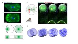
Figure 1: A. Chez le zygote d’oursins comme dans de nombreuses cellules, de larges astres de microtubules permettent de sentir la forme cellulaire et d’orienter le noyau et l’axe de division avec la forme de lacellule (Minc et al. 2011). B. Utilisation de pinces magnétiques pour appliquer des forces aux seins de cellules intactes et de contrôler la division cellulaire (Xie et al. 2021). C. Simulation numérique des forces exercées par les microtubules pour center le noyau en fonction de la forme cellulaire (Tanimoto et al. 2016). D. Simulation des géométries successives d’un embryon d’oursins (Pierre et al. 2016).


Video1

Légende : Embryons d’oursins se développant, dans lesquels l’ADN est marqué et suivi par fluorescence


2-      FORME ET CROISSANCE CELLULAIRE.

La morphologie des cellules individuelles ainsi que celles formant les tissus sous-tendent bon nombre de leurs fonctions. Nous cherchons à expliquer comment les cellules développent leur forme particulière et leur mode de croissance. Par exemple, de nombreuses cellules ne se développent qu’à un certain endroit, un processus appelé croissance polarisée, qui implique des réactions biochimiques complexes qui permettent de regrouper un domaine d’activité polarisé sous-cellulaire (Vidéo 2). Pour étudier ces aspects, nous utilisons des modèles de levures et de champignons, comme modèles de cellules très polarisées, qui grandissent rapidement et qui sont facilement manipulable génétiquement (Figure 2). Nous développons une recherche visant à comprendre comment les forces et les propriétés mécanique à la surface et au sein de la cellule interagissent avec des régulateurs biochimique pour  définir morphogénèse et croissance.

Figure 2: A. Images de fluorescence de levures exprimant des marqueurs pour localiser et étudier le cytosquelette, la polarité la croissance et la division cellulaire (Bonazzi et al. 2015). B. Images super-résolues de l’épaisseur de la paroi cellulaire autour d’un hyphe grandissant de champignon de l’espèce aspergillus (Chevallier et al).    C. Simulation numérique d’une spore de levure et de sa paroi (couleur) et polarité (blanc). D. Simulation de la vitesse de croissance.


Video2

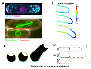
Croissance et division de levures exprimant des marqueurs fluorescents d’enveloppe nucléaire et de synthèse de paroi.

Responsable :

Nicolas MINC
Téléphone : +33 (0)157278052
Email : nicolas.minc (at) ijm.fr


Membres de l’équipe :

Serge DMITRIEFF Chercheur
Amir KHOSRAVANIZADEH Postdoctorant
Javad NAJAFI Postdoctorant
Yannis REIGNIER Assistant ingénieur
Charlotte MALLART Postdoctorante
Célia MUNICIO-DIAZ Ingénieure de recherche
Maria Isabel ARJONA HIDALGO Postdoctorante
Camille BOUCHER Postdoctorant
Aude NOMMICK Postdoctorante
Jeremy SALLÉ Chercheur
Laura RUIZ Ingénieur d’étude
Elyn MANSOUR ROBIC Master 2
Jiawei XU Master 2
  1. Xie J, Najafi J, Le Borgne R, Verbavatz J-M, Durieu C, Sallé J, and Minc N (2022) “Contribution of cytoplasm viscoelastic properties to mitotic spindle positioning” , Proc Natl Acad Sci U S A, 119 (8) e2115593119.
  2. Neeli-Venkata R, Municio Diaz C, Celador R, Sanchez Y, Minc N (2021) “Detection of surface forces by the cell-wall mechanosensor Wsc1 in yeast” , Developmental Cell 56, 1–15
  3. Palenzuela H, Lacroix B, Sallé J, Minami K, Shima T, Jegou A, Romet-Lemonne#G and Minc# N (2020) “In Vitro Reconstitution of Dynein Force Exertion in a Bulk Viscous Medium” , Current Biology, 30, 1–7
  4. Davi V, Chevalier L, Guo H, Tanimoto H, Barrett K, Couturier E, Boudaoud#A, and Minc# N, (2019) “Systematic mapping of cell wall mechanics in the regulation of cell morphogenesis” , Proc. Acad. Sci. USA, 116(28):13833-13838
  5. Sallé J, Xie J, Ershov D, Lacassin M, Dmitrieff S and Minc N, (2019) “Asymmetric division through a reduction of microtubule centering forces” J Cell Biol. 218(3):771-782 .
  6. Tanimoto H, Sallé J, Dodin L and Minc N (2018) “Physical forces determining the persistency and centring precision of microtubule asters” Nature Physics, 114, 848–854.
  7. Davì V, Tanimoto H, Ershov D, Haupt A, De Belly H, Le Borgne R, Couturier E, Boudaoud#A and Minc#, (2018) “Mechanosensation Dynamically Coordinates Polar Growth and Cell Wall Assembly to Promote Cell Survival” Developmental Cell, 45, 2, p170–182.
  8. Pierre A, Sallé J, Wühr M, Minc N., (2016) “Generic Theoretical Models to Predict Division Patterns of Cleaving Embryos.” Developmental Cell. 39(6):667-682
  9. Bonazzi* D, Julien* JD, Seddiki R, Romao M, Piel M, Boudaoud#A, Minc# N, (2014) “Symmetry breaking in spore germination relies on an interplay between polar cap stability and spore wall mechanics.” Developmental Cell, 28 (5):534-546
  10. Minc#, Burgess, D. and Chang F. (2011), “Influence of cell geometry on division plane positioning” Cell., 144 (3): 414-426.

Publications

2913254 EWC7IWVP items 1 0 date desc 8922 https://www.ijm.fr/wp-content/plugins/zotpress/
%7B%22status%22%3A%22success%22%2C%22updateneeded%22%3Afalse%2C%22instance%22%3A%22zotpress-a3c8fd4535f985b77e7fbf9c73816652%22%2C%22meta%22%3A%7B%22request_last%22%3A0%2C%22request_next%22%3A0%2C%22used_cache%22%3Atrue%7D%2C%22data%22%3A%5B%7B%22key%22%3A%22YNSD6AFM%22%2C%22library%22%3A%7B%22id%22%3A2913254%7D%2C%22meta%22%3A%7B%22creatorSummary%22%3A%22Mallart%20et%20al.%22%2C%22parsedDate%22%3A%222024-02-22%22%2C%22numChildren%22%3A1%7D%2C%22bib%22%3A%22%3Cdiv%20class%3D%5C%22csl-bib-body%5C%22%20style%3D%5C%22line-height%3A%202%3B%20padding-left%3A%201em%3B%20text-indent%3A-1em%3B%5C%22%3E%5Cn%20%20%3Cdiv%20class%3D%5C%22csl-entry%5C%22%3EMallart%2C%20C.%2C%20Netter%2C%20S.%2C%20Chalvet%2C%20F.%2C%20Claret%2C%20S.%2C%20Guichet%2C%20A.%2C%20Montagne%2C%20J.%2C%20Pret%2C%20A.-M.%2C%20%26amp%3B%20Malartre%2C%20M.%20%282024%29.%20JAK-STAT-dependent%20contact%20between%20follicle%20cells%20and%20the%20oocyte%20controls%20Drosophila%20anterior-posterior%20polarity%20and%20germline%20development.%20%3Ci%3ENature%20Communications%3C%5C%2Fi%3E%2C%20%3Ci%3E15%3C%5C%2Fi%3E%281%29%2C%201627.%20%3Ca%20href%3D%27https%3A%5C%2F%5C%2Fdoi.org%5C%2F10.1038%5C%2Fs41467-024-45963-z%27%3Ehttps%3A%5C%2F%5C%2Fdoi.org%5C%2F10.1038%5C%2Fs41467-024-45963-z%3C%5C%2Fa%3E%3C%5C%2Fdiv%3E%5Cn%3C%5C%2Fdiv%3E%22%2C%22data%22%3A%7B%22itemType%22%3A%22journalArticle%22%2C%22title%22%3A%22JAK-STAT-dependent%20contact%20between%20follicle%20cells%20and%20the%20oocyte%20controls%20Drosophila%20anterior-posterior%20polarity%20and%20germline%20development%22%2C%22creators%22%3A%5B%7B%22creatorType%22%3A%22author%22%2C%22firstName%22%3A%22Charlotte%22%2C%22lastName%22%3A%22Mallart%22%7D%2C%7B%22creatorType%22%3A%22author%22%2C%22firstName%22%3A%22Sophie%22%2C%22lastName%22%3A%22Netter%22%7D%2C%7B%22creatorType%22%3A%22author%22%2C%22firstName%22%3A%22Fabienne%22%2C%22lastName%22%3A%22Chalvet%22%7D%2C%7B%22creatorType%22%3A%22author%22%2C%22firstName%22%3A%22Sandra%22%2C%22lastName%22%3A%22Claret%22%7D%2C%7B%22creatorType%22%3A%22author%22%2C%22firstName%22%3A%22Antoine%22%2C%22lastName%22%3A%22Guichet%22%7D%2C%7B%22creatorType%22%3A%22author%22%2C%22firstName%22%3A%22Jacques%22%2C%22lastName%22%3A%22Montagne%22%7D%2C%7B%22creatorType%22%3A%22author%22%2C%22firstName%22%3A%22Anne-Marie%22%2C%22lastName%22%3A%22Pret%22%7D%2C%7B%22creatorType%22%3A%22author%22%2C%22firstName%22%3A%22Marianne%22%2C%22lastName%22%3A%22Malartre%22%7D%5D%2C%22abstractNote%22%3A%22The%20number%20of%20embryonic%20primordial%20germ%20cells%20in%20Drosophila%20is%20determined%20by%20the%20quantity%20of%20germ%20plasm%2C%20whose%20assembly%20starts%20in%20the%20posterior%20region%20of%20the%20oocyte%20during%20oogenesis.%20Here%2C%20we%20report%20that%20extending%20JAK-STAT%20activity%20in%20the%20posterior%20somatic%20follicular%20epithelium%20leads%20to%20an%20excess%20of%20primordial%20germ%20cells%20in%20the%20future%20embryo.%20We%20show%20that%20JAK-STAT%20signaling%20is%20necessary%20for%20the%20differentiation%20of%20approximately%2020%20specialized%20follicle%20cells%20maintaining%20tight%20contact%20with%20the%20oocyte.%20These%20cells%20define%2C%20in%20the%20underlying%20posterior%20oocyte%20cortex%2C%20the%20anchoring%20of%20the%20germ%20cell%20determinant%20oskar%20mRNA.%20We%20reveal%20that%20the%20apical%20surface%20of%20these%20posterior%20anchoring%20cells%20extends%20long%20filopodia%20penetrating%20the%20oocyte.%20We%20identify%20two%20JAK-STAT%20targets%20in%20these%20cells%20that%20are%20each%20sufficient%20to%20extend%20the%20zone%20of%20contact%20with%20the%20oocyte%2C%20thereby%20leading%20to%20production%20of%20extra%20primordial%20germ%20cells.%20JAK-STAT%20signaling%20thus%20determines%20a%20fixed%20number%20of%20posterior%20anchoring%20cells%20required%20for%20anterior-posterior%20oocyte%20polarity%20and%20for%20the%20development%20of%20the%20future%20germline.%22%2C%22date%22%3A%222024-02-22%22%2C%22language%22%3A%22en%22%2C%22DOI%22%3A%2210.1038%5C%2Fs41467-024-45963-z%22%2C%22ISSN%22%3A%222041-1723%22%2C%22url%22%3A%22https%3A%5C%2F%5C%2Fwww.nature.com%5C%2Farticles%5C%2Fs41467-024-45963-z%22%2C%22collections%22%3A%5B%22EWC7IWVP%22%2C%22UAHRRGCT%22%5D%2C%22dateModified%22%3A%222024-02-27T09%3A34%3A56Z%22%7D%7D%2C%7B%22key%22%3A%225ETEHECP%22%2C%22library%22%3A%7B%22id%22%3A2913254%7D%2C%22meta%22%3A%7B%22creatorSummary%22%3A%22Arjona%20et%20al.%22%2C%22parsedDate%22%3A%222023-12-01%22%2C%22numChildren%22%3A2%7D%2C%22bib%22%3A%22%3Cdiv%20class%3D%5C%22csl-bib-body%5C%22%20style%3D%5C%22line-height%3A%202%3B%20padding-left%3A%201em%3B%20text-indent%3A-1em%3B%5C%22%3E%5Cn%20%20%3Cdiv%20class%3D%5C%22csl-entry%5C%22%3EArjona%2C%20M.%20I.%2C%20Najafi%2C%20J.%2C%20%26amp%3B%20Minc%2C%20N.%20%282023%29.%20Cytoplasm%20mechanics%20and%20cellular%20organization.%20%3Ci%3ECurrent%20Opinion%20in%20Cell%20Biology%3C%5C%2Fi%3E%2C%20%3Ci%3E85%3C%5C%2Fi%3E%2C%20102278.%20%3Ca%20href%3D%27https%3A%5C%2F%5C%2Fdoi.org%5C%2F10.1016%5C%2Fj.ceb.2023.102278%27%3Ehttps%3A%5C%2F%5C%2Fdoi.org%5C%2F10.1016%5C%2Fj.ceb.2023.102278%3C%5C%2Fa%3E%3C%5C%2Fdiv%3E%5Cn%3C%5C%2Fdiv%3E%22%2C%22data%22%3A%7B%22itemType%22%3A%22journalArticle%22%2C%22title%22%3A%22Cytoplasm%20mechanics%20and%20cellular%20organization%22%2C%22creators%22%3A%5B%7B%22creatorType%22%3A%22author%22%2C%22firstName%22%3A%22Mar%5Cu00eda%20Isabel%22%2C%22lastName%22%3A%22Arjona%22%7D%2C%7B%22creatorType%22%3A%22author%22%2C%22firstName%22%3A%22Javad%22%2C%22lastName%22%3A%22Najafi%22%7D%2C%7B%22creatorType%22%3A%22author%22%2C%22firstName%22%3A%22Nicolas%22%2C%22lastName%22%3A%22Minc%22%7D%5D%2C%22abstractNote%22%3A%22As%20cells%20organize%20spatially%20or%20divide%2C%20they%20translocate%20many%20micron-scale%20organelles%20in%20their%20cytoplasm.%20These%20include%20endomembrane%20vesicles%2C%20nuclei%2C%20microtubule%20asters%2C%20mitotic%20spindles%2C%20or%20chromosomes.%20Organelle%20motion%20is%20powered%20by%20cytoskeleton%20forces%20but%20is%20opposed%20by%20viscoelastic%20forces%20imparted%20by%20the%20surrounding%20crowded%20cytoplasm%20medium.%20These%20resistive%20forces%20associated%20to%20cytoplasm%20physcial%20properties%20remain%20generally%20underappreciated%2C%20yet%20reach%20significant%20values%20to%20slow%20down%20organelle%20motion%20or%20even%20limit%20their%20displacement%20by%20springing%20them%20back%20towards%20their%20original%20position.%20The%20cytoplasm%20may%20also%20be%20itself%20organized%20in%20time%20and%20space%2C%20being%20for%20example%20stiffer%20or%20more%20fluid%20at%20certain%20locations%20or%20during%20particular%20cell%20cycle%20phases.%20Thus%2C%20cytoplasm%20mechanics%20may%20be%20viewed%20as%20a%20labile%20module%20that%20contributes%20to%20organize%20cells.%20We%20here%20review%20emerging%20methods%2C%20mechanisms%2C%20and%20concepts%20to%20study%20cytoplasm%20mechanical%20properties%20and%20their%20function%20in%20organelle%20positioning%2C%20cellular%20organization%20and%20division.%22%2C%22date%22%3A%222023-12-01%22%2C%22language%22%3A%22%22%2C%22DOI%22%3A%2210.1016%5C%2Fj.ceb.2023.102278%22%2C%22ISSN%22%3A%220955-0674%22%2C%22url%22%3A%22https%3A%5C%2F%5C%2Fwww.sciencedirect.com%5C%2Fscience%5C%2Farticle%5C%2Fpii%5C%2FS0955067423001278%22%2C%22collections%22%3A%5B%22EWC7IWVP%22%5D%2C%22dateModified%22%3A%222023-11-23T09%3A57%3A20Z%22%7D%7D%2C%7B%22key%22%3A%22P2SIXW2J%22%2C%22library%22%3A%7B%22id%22%3A2913254%7D%2C%22meta%22%3A%7B%22creatorSummary%22%3A%22Saleh%20et%20al.%22%2C%22parsedDate%22%3A%222023-07-06%22%2C%22numChildren%22%3A1%7D%2C%22bib%22%3A%22%3Cdiv%20class%3D%5C%22csl-bib-body%5C%22%20style%3D%5C%22line-height%3A%202%3B%20padding-left%3A%201em%3B%20text-indent%3A-1em%3B%5C%22%3E%5Cn%20%20%3Cdiv%20class%3D%5C%22csl-entry%5C%22%3ESaleh%2C%20J.%2C%20Fardin%2C%20M.-A.%2C%20Barai%2C%20A.%2C%20Soleilhac%2C%20M.%2C%20Frenoy%2C%20O.%2C%20Gaston%2C%20C.%2C%20Cui%2C%20H.%2C%20Dang%2C%20T.%2C%20Gaudin%2C%20N.%2C%20Vincent%2C%20A.%2C%20Minc%2C%20N.%2C%20%26amp%3B%20Delacour%2C%20D.%20%282023%29.%20Length%20limitation%20of%20astral%20microtubules%20orients%20cell%20divisions%20in%20murine%20intestinal%20crypts.%20%3Ci%3EDevelopmental%20Cell%3C%5C%2Fi%3E.%20%3Ca%20href%3D%27https%3A%5C%2F%5C%2Fdoi.org%5C%2F10.1016%5C%2Fj.devcel.2023.06.004%27%3Ehttps%3A%5C%2F%5C%2Fdoi.org%5C%2F10.1016%5C%2Fj.devcel.2023.06.004%3C%5C%2Fa%3E%3C%5C%2Fdiv%3E%5Cn%3C%5C%2Fdiv%3E%22%2C%22data%22%3A%7B%22itemType%22%3A%22journalArticle%22%2C%22title%22%3A%22Length%20limitation%20of%20astral%20microtubules%20orients%20cell%20divisions%20in%20murine%20intestinal%20crypts%22%2C%22creators%22%3A%5B%7B%22creatorType%22%3A%22author%22%2C%22firstName%22%3A%22Jad%22%2C%22lastName%22%3A%22Saleh%22%7D%2C%7B%22creatorType%22%3A%22author%22%2C%22firstName%22%3A%22Marc-Antoine%22%2C%22lastName%22%3A%22Fardin%22%7D%2C%7B%22creatorType%22%3A%22author%22%2C%22firstName%22%3A%22Amlan%22%2C%22lastName%22%3A%22Barai%22%7D%2C%7B%22creatorType%22%3A%22author%22%2C%22firstName%22%3A%22Matis%22%2C%22lastName%22%3A%22Soleilhac%22%7D%2C%7B%22creatorType%22%3A%22author%22%2C%22firstName%22%3A%22Olivia%22%2C%22lastName%22%3A%22Frenoy%22%7D%2C%7B%22creatorType%22%3A%22author%22%2C%22firstName%22%3A%22C%5Cu00e9cile%22%2C%22lastName%22%3A%22Gaston%22%7D%2C%7B%22creatorType%22%3A%22author%22%2C%22firstName%22%3A%22Hongyue%22%2C%22lastName%22%3A%22Cui%22%7D%2C%7B%22creatorType%22%3A%22author%22%2C%22firstName%22%3A%22Tien%22%2C%22lastName%22%3A%22Dang%22%7D%2C%7B%22creatorType%22%3A%22author%22%2C%22firstName%22%3A%22No%5Cu00e9mie%22%2C%22lastName%22%3A%22Gaudin%22%7D%2C%7B%22creatorType%22%3A%22author%22%2C%22firstName%22%3A%22Audrey%22%2C%22lastName%22%3A%22Vincent%22%7D%2C%7B%22creatorType%22%3A%22author%22%2C%22firstName%22%3A%22Nicolas%22%2C%22lastName%22%3A%22Minc%22%7D%2C%7B%22creatorType%22%3A%22author%22%2C%22firstName%22%3A%22Delphine%22%2C%22lastName%22%3A%22Delacour%22%7D%5D%2C%22abstractNote%22%3A%22Planar%20spindle%20orientation%20is%20critical%20for%20epithelial%20tissue%20organization%20and%20is%20generally%20instructed%20by%20the%20long%20cell-shape%20axis%20or%20cortical%20polarity%20domains.%20We%20introduced%20mouse%20intestinal%20organoids%20in%20order%20to%20study%20spindle%20orientation%20in%20a%20monolayered%20mammalian%20epithelium.%20Although%20spindles%20were%20planar%2C%20mitotic%20cells%20remained%20elongated%20along%20the%20apico-basal%20%28A-B%29%20axis%2C%20and%20polarity%20complexes%20were%20segregated%20to%20basal%20poles%2C%20so%20that%20spindles%20oriented%20in%20an%20unconventional%20manner%2C%20orthogonal%20to%20both%20polarity%20and%20geometric%20cues.%20Using%20high-resolution%203D%20imaging%2C%20simulations%2C%20and%20cell-shape%20and%20cytoskeleton%20manipulations%2C%20we%20show%20that%20planar%20divisions%20resulted%20from%20a%20length%20limitation%20in%20astral%20microtubules%20%28MTs%29%20which%20precludes%20them%20from%20interacting%20with%20basal%20polarity%2C%20and%20orient%20spindles%20from%20the%20local%20geometry%20of%20apical%20domains.%20Accordingly%2C%20lengthening%20MTs%20affected%20spindle%20planarity%2C%20cell%20positioning%2C%20and%20crypt%20arrangement.%20We%20conclude%20that%20MT%20length%20regulation%20may%20serve%20as%20a%20key%20mechanism%20for%20spindles%20to%20sense%20local%20cell%20shapes%20and%20tissue%20forces%20to%20preserve%20mammalian%20epithelial%20architecture.%22%2C%22date%22%3A%222023-07-06%22%2C%22language%22%3A%22en%22%2C%22DOI%22%3A%2210.1016%5C%2Fj.devcel.2023.06.004%22%2C%22ISSN%22%3A%221534-5807%22%2C%22url%22%3A%22https%3A%5C%2F%5C%2Fwww.sciencedirect.com%5C%2Fscience%5C%2Farticle%5C%2Fpii%5C%2FS1534580723003027%22%2C%22collections%22%3A%5B%22EWC7IWVP%22%5D%2C%22dateModified%22%3A%222023-07-10T13%3A25%3A49Z%22%7D%7D%2C%7B%22key%22%3A%226GG9HRIN%22%2C%22library%22%3A%7B%22id%22%3A2913254%7D%2C%22meta%22%3A%7B%22creatorSummary%22%3A%22Guevara-Garcia%20et%20al.%22%2C%22parsedDate%22%3A%222023-01-24%22%2C%22numChildren%22%3A2%7D%2C%22bib%22%3A%22%3Cdiv%20class%3D%5C%22csl-bib-body%5C%22%20style%3D%5C%22line-height%3A%202%3B%20padding-left%3A%201em%3B%20text-indent%3A-1em%3B%5C%22%3E%5Cn%20%20%3Cdiv%20class%3D%5C%22csl-entry%5C%22%3EGuevara-Garcia%2C%20A.%2C%20Soleilhac%2C%20M.%2C%20Minc%2C%20N.%2C%20%26amp%3B%20Delacour%2C%20D.%20%282023%29.%20Regulation%20and%20functions%20of%20cell%20division%20in%20the%20intestinal%20tissue.%20%3Ci%3ESeminars%20in%20Cell%20%26amp%3B%20Developmental%20Biology%3C%5C%2Fi%3E%2C%20S1084-9521%2823%2900004-6.%20%3Ca%20href%3D%27https%3A%5C%2F%5C%2Fdoi.org%5C%2F10.1016%5C%2Fj.semcdb.2023.01.004%27%3Ehttps%3A%5C%2F%5C%2Fdoi.org%5C%2F10.1016%5C%2Fj.semcdb.2023.01.004%3C%5C%2Fa%3E%3C%5C%2Fdiv%3E%5Cn%3C%5C%2Fdiv%3E%22%2C%22data%22%3A%7B%22itemType%22%3A%22journalArticle%22%2C%22title%22%3A%22Regulation%20and%20functions%20of%20cell%20division%20in%20the%20intestinal%20tissue%22%2C%22creators%22%3A%5B%7B%22creatorType%22%3A%22author%22%2C%22firstName%22%3A%22Amaris%22%2C%22lastName%22%3A%22Guevara-Garcia%22%7D%2C%7B%22creatorType%22%3A%22author%22%2C%22firstName%22%3A%22Matis%22%2C%22lastName%22%3A%22Soleilhac%22%7D%2C%7B%22creatorType%22%3A%22author%22%2C%22firstName%22%3A%22Nicolas%22%2C%22lastName%22%3A%22Minc%22%7D%2C%7B%22creatorType%22%3A%22author%22%2C%22firstName%22%3A%22Delphine%22%2C%22lastName%22%3A%22Delacour%22%7D%5D%2C%22abstractNote%22%3A%22In%20multicellular%20organisms%2C%20epithelial%20cells%20are%20key%20elements%20of%20tissue%20organization.%20In%20developing%20epithelial%20tissues%2C%20cellular%20proliferation%20and%20differentiation%20are%20under%20the%20tight%20regulation%20of%20morphogenetic%20programs%20to%20ensure%20correct%20organ%20formation%20and%20functioning.%20In%20these%20processes%2C%20proliferation%20rates%20and%20division%20orientation%20regulate%20the%20speed%2C%20timing%20and%20direction%20of%20tissue%20expansion%20but%20also%20its%20proper%20patterning.%20Moreover%2C%20tissue%20homeostasis%20relies%20on%20spatio-temporal%20modulations%20of%20daughter%20cell%20behavior%20and%20arrangement.%20These%20aspects%20are%20particularly%20crucial%20in%20the%20intestine%2C%20which%20is%20one%20of%20the%20most%20proliferative%20tissues%20in%20adults%2C%20making%20it%20a%20very%20attractive%20adult%20organ%20system%20to%20study%20the%20role%20of%20cell%20division%20on%20epithelial%20morphogenesis%20and%20organ%20function.%20Although%20epithelial%20cell%20division%20has%20been%20the%20subject%20of%20intense%20research%20for%20many%20years%20in%20multiple%20models%2C%20it%20still%20remains%20in%20its%20infancy%20in%20the%20context%20of%20the%20intestinal%20tissue.%20In%20this%20review%2C%20we%20focus%20on%20the%20current%20knowledge%20on%20cell%20division%20and%20regulatory%20mechanisms%20at%20play%20in%20the%20intestinal%20epithelial%20tissue%2C%20as%20well%20as%20their%20importance%20in%20developmental%20biology%20and%20physiopathology.%22%2C%22date%22%3A%222023-01-24%22%2C%22language%22%3A%22eng%22%2C%22DOI%22%3A%2210.1016%5C%2Fj.semcdb.2023.01.004%22%2C%22ISSN%22%3A%221096-3634%22%2C%22url%22%3A%22%22%2C%22collections%22%3A%5B%22EWC7IWVP%22%5D%2C%22dateModified%22%3A%222023-05-03T12%3A50%3A45Z%22%7D%7D%2C%7B%22key%22%3A%22G6QW74PY%22%2C%22library%22%3A%7B%22id%22%3A2913254%7D%2C%22meta%22%3A%7B%22lastModifiedByUser%22%3A%7B%22id%22%3A11274337%2C%22username%22%3A%22Charlotte_Brancaz%22%2C%22name%22%3A%22%22%2C%22links%22%3A%7B%22alternate%22%3A%7B%22href%22%3A%22https%3A%5C%2F%5C%2Fwww.zotero.org%5C%2Fcharlotte_brancaz%22%2C%22type%22%3A%22text%5C%2Fhtml%22%7D%7D%7D%2C%22creatorSummary%22%3A%22Jasnin%20et%20al.%22%2C%22parsedDate%22%3A%222022-07-04%22%2C%22numChildren%22%3A3%7D%2C%22bib%22%3A%22%3Cdiv%20class%3D%5C%22csl-bib-body%5C%22%20style%3D%5C%22line-height%3A%202%3B%20padding-left%3A%201em%3B%20text-indent%3A-1em%3B%5C%22%3E%5Cn%20%20%3Cdiv%20class%3D%5C%22csl-entry%5C%22%3EJasnin%2C%20M.%2C%20Hervy%2C%20J.%2C%20Balor%2C%20S.%2C%20Bouissou%2C%20A.%2C%20Proag%2C%20A.%2C%20Voituriez%2C%20R.%2C%20Schneider%2C%20J.%2C%20Mangeat%2C%20T.%2C%20Maridonneau-Parini%2C%20I.%2C%20Baumeister%2C%20W.%2C%20Dmitrieff%2C%20S.%2C%20%26amp%3B%20Poincloux%2C%20R.%20%282022%29.%20Elasticity%20of%20podosome%20actin%20networks%20produces%20nanonewton%20protrusive%20forces.%20%3Ci%3ENature%20Communications%3C%5C%2Fi%3E%2C%20%3Ci%3E13%3C%5C%2Fi%3E%281%29%2C%203842.%20%3Ca%20href%3D%27https%3A%5C%2F%5C%2Fdoi.org%5C%2F10.1038%5C%2Fs41467-022-30652-6%27%3Ehttps%3A%5C%2F%5C%2Fdoi.org%5C%2F10.1038%5C%2Fs41467-022-30652-6%3C%5C%2Fa%3E%3C%5C%2Fdiv%3E%5Cn%3C%5C%2Fdiv%3E%22%2C%22data%22%3A%7B%22itemType%22%3A%22journalArticle%22%2C%22title%22%3A%22Elasticity%20of%20podosome%20actin%20networks%20produces%20nanonewton%20protrusive%20forces%22%2C%22creators%22%3A%5B%7B%22creatorType%22%3A%22author%22%2C%22firstName%22%3A%22Marion%22%2C%22lastName%22%3A%22Jasnin%22%7D%2C%7B%22creatorType%22%3A%22author%22%2C%22firstName%22%3A%22Jordan%22%2C%22lastName%22%3A%22Hervy%22%7D%2C%7B%22creatorType%22%3A%22author%22%2C%22firstName%22%3A%22St%5Cu00e9phanie%22%2C%22lastName%22%3A%22Balor%22%7D%2C%7B%22creatorType%22%3A%22author%22%2C%22firstName%22%3A%22Ana%5Cu00efs%22%2C%22lastName%22%3A%22Bouissou%22%7D%2C%7B%22creatorType%22%3A%22author%22%2C%22firstName%22%3A%22Amsha%22%2C%22lastName%22%3A%22Proag%22%7D%2C%7B%22creatorType%22%3A%22author%22%2C%22firstName%22%3A%22Rapha%5Cu00ebl%22%2C%22lastName%22%3A%22Voituriez%22%7D%2C%7B%22creatorType%22%3A%22author%22%2C%22firstName%22%3A%22Jonathan%22%2C%22lastName%22%3A%22Schneider%22%7D%2C%7B%22creatorType%22%3A%22author%22%2C%22firstName%22%3A%22Thomas%22%2C%22lastName%22%3A%22Mangeat%22%7D%2C%7B%22creatorType%22%3A%22author%22%2C%22firstName%22%3A%22Isabelle%22%2C%22lastName%22%3A%22Maridonneau-Parini%22%7D%2C%7B%22creatorType%22%3A%22author%22%2C%22firstName%22%3A%22Wolfgang%22%2C%22lastName%22%3A%22Baumeister%22%7D%2C%7B%22creatorType%22%3A%22author%22%2C%22firstName%22%3A%22Serge%22%2C%22lastName%22%3A%22Dmitrieff%22%7D%2C%7B%22creatorType%22%3A%22author%22%2C%22firstName%22%3A%22Renaud%22%2C%22lastName%22%3A%22Poincloux%22%7D%5D%2C%22abstractNote%22%3A%22Actin%20filaments%20assemble%20into%20force-generating%20systems%20involved%20in%20diverse%20cellular%20functions%2C%20including%20cell%20motility%2C%20adhesion%2C%20contractility%20and%20division.%20It%20remains%20unclear%20how%20networks%20of%20actin%20filaments%2C%20which%20individually%20generate%20piconewton%20forces%2C%20can%20produce%20forces%20reaching%20tens%20of%20nanonewtons.%20Here%20we%20use%20in%20situ%20cryo-electron%20tomography%20to%20unveil%20how%20the%20nanoscale%20architecture%20of%20macrophage%20podosomes%20enables%20basal%20membrane%20protrusion.%20We%20show%20that%20the%20sum%20of%20the%20actin%20polymerization%20forces%20at%20the%20membrane%20is%20not%20sufficient%20to%20explain%20podosome%20protrusive%20forces.%20Quantitative%20analysis%20of%20podosome%20organization%20demonstrates%20that%20the%20core%20is%20composed%20of%20a%20dense%20network%20of%20bent%20actin%20filaments%20storing%20elastic%20energy.%20Theoretical%20modelling%20of%20the%20network%20as%20a%20spring-loaded%20elastic%20material%20reveals%20that%20it%20exerts%20forces%20of%20a%20few%20tens%20of%20nanonewtons%2C%20in%20a%20range%20similar%20to%20that%20evaluated%20experimentally.%20Thus%2C%20taking%20into%20account%20not%20only%20the%20interface%20with%20the%20membrane%20but%20also%20the%20bulk%20of%20the%20network%2C%20is%20crucial%20to%20understand%20force%20generation%20by%20actin%20machineries.%20Our%20integrative%20approach%20sheds%20light%20on%20the%20elastic%20behavior%20of%20dense%20actin%20networks%20and%20opens%20new%20avenues%20to%20understand%20force%20production%20inside%20cells.%22%2C%22date%22%3A%222022-07-04%22%2C%22language%22%3A%22eng%22%2C%22DOI%22%3A%2210.1038%5C%2Fs41467-022-30652-6%22%2C%22ISSN%22%3A%222041-1723%22%2C%22url%22%3A%22%22%2C%22collections%22%3A%5B%22EWC7IWVP%22%5D%2C%22dateModified%22%3A%222022-09-05T13%3A25%3A09Z%22%7D%7D%2C%7B%22key%22%3A%2255V5SURA%22%2C%22library%22%3A%7B%22id%22%3A2913254%7D%2C%22meta%22%3A%7B%22lastModifiedByUser%22%3A%7B%22id%22%3A11274337%2C%22username%22%3A%22Charlotte_Brancaz%22%2C%22name%22%3A%22%22%2C%22links%22%3A%7B%22alternate%22%3A%7B%22href%22%3A%22https%3A%5C%2F%5C%2Fwww.zotero.org%5C%2Fcharlotte_brancaz%22%2C%22type%22%3A%22text%5C%2Fhtml%22%7D%7D%7D%2C%22creatorSummary%22%3A%22Letort%20et%20al.%22%2C%22parsedDate%22%3A%222022-07-01%22%2C%22numChildren%22%3A3%7D%2C%22bib%22%3A%22%3Cdiv%20class%3D%5C%22csl-bib-body%5C%22%20style%3D%5C%22line-height%3A%202%3B%20padding-left%3A%201em%3B%20text-indent%3A-1em%3B%5C%22%3E%5Cn%20%20%3Cdiv%20class%3D%5C%22csl-entry%5C%22%3ELetort%2C%20G.%2C%20Eichmuller%2C%20A.%2C%20Da%20Silva%2C%20C.%2C%20Nikalayevich%2C%20E.%2C%20Crozet%2C%20F.%2C%20Salle%2C%20J.%2C%20Minc%2C%20N.%2C%20Labrune%2C%20E.%2C%20Wolf%2C%20J.-P.%2C%20Terret%2C%20M.-E.%2C%20%26amp%3B%20Verlhac%2C%20M.-H.%20%282022%29.%20An%20interpretable%20and%20versatile%20machine%20learning%20approach%20for%20oocyte%20phenotyping.%20%3Ci%3EJournal%20of%20Cell%20Science%3C%5C%2Fi%3E%2C%20%3Ci%3E135%3C%5C%2Fi%3E%2813%29%2C%20jcs260281.%20%3Ca%20href%3D%27https%3A%5C%2F%5C%2Fdoi.org%5C%2F10.1242%5C%2Fjcs.260281%27%3Ehttps%3A%5C%2F%5C%2Fdoi.org%5C%2F10.1242%5C%2Fjcs.260281%3C%5C%2Fa%3E%3C%5C%2Fdiv%3E%5Cn%3C%5C%2Fdiv%3E%22%2C%22data%22%3A%7B%22itemType%22%3A%22journalArticle%22%2C%22title%22%3A%22An%20interpretable%20and%20versatile%20machine%20learning%20approach%20for%20oocyte%20phenotyping%22%2C%22creators%22%3A%5B%7B%22creatorType%22%3A%22author%22%2C%22firstName%22%3A%22Gaelle%22%2C%22lastName%22%3A%22Letort%22%7D%2C%7B%22creatorType%22%3A%22author%22%2C%22firstName%22%3A%22Adrien%22%2C%22lastName%22%3A%22Eichmuller%22%7D%2C%7B%22creatorType%22%3A%22author%22%2C%22firstName%22%3A%22Christelle%22%2C%22lastName%22%3A%22Da%20Silva%22%7D%2C%7B%22creatorType%22%3A%22author%22%2C%22firstName%22%3A%22Elvira%22%2C%22lastName%22%3A%22Nikalayevich%22%7D%2C%7B%22creatorType%22%3A%22author%22%2C%22firstName%22%3A%22Flora%22%2C%22lastName%22%3A%22Crozet%22%7D%2C%7B%22creatorType%22%3A%22author%22%2C%22firstName%22%3A%22Jeremy%22%2C%22lastName%22%3A%22Salle%22%7D%2C%7B%22creatorType%22%3A%22author%22%2C%22firstName%22%3A%22Nicolas%22%2C%22lastName%22%3A%22Minc%22%7D%2C%7B%22creatorType%22%3A%22author%22%2C%22firstName%22%3A%22Elsa%22%2C%22lastName%22%3A%22Labrune%22%7D%2C%7B%22creatorType%22%3A%22author%22%2C%22firstName%22%3A%22Jean-Philippe%22%2C%22lastName%22%3A%22Wolf%22%7D%2C%7B%22creatorType%22%3A%22author%22%2C%22firstName%22%3A%22Marie-Emilie%22%2C%22lastName%22%3A%22Terret%22%7D%2C%7B%22creatorType%22%3A%22author%22%2C%22firstName%22%3A%22Marie-H%5Cu00e9l%5Cu00e8ne%22%2C%22lastName%22%3A%22Verlhac%22%7D%5D%2C%22abstractNote%22%3A%22Meiotic%20maturation%20is%20a%20crucial%20step%20of%20oocyte%20formation%2C%20allowing%20its%20potential%20fertilization%20and%20embryo%20development.%20Elucidating%20this%20process%20is%20important%20for%20both%20fundamental%20research%20and%20assisted%20reproductive%20technology.%20However%2C%20few%20computational%20tools%20based%20on%20non-invasive%20measurements%20are%20available%20to%20characterize%20oocyte%20meiotic%20maturation.%20Here%2C%20we%20develop%20a%20computational%20framework%20to%20phenotype%20oocytes%20based%20on%20images%20acquired%20in%20transmitted%20light.%20We%20trained%20neural%20networks%20to%20segment%20the%20contour%20of%20oocytes%20and%20their%20zona%20pellucida%20using%20oocytes%20from%20diverse%20species.%20We%20defined%20a%20comprehensive%20set%20of%20morphological%20features%20to%20describe%20an%20oocyte.%20These%20steps%20were%20implemented%20in%20an%20open-source%20Fiji%20plugin.%20We%20present%20a%20feature-based%20machine%20learning%20pipeline%20to%20recognize%20oocyte%20populations%20and%20determine%20morphological%20differences%20between%20them.%20We%20first%20demonstrate%20its%20potential%20to%20screen%20oocytes%20from%20different%20strains%20and%20automatically%20identify%20their%20morphological%20characteristics.%20Its%20second%20application%20is%20to%20predict%20and%20characterize%20the%20maturation%20potential%20of%20oocytes.%20We%20identify%20the%20texture%20of%20the%20zona%20pellucida%20and%20cytoplasmic%20particle%20size%20as%20features%20to%20assess%20mouse%20oocyte%20maturation%20potential%20and%20tested%20whether%20these%20features%20were%20applicable%20to%20the%20developmental%20potential%20of%20human%20oocytes.%20This%20article%20has%20an%20associated%20First%20Person%20interview%20with%20the%20first%20author%20of%20the%20paper.%22%2C%22date%22%3A%222022-07-01%22%2C%22language%22%3A%22eng%22%2C%22DOI%22%3A%2210.1242%5C%2Fjcs.260281%22%2C%22ISSN%22%3A%221477-9137%22%2C%22url%22%3A%22%22%2C%22collections%22%3A%5B%22EWC7IWVP%22%5D%2C%22dateModified%22%3A%222022-09-05T13%3A25%3A09Z%22%7D%7D%2C%7B%22key%22%3A%22U4LMMFV6%22%2C%22library%22%3A%7B%22id%22%3A2913254%7D%2C%22meta%22%3A%7B%22lastModifiedByUser%22%3A%7B%22id%22%3A11274337%2C%22username%22%3A%22Charlotte_Brancaz%22%2C%22name%22%3A%22%22%2C%22links%22%3A%7B%22alternate%22%3A%7B%22href%22%3A%22https%3A%5C%2F%5C%2Fwww.zotero.org%5C%2Fcharlotte_brancaz%22%2C%22type%22%3A%22text%5C%2Fhtml%22%7D%7D%7D%2C%22creatorSummary%22%3A%22Darnat%20et%20al.%22%2C%22parsedDate%22%3A%222022-05-17%22%2C%22numChildren%22%3A3%7D%2C%22bib%22%3A%22%3Cdiv%20class%3D%5C%22csl-bib-body%5C%22%20style%3D%5C%22line-height%3A%202%3B%20padding-left%3A%201em%3B%20text-indent%3A-1em%3B%5C%22%3E%5Cn%20%20%3Cdiv%20class%3D%5C%22csl-entry%5C%22%3EDarnat%2C%20P.%2C%20Burg%2C%20A.%2C%20Sall%26%23xE9%3B%2C%20J.%2C%20Lacoste%2C%20J.%2C%20Louvet-Vall%26%23xE9%3Be%2C%20S.%2C%20Gho%2C%20M.%2C%20%26amp%3B%20Audibert%2C%20A.%20%282022%29.%20Cortical%20Cyclin%20A%20controls%20spindle%20orientation%20during%20asymmetric%20cell%20divisions%20in%20Drosophila.%20%3Ci%3ENature%20Communications%3C%5C%2Fi%3E%2C%20%3Ci%3E13%3C%5C%2Fi%3E%281%29%2C%202723.%20%3Ca%20href%3D%27https%3A%5C%2F%5C%2Fdoi.org%5C%2F10.1038%5C%2Fs41467-022-30182-1%27%3Ehttps%3A%5C%2F%5C%2Fdoi.org%5C%2F10.1038%5C%2Fs41467-022-30182-1%3C%5C%2Fa%3E%3C%5C%2Fdiv%3E%5Cn%3C%5C%2Fdiv%3E%22%2C%22data%22%3A%7B%22itemType%22%3A%22journalArticle%22%2C%22title%22%3A%22Cortical%20Cyclin%20A%20controls%20spindle%20orientation%20during%20asymmetric%20cell%20divisions%20in%20Drosophila%22%2C%22creators%22%3A%5B%7B%22creatorType%22%3A%22author%22%2C%22firstName%22%3A%22P%5Cu00e9n%5Cu00e9lope%22%2C%22lastName%22%3A%22Darnat%22%7D%2C%7B%22creatorType%22%3A%22author%22%2C%22firstName%22%3A%22Ang%5Cu00e9lique%22%2C%22lastName%22%3A%22Burg%22%7D%2C%7B%22creatorType%22%3A%22author%22%2C%22firstName%22%3A%22J%5Cu00e9r%5Cu00e9my%22%2C%22lastName%22%3A%22Sall%5Cu00e9%22%7D%2C%7B%22creatorType%22%3A%22author%22%2C%22firstName%22%3A%22J%5Cu00e9r%5Cu00f4me%22%2C%22lastName%22%3A%22Lacoste%22%7D%2C%7B%22creatorType%22%3A%22author%22%2C%22firstName%22%3A%22Sophie%22%2C%22lastName%22%3A%22Louvet-Vall%5Cu00e9e%22%7D%2C%7B%22creatorType%22%3A%22author%22%2C%22firstName%22%3A%22Michel%22%2C%22lastName%22%3A%22Gho%22%7D%2C%7B%22creatorType%22%3A%22author%22%2C%22firstName%22%3A%22Agn%5Cu00e8s%22%2C%22lastName%22%3A%22Audibert%22%7D%5D%2C%22abstractNote%22%3A%22The%20coordination%20between%20cell%20proliferation%20and%20cell%20polarity%20is%20crucial%20to%20orient%20the%20asymmetric%20cell%20divisions%20to%20generate%20cell%20diversity%20in%20epithelia.%20In%20many%20instances%2C%20the%20Frizzled%5C%2FDishevelled%20planar%20cell%20polarity%20pathway%20is%20involved%20in%20mitotic%20spindle%20orientation%2C%20but%20how%20this%20is%20spatially%20and%20temporally%20coordinated%20with%20cell%20cycle%20progression%20has%20remained%20elusive.%20Using%20Drosophila%20sensory%20organ%20precursor%20cells%20as%20a%20model%20system%2C%20we%20show%20that%20Cyclin%20A%2C%20the%20main%20Cyclin%20driving%20the%20transition%20to%20M-phase%20of%20the%20cell%20cycle%2C%20is%20recruited%20to%20the%20apical-posterior%20cortex%20in%20prophase%20by%20the%20Frizzled%5C%2FDishevelled%20complex.%20This%20cortically%20localized%20Cyclin%20A%20then%20regulates%20the%20orientation%20of%20the%20division%20by%20recruiting%20Mud%2C%20a%20homologue%20of%20NuMA%2C%20the%20well-known%20spindle-associated%20protein.%20The%20observed%20non-canonical%20subcellular%20localization%20of%20Cyclin%20A%20reveals%20this%20mitotic%20factor%20as%20a%20direct%20link%20between%20cell%20proliferation%2C%20cell%20polarity%20and%20spindle%20orientation.%22%2C%22date%22%3A%222022-05-17%22%2C%22language%22%3A%22eng%22%2C%22DOI%22%3A%2210.1038%5C%2Fs41467-022-30182-1%22%2C%22ISSN%22%3A%222041-1723%22%2C%22url%22%3A%22%22%2C%22collections%22%3A%5B%22EWC7IWVP%22%5D%2C%22dateModified%22%3A%222022-09-05T13%3A25%3A09Z%22%7D%7D%2C%7B%22key%22%3A%22D5T75ZNL%22%2C%22library%22%3A%7B%22id%22%3A2913254%7D%2C%22meta%22%3A%7B%22creatorSummary%22%3A%22Xie%20et%20al.%22%2C%22parsedDate%22%3A%222022-02-22%22%2C%22numChildren%22%3A2%7D%2C%22bib%22%3A%22%3Cdiv%20class%3D%5C%22csl-bib-body%5C%22%20style%3D%5C%22line-height%3A%202%3B%20padding-left%3A%201em%3B%20text-indent%3A-1em%3B%5C%22%3E%5Cn%20%20%3Cdiv%20class%3D%5C%22csl-entry%5C%22%3EXie%2C%20J.%2C%20Najafi%2C%20J.%2C%20Le%20Borgne%2C%20R.%2C%20Verbavatz%2C%20J.-M.%2C%20Durieu%2C%20C.%2C%20Sall%26%23xE9%3B%2C%20J.%2C%20%26amp%3B%20Minc%2C%20N.%20%282022%29.%20Contribution%20of%20cytoplasm%20viscoelastic%20properties%20to%20mitotic%20spindle%20positioning.%20%3Ci%3EProceedings%20of%20the%20National%20Academy%20of%20Sciences%20of%20the%20United%20States%20of%20America%3C%5C%2Fi%3E%2C%20%3Ci%3E119%3C%5C%2Fi%3E%288%29%2C%20e2115593119.%20%3Ca%20href%3D%27https%3A%5C%2F%5C%2Fdoi.org%5C%2F10.1073%5C%2Fpnas.2115593119%27%3Ehttps%3A%5C%2F%5C%2Fdoi.org%5C%2F10.1073%5C%2Fpnas.2115593119%3C%5C%2Fa%3E%3C%5C%2Fdiv%3E%5Cn%3C%5C%2Fdiv%3E%22%2C%22data%22%3A%7B%22itemType%22%3A%22journalArticle%22%2C%22title%22%3A%22Contribution%20of%20cytoplasm%20viscoelastic%20properties%20to%20mitotic%20spindle%20positioning%22%2C%22creators%22%3A%5B%7B%22creatorType%22%3A%22author%22%2C%22firstName%22%3A%22Jing%22%2C%22lastName%22%3A%22Xie%22%7D%2C%7B%22creatorType%22%3A%22author%22%2C%22firstName%22%3A%22Javad%22%2C%22lastName%22%3A%22Najafi%22%7D%2C%7B%22creatorType%22%3A%22author%22%2C%22firstName%22%3A%22R%5Cu00e9mi%22%2C%22lastName%22%3A%22Le%20Borgne%22%7D%2C%7B%22creatorType%22%3A%22author%22%2C%22firstName%22%3A%22Jean-Marc%22%2C%22lastName%22%3A%22Verbavatz%22%7D%2C%7B%22creatorType%22%3A%22author%22%2C%22firstName%22%3A%22Catherine%22%2C%22lastName%22%3A%22Durieu%22%7D%2C%7B%22creatorType%22%3A%22author%22%2C%22firstName%22%3A%22Jeremy%22%2C%22lastName%22%3A%22Sall%5Cu00e9%22%7D%2C%7B%22creatorType%22%3A%22author%22%2C%22firstName%22%3A%22Nicolas%22%2C%22lastName%22%3A%22Minc%22%7D%5D%2C%22abstractNote%22%3A%22Cells%20are%20filled%20with%20macromolecules%20and%20polymer%20networks%20that%20set%20scale-dependent%20viscous%20and%20elastic%20properties%20to%20the%20cytoplasm.%20Although%20the%20role%20of%20these%20parameters%20in%20molecular%20diffusion%2C%20reaction%20kinetics%2C%20and%20cellular%20biochemistry%20is%20being%20increasingly%20recognized%2C%20their%20contributions%20to%20the%20motion%20and%20positioning%20of%20larger%20organelles%2C%20such%20as%20mitotic%20spindles%20for%20cell%20division%2C%20remain%20unknown.%20Here%2C%20using%20magnetic%20tweezers%20to%20displace%20and%20rotate%20mitotic%20spindles%20in%20living%20embryos%2C%20we%20uncovered%20that%20the%20cytoplasm%20can%20impart%20viscoelastic%20reactive%20forces%20that%20move%20spindles%2C%20or%20passive%20objects%20with%20similar%20size%2C%20back%20to%20their%20original%20positions.%20These%20forces%20are%20independent%20of%20cytoskeletal%20force%20generators%20yet%20reach%20hundreds%20of%20piconewtons%20and%20scale%20with%20cytoplasm%20crowding.%20Spindle%20motion%20shears%20and%20fluidizes%20the%20cytoplasm%2C%20dissipating%20elastic%20energy%20and%20limiting%20spindle%20recoils%20with%20functional%20implications%20for%20asymmetric%20and%20oriented%20divisions.%20These%20findings%20suggest%20that%20bulk%20cytoplasm%20material%20properties%20may%20constitute%20important%20control%20elements%20for%20the%20regulation%20of%20division%20positioning%20and%20cellular%20organization.%22%2C%22date%22%3A%222022-02-22%22%2C%22language%22%3A%22eng%22%2C%22DOI%22%3A%2210.1073%5C%2Fpnas.2115593119%22%2C%22ISSN%22%3A%221091-6490%22%2C%22url%22%3A%22%22%2C%22collections%22%3A%5B%22EWC7IWVP%22%5D%2C%22dateModified%22%3A%222023-05-02T12%3A26%3A31Z%22%7D%7D%2C%7B%22key%22%3A%22682HLBIB%22%2C%22library%22%3A%7B%22id%22%3A2913254%7D%2C%22meta%22%3A%7B%22lastModifiedByUser%22%3A%7B%22id%22%3A11274337%2C%22username%22%3A%22Charlotte_Brancaz%22%2C%22name%22%3A%22%22%2C%22links%22%3A%7B%22alternate%22%3A%7B%22href%22%3A%22https%3A%5C%2F%5C%2Fwww.zotero.org%5C%2Fcharlotte_brancaz%22%2C%22type%22%3A%22text%5C%2Fhtml%22%7D%7D%7D%2C%22creatorSummary%22%3A%22Neeli-Venkata%20et%20al.%22%2C%22parsedDate%22%3A%222021-10-25%22%2C%22numChildren%22%3A2%7D%2C%22bib%22%3A%22%3Cdiv%20class%3D%5C%22csl-bib-body%5C%22%20style%3D%5C%22line-height%3A%202%3B%20padding-left%3A%201em%3B%20text-indent%3A-1em%3B%5C%22%3E%5Cn%20%20%3Cdiv%20class%3D%5C%22csl-entry%5C%22%3ENeeli-Venkata%2C%20R.%2C%20Diaz%2C%20C.%20M.%2C%20Celador%2C%20R.%2C%20Sanchez%2C%20Y.%2C%20%26amp%3B%20Minc%2C%20N.%20%282021%29.%20Detection%20of%20surface%20forces%20by%20the%20cell-wall%20mechanosensor%20Wsc1%20in%20yeast.%20%3Ci%3EDevelopmental%20Cell%3C%5C%2Fi%3E%2C%20%3Ci%3E56%3C%5C%2Fi%3E%2820%29%2C%202856-2870.e7.%20%3Ca%20href%3D%27https%3A%5C%2F%5C%2Fdoi.org%5C%2F10.1016%5C%2Fj.devcel.2021.09.024%27%3Ehttps%3A%5C%2F%5C%2Fdoi.org%5C%2F10.1016%5C%2Fj.devcel.2021.09.024%3C%5C%2Fa%3E%3C%5C%2Fdiv%3E%5Cn%3C%5C%2Fdiv%3E%22%2C%22data%22%3A%7B%22itemType%22%3A%22journalArticle%22%2C%22title%22%3A%22Detection%20of%20surface%20forces%20by%20the%20cell-wall%20mechanosensor%20Wsc1%20in%20yeast%22%2C%22creators%22%3A%5B%7B%22creatorType%22%3A%22author%22%2C%22firstName%22%3A%22Ramakanth%22%2C%22lastName%22%3A%22Neeli-Venkata%22%7D%2C%7B%22creatorType%22%3A%22author%22%2C%22firstName%22%3A%22Celia%20Municio%22%2C%22lastName%22%3A%22Diaz%22%7D%2C%7B%22creatorType%22%3A%22author%22%2C%22firstName%22%3A%22Ruben%22%2C%22lastName%22%3A%22Celador%22%7D%2C%7B%22creatorType%22%3A%22author%22%2C%22firstName%22%3A%22Yolanda%22%2C%22lastName%22%3A%22Sanchez%22%7D%2C%7B%22creatorType%22%3A%22author%22%2C%22firstName%22%3A%22Nicolas%22%2C%22lastName%22%3A%22Minc%22%7D%5D%2C%22abstractNote%22%3A%22Surface%20receptors%20of%20animal%20cells%2C%20such%20as%20integrins%2C%20promote%20mechanosensation%20by%20forming%20clusters%20as%20signaling%20hubs%20that%20transduce%20tensile%20forces.%20Walled%20cells%20of%20plants%20and%20fungi%20also%20feature%20surface%20sensors%2C%20with%20long%20extracellular%20domains%20that%20are%20embedded%20in%20their%20cell%20walls%20%28CWs%29%20and%20are%20thought%20to%20detect%20injuries%20and%20promote%20repair.%20How%20these%20sensors%20probe%20surface%20forces%20remains%20unknown.%20By%20studying%20the%20conserved%20CW%20sensor%20Wsc1%20in%20fission%20yeast%2C%20we%20uncovered%20the%20formation%20of%20micrometer-sized%20clusters%20at%20sites%20of%20force%20application%20onto%20the%20CW.%20Clusters%20assembled%20within%20minutes%20of%20CW%20compression%2C%20in%20dose%20dependence%20with%20mechanical%20stress%20and%20disassembled%20upon%20relaxation.%20Our%20data%20support%20that%20Wsc1%20accumulates%20to%20sites%20of%20enhanced%20mechanical%20stress%20through%20reduced%20lateral%20diffusivity%2C%20mediated%20by%20the%20binding%20of%20its%20extracellular%20WSC%20domain%20to%20CW%20polysaccharides%2C%20independent%20of%20canonical%20polarity%2C%20trafficking%2C%20and%20downstream%20CW%20regulatory%20pathways.%20Wsc1%20may%20represent%20an%20autonomous%20module%20to%20detect%20and%20transduce%20local%20surface%20forces%20onto%20the%20CW.%22%2C%22date%22%3A%222021-10-25%22%2C%22language%22%3A%22eng%22%2C%22DOI%22%3A%2210.1016%5C%2Fj.devcel.2021.09.024%22%2C%22ISSN%22%3A%221878-1551%22%2C%22url%22%3A%22%22%2C%22collections%22%3A%5B%22EWC7IWVP%22%5D%2C%22dateModified%22%3A%222022-09-05T13%3A26%3A09Z%22%7D%7D%2C%7B%22key%22%3A%224MYEZ7MF%22%2C%22library%22%3A%7B%22id%22%3A2913254%7D%2C%22meta%22%3A%7B%22lastModifiedByUser%22%3A%7B%22id%22%3A11274337%2C%22username%22%3A%22Charlotte_Brancaz%22%2C%22name%22%3A%22%22%2C%22links%22%3A%7B%22alternate%22%3A%7B%22href%22%3A%22https%3A%5C%2F%5C%2Fwww.zotero.org%5C%2Fcharlotte_brancaz%22%2C%22type%22%3A%22text%5C%2Fhtml%22%7D%7D%7D%2C%22creatorSummary%22%3A%22Fukui%20et%20al.%22%2C%22parsedDate%22%3A%222021-10-15%22%2C%22numChildren%22%3A2%7D%2C%22bib%22%3A%22%3Cdiv%20class%3D%5C%22csl-bib-body%5C%22%20style%3D%5C%22line-height%3A%202%3B%20padding-left%3A%201em%3B%20text-indent%3A-1em%3B%5C%22%3E%5Cn%20%20%3Cdiv%20class%3D%5C%22csl-entry%5C%22%3EFukui%2C%20H.%2C%20Chow%2C%20R.%20W.-Y.%2C%20Xie%2C%20J.%2C%20Foo%2C%20Y.%20Y.%2C%20Yap%2C%20C.%20H.%2C%20Minc%2C%20N.%2C%20Mochizuki%2C%20N.%2C%20%26amp%3B%20Vermot%2C%20J.%20%282021%29.%20Bioelectric%20signaling%20and%20the%20control%20of%20cardiac%20cell%20identity%20in%20response%20to%20mechanical%20forces.%20%3Ci%3EScience%20%28New%20York%2C%20N.Y.%29%3C%5C%2Fi%3E%2C%20%3Ci%3E374%3C%5C%2Fi%3E%286565%29%2C%20351%26%23x2013%3B354.%20%3Ca%20href%3D%27https%3A%5C%2F%5C%2Fdoi.org%5C%2F10.1126%5C%2Fscience.abc6229%27%3Ehttps%3A%5C%2F%5C%2Fdoi.org%5C%2F10.1126%5C%2Fscience.abc6229%3C%5C%2Fa%3E%3C%5C%2Fdiv%3E%5Cn%3C%5C%2Fdiv%3E%22%2C%22data%22%3A%7B%22itemType%22%3A%22journalArticle%22%2C%22title%22%3A%22Bioelectric%20signaling%20and%20the%20control%20of%20cardiac%20cell%20identity%20in%20response%20to%20mechanical%20forces%22%2C%22creators%22%3A%5B%7B%22creatorType%22%3A%22author%22%2C%22firstName%22%3A%22Hajime%22%2C%22lastName%22%3A%22Fukui%22%7D%2C%7B%22creatorType%22%3A%22author%22%2C%22firstName%22%3A%22Renee%20Wei-Yan%22%2C%22lastName%22%3A%22Chow%22%7D%2C%7B%22creatorType%22%3A%22author%22%2C%22firstName%22%3A%22Jing%22%2C%22lastName%22%3A%22Xie%22%7D%2C%7B%22creatorType%22%3A%22author%22%2C%22firstName%22%3A%22Yoke%20Yin%22%2C%22lastName%22%3A%22Foo%22%7D%2C%7B%22creatorType%22%3A%22author%22%2C%22firstName%22%3A%22Choon%20Hwai%22%2C%22lastName%22%3A%22Yap%22%7D%2C%7B%22creatorType%22%3A%22author%22%2C%22firstName%22%3A%22Nicolas%22%2C%22lastName%22%3A%22Minc%22%7D%2C%7B%22creatorType%22%3A%22author%22%2C%22firstName%22%3A%22Naoki%22%2C%22lastName%22%3A%22Mochizuki%22%7D%2C%7B%22creatorType%22%3A%22author%22%2C%22firstName%22%3A%22Julien%22%2C%22lastName%22%3A%22Vermot%22%7D%5D%2C%22abstractNote%22%3A%22Developing%20cardiovascular%20systems%20use%20mechanical%20forces%20to%20take%20shape%2C%20but%20how%20ubiquitous%20blood%20flow%20forces%20instruct%20local%20cardiac%20cell%20identity%20is%20still%20unclear.%20By%20manipulating%20mechanical%20forces%20in%20vivo%2C%20we%20show%20here%20that%20shear%20stress%20is%20necessary%20and%20sufficient%20to%20promote%20valvulogenesis.%20We%20found%20that%20valve%20formation%20is%20associated%20with%20the%20activation%20of%20an%20extracellular%20adenosine%20triphosphate%20%28ATP%29%5Cu2013dependent%20purinergic%20receptor%20pathway%2C%20specifically%20triggering%20calcium%20ion%20%28Ca2%2B%29%20pulses%20and%20nuclear%20factor%20of%20activated%20T%20cells%201%20%28Nfatc1%29%20activation.%20Thus%2C%20mechanical%20forces%20are%20converted%20into%20discrete%20bioelectric%20signals%20by%20an%20ATP-Ca2%2B-Nfatc1%5Cu2013mechanosensitive%20pathway%20to%20generate%20positional%20information%20and%20control%20valve%20formation.%22%2C%22date%22%3A%222021-10-15%22%2C%22language%22%3A%22eng%22%2C%22DOI%22%3A%2210.1126%5C%2Fscience.abc6229%22%2C%22ISSN%22%3A%221095-9203%22%2C%22url%22%3A%22%22%2C%22collections%22%3A%5B%22EWC7IWVP%22%5D%2C%22dateModified%22%3A%222022-09-05T13%3A26%3A09Z%22%7D%7D%2C%7B%22key%22%3A%22ZFWKLN7R%22%2C%22library%22%3A%7B%22id%22%3A2913254%7D%2C%22meta%22%3A%7B%22lastModifiedByUser%22%3A%7B%22id%22%3A11274337%2C%22username%22%3A%22Charlotte_Brancaz%22%2C%22name%22%3A%22%22%2C%22links%22%3A%7B%22alternate%22%3A%7B%22href%22%3A%22https%3A%5C%2F%5C%2Fwww.zotero.org%5C%2Fcharlotte_brancaz%22%2C%22type%22%3A%22text%5C%2Fhtml%22%7D%7D%7D%2C%22creatorSummary%22%3A%22Taheraly%20et%20al.%22%2C%22parsedDate%22%3A%222020-11-30%22%2C%22numChildren%22%3A3%7D%2C%22bib%22%3A%22%3Cdiv%20class%3D%5C%22csl-bib-body%5C%22%20style%3D%5C%22line-height%3A%202%3B%20padding-left%3A%201em%3B%20text-indent%3A-1em%3B%5C%22%3E%5Cn%20%20%3Cdiv%20class%3D%5C%22csl-entry%5C%22%3ETaheraly%2C%20S.%2C%20Ershov%2C%20D.%2C%20Dmitrieff%2C%20S.%2C%20%26amp%3B%20Minc%2C%20N.%20%282020%29.%20An%20image%20analysis%20method%20to%20survey%20the%20dynamics%20of%20polar%20protein%20abundance%20in%20the%20regulation%20of%20tip%20growth.%20%3Ci%3EJournal%20of%20Cell%20Science%3C%5C%2Fi%3E%2C%20%3Ci%3E133%3C%5C%2Fi%3E%2822%29%2C%20jcs252064.%20%3Ca%20href%3D%27https%3A%5C%2F%5C%2Fdoi.org%5C%2F10.1242%5C%2Fjcs.252064%27%3Ehttps%3A%5C%2F%5C%2Fdoi.org%5C%2F10.1242%5C%2Fjcs.252064%3C%5C%2Fa%3E%3C%5C%2Fdiv%3E%5Cn%3C%5C%2Fdiv%3E%22%2C%22data%22%3A%7B%22itemType%22%3A%22journalArticle%22%2C%22title%22%3A%22An%20image%20analysis%20method%20to%20survey%20the%20dynamics%20of%20polar%20protein%20abundance%20in%20the%20regulation%20of%20tip%20growth%22%2C%22creators%22%3A%5B%7B%22creatorType%22%3A%22author%22%2C%22firstName%22%3A%22Sarah%22%2C%22lastName%22%3A%22Taheraly%22%7D%2C%7B%22creatorType%22%3A%22author%22%2C%22firstName%22%3A%22Dmitry%22%2C%22lastName%22%3A%22Ershov%22%7D%2C%7B%22creatorType%22%3A%22author%22%2C%22firstName%22%3A%22Serge%22%2C%22lastName%22%3A%22Dmitrieff%22%7D%2C%7B%22creatorType%22%3A%22author%22%2C%22firstName%22%3A%22Nicolas%22%2C%22lastName%22%3A%22Minc%22%7D%5D%2C%22abstractNote%22%3A%22Tip%20growth%20is%20critical%20for%20the%20lifestyle%20of%20many%20walled%20cells.%20In%20yeast%20and%20fungi%2C%20this%20process%20is%20typically%20associated%20with%20the%20polarized%20deposition%20of%20conserved%20tip%20factors%2C%20including%20landmarks%2C%20Rho%20GTPases%2C%20cytoskeleton%20regulators%2C%20and%20membrane%20and%20cell%20wall%20remodelers.%20Because%20tip%20growth%20speeds%20may%20vary%20extensively%20between%20life%20cycles%20or%20species%2C%20we%20asked%20whether%20the%20local%20amount%20of%20specific%20polar%20elements%20could%20determine%20or%20limit%20tip%20growth%20speeds.%20Using%20the%20model%20fission%20yeast%2C%20we%20developed%20a%20quantitative%20image%20analysis%20pipeline%20to%20dynamically%20correlate%20single%20tip%20elongation%20speeds%20and%20polar%20protein%20abundance%20in%20large%20data%20sets.%20We%20found%20that%20polarity%20landmarks%20are%20typically%20diluted%20by%20growth.%20In%20contrast%2C%20tip%20growth%20speed%20is%20positively%20correlated%20with%20the%20local%20amount%20of%20factors%20related%20to%20actin%2C%20secretion%20or%20cell%20wall%20remodeling%2C%20but%2C%20surprisingly%2C%20exhibits%20long%20saturation%20plateaus%20above%20certain%20concentrations%20of%20those%20factors.%20Similar%20saturation%20observed%20for%20Spitzenk%5Cu00f6rper%20components%20in%20much%20faster%20growing%20fungal%20hyphae%20suggests%20that%20elements%20independent%20of%20canonical%20surface%20remodelers%20may%20limit%20single%20tip%20growth.%20This%20work%20provides%20standardized%20methods%20and%20resources%20to%20decipher%20the%20complex%20mechanisms%20that%20control%20cell%20growth.This%20article%20has%20an%20associated%20First%20Person%20interview%20with%20Sarah%20Taheraly%2C%20joint%20first%20author%20of%20the%20paper.%22%2C%22date%22%3A%222020-11-30%22%2C%22language%22%3A%22eng%22%2C%22DOI%22%3A%2210.1242%5C%2Fjcs.252064%22%2C%22ISSN%22%3A%221477-9137%22%2C%22url%22%3A%22%22%2C%22collections%22%3A%5B%22EWC7IWVP%22%5D%2C%22dateModified%22%3A%222022-09-05T13%3A26%3A39Z%22%7D%7D%2C%7B%22key%22%3A%22P9QDQTEP%22%2C%22library%22%3A%7B%22id%22%3A2913254%7D%2C%22meta%22%3A%7B%22creatorSummary%22%3A%22Palenzuela%20et%20al.%22%2C%22parsedDate%22%3A%222020-11-16%22%2C%22numChildren%22%3A2%7D%2C%22bib%22%3A%22%3Cdiv%20class%3D%5C%22csl-bib-body%5C%22%20style%3D%5C%22line-height%3A%202%3B%20padding-left%3A%201em%3B%20text-indent%3A-1em%3B%5C%22%3E%5Cn%20%20%3Cdiv%20class%3D%5C%22csl-entry%5C%22%3EPalenzuela%2C%20H.%2C%20Lacroix%2C%20B.%2C%20Sall%26%23xE9%3B%2C%20J.%2C%20Minami%2C%20K.%2C%20Shima%2C%20T.%2C%20Jegou%2C%20A.%2C%20Romet-Lemonne%2C%20G.%2C%20%26amp%3B%20Minc%2C%20N.%20%282020%29.%20In%26%23xA0%3BVitro%20Reconstitution%20of%20Dynein%20Force%20Exertion%20in%20a%20Bulk%20Viscous%20Medium.%20%3Ci%3ECurrent%20Biology%3A%20CB%3C%5C%2Fi%3E%2C%20%3Ci%3E30%3C%5C%2Fi%3E%2822%29%2C%204534-4540.e7.%20%3Ca%20href%3D%27https%3A%5C%2F%5C%2Fdoi.org%5C%2F10.1016%5C%2Fj.cub.2020.08.078%27%3Ehttps%3A%5C%2F%5C%2Fdoi.org%5C%2F10.1016%5C%2Fj.cub.2020.08.078%3C%5C%2Fa%3E%3C%5C%2Fdiv%3E%5Cn%3C%5C%2Fdiv%3E%22%2C%22data%22%3A%7B%22itemType%22%3A%22journalArticle%22%2C%22title%22%3A%22In%5Cu00a0Vitro%20Reconstitution%20of%20Dynein%20Force%20Exertion%20in%20a%20Bulk%20Viscous%20Medium%22%2C%22creators%22%3A%5B%7B%22creatorType%22%3A%22author%22%2C%22firstName%22%3A%22H%5Cu00e9liciane%22%2C%22lastName%22%3A%22Palenzuela%22%7D%2C%7B%22creatorType%22%3A%22author%22%2C%22firstName%22%3A%22Benjamin%22%2C%22lastName%22%3A%22Lacroix%22%7D%2C%7B%22creatorType%22%3A%22author%22%2C%22firstName%22%3A%22J%5Cu00e9r%5Cu00e9my%22%2C%22lastName%22%3A%22Sall%5Cu00e9%22%7D%2C%7B%22creatorType%22%3A%22author%22%2C%22firstName%22%3A%22Katsuhiko%22%2C%22lastName%22%3A%22Minami%22%7D%2C%7B%22creatorType%22%3A%22author%22%2C%22firstName%22%3A%22Tomohiro%22%2C%22lastName%22%3A%22Shima%22%7D%2C%7B%22creatorType%22%3A%22author%22%2C%22firstName%22%3A%22Antoine%22%2C%22lastName%22%3A%22Jegou%22%7D%2C%7B%22creatorType%22%3A%22author%22%2C%22firstName%22%3A%22Guillaume%22%2C%22lastName%22%3A%22Romet-Lemonne%22%7D%2C%7B%22creatorType%22%3A%22author%22%2C%22firstName%22%3A%22Nicolas%22%2C%22lastName%22%3A%22Minc%22%7D%5D%2C%22abstractNote%22%3A%22The%20forces%20generated%20by%20microtubules%20%28MTs%29%20and%20their%20associated%20motors%20orchestrate%20essential%20cellular%20processes%20ranging%20from%20vesicular%20trafficking%20to%20centrosome%20positioning%20%5B1%2C%202%5D.%20To%20date%2C%20most%20studies%20have%20focused%20on%20MT%20force%20exertion%20by%20motors%20anchored%20to%20a%20static%20surface%2C%20such%20as%20the%20cell%20cortex%20in%5Cu00a0vivo%20or%20glass%20surfaces%20in%5Cu00a0vitro%20%5B2-4%5D.%20However%2C%20motors%20also%20transport%20large%20cargos%20and%20endomembrane%20networks%2C%20whose%20hydrodynamic%20interactions%20with%20the%20viscous%20cytoplasm%20should%20generate%20sizable%20forces%20in%20bulk.%20Such%20forces%20may%20contribute%20to%20MT%20aster%20centration%2C%20organization%2C%20and%20orientation%20%5B5-14%5D%20but%20have%20yet%20to%20be%20evidenced%20and%20studied%20in%20a%20minimal%20reconstituted%20system.%20By%20developing%20a%20bulk%20motility%20assay%2C%20based%20on%20stabilized%20MTs%20and%20dynein-coated%20beads%20freely%20floating%20in%20a%20viscous%20medium%20away%20from%20any%20surface%2C%20we%20demonstrate%20that%20the%20motion%20of%20a%20cargo%20exerts%20a%20pulling%20force%20on%20the%20MT%20and%20propels%20it%20in%20opposite%20direction.%20Quantification%20of%20resulting%20MT%20movements%20for%20different%20motors%2C%20motor%20velocities%2C%20over%20a%20range%20of%20cargo%20sizes%20and%20medium%20viscosities%20shows%20that%20the%20efficiency%20of%20this%20mechanism%20is%20primarily%20determined%20by%20cargo%20size%20and%20MT%20length.%20Forces%20exerted%20by%20cargos%20are%20additive%2C%20allowing%20us%20to%20recapitulate%20tug-of-war%20situations%20or%20bi-dimensional%20motions%20of%20minimal%20asters.%20These%20data%20also%20reveal%20unappreciated%20effects%20of%20the%20nature%20of%20viscous%20crowders%20and%20hydrodynamic%20interactions%20between%20cargos%20and%20MTs%2C%20likely%20relevant%20to%20understand%20this%20mode%20of%20force%20exertion%20in%20living%20cells.%20This%20study%20reinforces%20the%20notion%20that%20endomembrane%20transport%20can%20exert%20significant%20forces%20on%20MTs.%22%2C%22date%22%3A%222020-11-16%22%2C%22language%22%3A%22eng%22%2C%22DOI%22%3A%2210.1016%5C%2Fj.cub.2020.08.078%22%2C%22ISSN%22%3A%221879-0445%22%2C%22url%22%3A%22%22%2C%22collections%22%3A%5B%22EWC7IWVP%22%5D%2C%22dateModified%22%3A%222023-05-02T13%3A36%3A44Z%22%7D%7D%2C%7B%22key%22%3A%22CGI2HP3T%22%2C%22library%22%3A%7B%22id%22%3A2913254%7D%2C%22meta%22%3A%7B%22lastModifiedByUser%22%3A%7B%22id%22%3A11274337%2C%22username%22%3A%22Charlotte_Brancaz%22%2C%22name%22%3A%22%22%2C%22links%22%3A%7B%22alternate%22%3A%7B%22href%22%3A%22https%3A%5C%2F%5C%2Fwww.zotero.org%5C%2Fcharlotte_brancaz%22%2C%22type%22%3A%22text%5C%2Fhtml%22%7D%7D%7D%2C%22creatorSummary%22%3A%22Mukherjee%20et%20al.%22%2C%22parsedDate%22%3A%222020-08-10%22%2C%22numChildren%22%3A3%7D%2C%22bib%22%3A%22%3Cdiv%20class%3D%5C%22csl-bib-body%5C%22%20style%3D%5C%22line-height%3A%202%3B%20padding-left%3A%201em%3B%20text-indent%3A-1em%3B%5C%22%3E%5Cn%20%20%3Cdiv%20class%3D%5C%22csl-entry%5C%22%3EMukherjee%2C%20R.%20N.%2C%20Sall%26%23xE9%3B%2C%20J.%2C%20Dmitrieff%2C%20S.%2C%20Nelson%2C%20K.%20M.%2C%20Oakey%2C%20J.%2C%20Minc%2C%20N.%2C%20%26amp%3B%20Levy%2C%20D.%20L.%20%282020%29.%20The%20Perinuclear%20ER%20Scales%20Nuclear%20Size%20Independently%20of%20Cell%20Size%20in%20Early%20Embryos.%20%3Ci%3EDevelopmental%20Cell%3C%5C%2Fi%3E%2C%20%3Ci%3E54%3C%5C%2Fi%3E%283%29%2C%20395-409.e7.%20%3Ca%20href%3D%27https%3A%5C%2F%5C%2Fdoi.org%5C%2F10.1016%5C%2Fj.devcel.2020.05.003%27%3Ehttps%3A%5C%2F%5C%2Fdoi.org%5C%2F10.1016%5C%2Fj.devcel.2020.05.003%3C%5C%2Fa%3E%3C%5C%2Fdiv%3E%5Cn%3C%5C%2Fdiv%3E%22%2C%22data%22%3A%7B%22itemType%22%3A%22journalArticle%22%2C%22title%22%3A%22The%20Perinuclear%20ER%20Scales%20Nuclear%20Size%20Independently%20of%20Cell%20Size%20in%20Early%20Embryos%22%2C%22creators%22%3A%5B%7B%22creatorType%22%3A%22author%22%2C%22firstName%22%3A%22Richik%20Nilay%22%2C%22lastName%22%3A%22Mukherjee%22%7D%2C%7B%22creatorType%22%3A%22author%22%2C%22firstName%22%3A%22J%5Cu00e9r%5Cu00e9my%22%2C%22lastName%22%3A%22Sall%5Cu00e9%22%7D%2C%7B%22creatorType%22%3A%22author%22%2C%22firstName%22%3A%22Serge%22%2C%22lastName%22%3A%22Dmitrieff%22%7D%2C%7B%22creatorType%22%3A%22author%22%2C%22firstName%22%3A%22Katherine%20M.%22%2C%22lastName%22%3A%22Nelson%22%7D%2C%7B%22creatorType%22%3A%22author%22%2C%22firstName%22%3A%22John%22%2C%22lastName%22%3A%22Oakey%22%7D%2C%7B%22creatorType%22%3A%22author%22%2C%22firstName%22%3A%22Nicolas%22%2C%22lastName%22%3A%22Minc%22%7D%2C%7B%22creatorType%22%3A%22author%22%2C%22firstName%22%3A%22Daniel%20L.%22%2C%22lastName%22%3A%22Levy%22%7D%5D%2C%22abstractNote%22%3A%22Nuclear%20size%20plays%20pivotal%20roles%20in%20gene%20expression%2C%20embryo%20development%2C%20and%20disease.%20A%20central%20hypothesis%20in%20organisms%20ranging%20from%20yeast%20to%20vertebrates%20is%20that%20nuclear%20size%20scales%20to%20cell%20size.%20This%20implies%20that%20nuclei%20may%20reach%20steady-state%20sizes%20set%20by%20limiting%20cytoplasmic%20pools%20of%20size-regulating%20components.%20By%20monitoring%20nuclear%20dynamics%20in%20early%20sea%20urchin%20embryos%2C%20we%20found%20that%20nuclei%20undergo%20substantial%20growth%20in%20each%20interphase%2C%20reaching%20a%20maximal%20size%20prior%20to%20mitosis%20that%20declined%20steadily%20over%20the%20course%20of%20development.%20Manipulations%20of%20cytoplasmic%20volume%20through%20multiple%20chemical%20and%20physical%20means%20ruled%20out%20cell%20size%20as%20a%20major%20determinant%20of%20nuclear%20size%20and%20growth.%20Rather%2C%20our%20data%20suggest%20that%20the%20perinuclear%20endoplasmic%20reticulum%2C%20accumulated%20through%20dynein%20activity%2C%20serves%20as%20a%20limiting%20membrane%20pool%20that%20sets%20nuclear%20surface%20growth%20rate.%20Partitioning%20of%20this%20local%20pool%20at%20each%20cell%20division%20modulates%20nuclear%20growth%20kinetics%20and%20dictates%20size%20scaling%20throughout%20early%20development.%22%2C%22date%22%3A%222020-08-10%22%2C%22language%22%3A%22eng%22%2C%22DOI%22%3A%2210.1016%5C%2Fj.devcel.2020.05.003%22%2C%22ISSN%22%3A%221878-1551%22%2C%22url%22%3A%22%22%2C%22collections%22%3A%5B%22EWC7IWVP%22%5D%2C%22dateModified%22%3A%222022-09-05T13%3A26%3A54Z%22%7D%7D%2C%7B%22key%22%3A%22H9G99J2P%22%2C%22library%22%3A%7B%22id%22%3A2913254%7D%2C%22meta%22%3A%7B%22creatorSummary%22%3A%22Suzuki%20et%20al.%22%2C%22parsedDate%22%3A%222020-01-08%22%2C%22numChildren%22%3A2%7D%2C%22bib%22%3A%22%3Cdiv%20class%3D%5C%22csl-bib-body%5C%22%20style%3D%5C%22line-height%3A%202%3B%20padding-left%3A%201em%3B%20text-indent%3A-1em%3B%5C%22%3E%5Cn%20%20%3Cdiv%20class%3D%5C%22csl-entry%5C%22%3ESuzuki%2C%20E.%20L.%2C%20Chikireddy%2C%20J.%2C%20Dmitrieff%2C%20S.%2C%20Guichard%2C%20B.%2C%20Romet-Lemonne%2C%20G.%2C%20%26amp%3B%20J%26%23xE9%3Bgou%2C%20A.%20%282020%29.%20Geometrical%20Constraints%20Greatly%20Hinder%20Formin%20mDia1%20Activity.%20%3Ci%3ENano%20Letters%3C%5C%2Fi%3E%2C%20%3Ci%3E20%3C%5C%2Fi%3E%281%29%2C%2022%26%23x2013%3B32.%20%3Ca%20href%3D%27https%3A%5C%2F%5C%2Fdoi.org%5C%2F10.1021%5C%2Facs.nanolett.9b02241%27%3Ehttps%3A%5C%2F%5C%2Fdoi.org%5C%2F10.1021%5C%2Facs.nanolett.9b02241%3C%5C%2Fa%3E%3C%5C%2Fdiv%3E%5Cn%3C%5C%2Fdiv%3E%22%2C%22data%22%3A%7B%22itemType%22%3A%22journalArticle%22%2C%22title%22%3A%22Geometrical%20Constraints%20Greatly%20Hinder%20Formin%20mDia1%20Activity%22%2C%22creators%22%3A%5B%7B%22creatorType%22%3A%22author%22%2C%22firstName%22%3A%22Emiko%20L.%22%2C%22lastName%22%3A%22Suzuki%22%7D%2C%7B%22creatorType%22%3A%22author%22%2C%22firstName%22%3A%22Jahnavi%22%2C%22lastName%22%3A%22Chikireddy%22%7D%2C%7B%22creatorType%22%3A%22author%22%2C%22firstName%22%3A%22Serge%22%2C%22lastName%22%3A%22Dmitrieff%22%7D%2C%7B%22creatorType%22%3A%22author%22%2C%22firstName%22%3A%22B%5Cu00e9reng%5Cu00e8re%22%2C%22lastName%22%3A%22Guichard%22%7D%2C%7B%22creatorType%22%3A%22author%22%2C%22firstName%22%3A%22Guillaume%22%2C%22lastName%22%3A%22Romet-Lemonne%22%7D%2C%7B%22creatorType%22%3A%22author%22%2C%22firstName%22%3A%22Antoine%22%2C%22lastName%22%3A%22J%5Cu00e9gou%22%7D%5D%2C%22abstractNote%22%3A%22Formins%20are%20one%20of%20the%20central%20players%20in%20the%20assembly%20of%20most%20actin%20networks%20in%20cells.%20The%20sensitivity%20of%20these%20processive%20molecular%20machines%20to%20mechanical%20tension%20is%20now%20well%20established.%20However%2C%20how%20the%20activity%20of%20formins%20is%20affected%20by%20geometrical%20constraints%20related%20to%20network%20architecture%2C%20such%20as%20filament%20cross-linking%20and%20formin%20spatial%20confinement%2C%20remains%20largely%20unknown.%20Combining%20microfluidics%20and%20micropatterning%2C%20we%20reconstituted%20in%20vitro%20mDia1%20formin-elongated%20filament%20bundles%20induced%20by%20fascin%2C%20with%20different%20geometrical%20constraints%20on%20the%20formins%2C%20and%20measured%20the%20impact%20of%20these%20constraints%20on%20formin%20elongation%20rate%20and%20processivity.%20When%20filaments%20are%20not%20bundled%2C%20the%20anchoring%20details%20of%20formins%20have%20only%20a%20mild%20impact%20on%20their%20processivity%20and%20do%20not%20affect%20their%20elongation%20rate.%20When%20formins%20are%20unanchored%2C%20we%20show%20that%20filament%20bundling%20by%20fascin%20reduces%20both%20their%20elongation%20rate%20and%20their%20processivity.%20Strikingly%2C%20when%20filaments%20elongated%20by%20surface-anchored%20formins%20are%20cross-linked%20together%2C%20formin%20elongation%20rate%20immediately%20decreases%20and%20processivity%20is%20reduced%20up%20to%2024-fold%20depending%20on%20the%20cumulative%20impact%20of%20formin%20rotational%20and%20translational%20freedom.%20Our%20results%20reveal%20an%20unexpected%20crosstalk%20between%20the%20constraints%20at%20the%20filament%20and%20the%20formin%20levels.%20We%20anticipate%20that%20in%20cells%20the%20molecular%20details%20of%20formin%20anchoring%20to%20the%20plasma%20membrane%20strongly%20modulate%20formin%20activity%20at%20actin%20filament%20barbed%20ends.%22%2C%22date%22%3A%222020-01-08%22%2C%22language%22%3A%22eng%22%2C%22DOI%22%3A%2210.1021%5C%2Facs.nanolett.9b02241%22%2C%22ISSN%22%3A%221530-6992%22%2C%22url%22%3A%22%22%2C%22collections%22%3A%5B%22EWC7IWVP%22%5D%2C%22dateModified%22%3A%222023-05-02T13%3A37%3A25Z%22%7D%7D%2C%7B%22key%22%3A%22BCJ2T5G6%22%2C%22library%22%3A%7B%22id%22%3A2913254%7D%2C%22meta%22%3A%7B%22lastModifiedByUser%22%3A%7B%22id%22%3A11274337%2C%22username%22%3A%22Charlotte_Brancaz%22%2C%22name%22%3A%22%22%2C%22links%22%3A%7B%22alternate%22%3A%7B%22href%22%3A%22https%3A%5C%2F%5C%2Fwww.zotero.org%5C%2Fcharlotte_brancaz%22%2C%22type%22%3A%22text%5C%2Fhtml%22%7D%7D%7D%2C%22creatorSummary%22%3A%22Dav%5Cu00ec%20et%20al.%22%2C%22parsedDate%22%3A%222019-07-09%22%2C%22numChildren%22%3A3%7D%2C%22bib%22%3A%22%3Cdiv%20class%3D%5C%22csl-bib-body%5C%22%20style%3D%5C%22line-height%3A%202%3B%20padding-left%3A%201em%3B%20text-indent%3A-1em%3B%5C%22%3E%5Cn%20%20%3Cdiv%20class%3D%5C%22csl-entry%5C%22%3EDav%26%23xEC%3B%2C%20V.%2C%20Chevalier%2C%20L.%2C%20Guo%2C%20H.%2C%20Tanimoto%2C%20H.%2C%20Barrett%2C%20K.%2C%20Couturier%2C%20E.%2C%20Boudaoud%2C%20A.%2C%20%26amp%3B%20Minc%2C%20N.%20%282019%29.%20Systematic%20mapping%20of%20cell%20wall%20mechanics%20in%20the%20regulation%20of%20cell%20morphogenesis.%20%3Ci%3EProceedings%20of%20the%20National%20Academy%20of%20Sciences%20of%20the%20United%20States%20of%20America%3C%5C%2Fi%3E%2C%20%3Ci%3E116%3C%5C%2Fi%3E%2828%29%2C%2013833%26%23x2013%3B13838.%20%3Ca%20href%3D%27https%3A%5C%2F%5C%2Fdoi.org%5C%2F10.1073%5C%2Fpnas.1820455116%27%3Ehttps%3A%5C%2F%5C%2Fdoi.org%5C%2F10.1073%5C%2Fpnas.1820455116%3C%5C%2Fa%3E%3C%5C%2Fdiv%3E%5Cn%3C%5C%2Fdiv%3E%22%2C%22data%22%3A%7B%22itemType%22%3A%22journalArticle%22%2C%22title%22%3A%22Systematic%20mapping%20of%20cell%20wall%20mechanics%20in%20the%20regulation%20of%20cell%20morphogenesis%22%2C%22creators%22%3A%5B%7B%22creatorType%22%3A%22author%22%2C%22firstName%22%3A%22Valeria%22%2C%22lastName%22%3A%22Dav%5Cu00ec%22%7D%2C%7B%22creatorType%22%3A%22author%22%2C%22firstName%22%3A%22Louis%22%2C%22lastName%22%3A%22Chevalier%22%7D%2C%7B%22creatorType%22%3A%22author%22%2C%22firstName%22%3A%22Haotian%22%2C%22lastName%22%3A%22Guo%22%7D%2C%7B%22creatorType%22%3A%22author%22%2C%22firstName%22%3A%22Hirokazu%22%2C%22lastName%22%3A%22Tanimoto%22%7D%2C%7B%22creatorType%22%3A%22author%22%2C%22firstName%22%3A%22Katia%22%2C%22lastName%22%3A%22Barrett%22%7D%2C%7B%22creatorType%22%3A%22author%22%2C%22firstName%22%3A%22Etienne%22%2C%22lastName%22%3A%22Couturier%22%7D%2C%7B%22creatorType%22%3A%22author%22%2C%22firstName%22%3A%22Arezki%22%2C%22lastName%22%3A%22Boudaoud%22%7D%2C%7B%22creatorType%22%3A%22author%22%2C%22firstName%22%3A%22Nicolas%22%2C%22lastName%22%3A%22Minc%22%7D%5D%2C%22abstractNote%22%3A%22Walled%20cells%20of%20plants%2C%20fungi%2C%20and%20bacteria%20come%20with%20a%20large%20range%20of%20shapes%20and%20sizes%2C%20which%20are%20ultimately%20dictated%20by%20the%20mechanics%20of%20their%20cell%20wall.%20This%20stiff%20and%20thin%20polymeric%20layer%20encases%20the%20plasma%20membrane%20and%20protects%20the%20cells%20mechanically%20by%20opposing%20large%20turgor%20pressure%20derived%20mechanical%20stresses.%20To%20date%2C%20however%2C%20we%20still%20lack%20a%20quantitative%20understanding%20for%20how%20local%20and%5C%2For%20global%20mechanical%20properties%20of%20the%20wall%20support%20cell%20morphogenesis.%20Here%2C%20we%20combine%20subresolution%20imaging%20and%20laser-mediated%20wall%20relaxation%20to%20quantitate%20subcellular%20values%20of%20wall%20thickness%20%28h%29%20and%20bulk%20elastic%20moduli%20%28Y%29%20in%20large%20populations%20of%20live%20mutant%20cells%20and%20in%20conditions%20affecting%20cell%20diameter%20in%20the%20rod-shaped%20model%20fission%20yeast.%20We%20find%20that%20lateral%20wall%20stiffness%2C%20defined%20by%20the%20surface%20modulus%2C%20%5Cu03c3%20%3D%20hY%2C%20robustly%20scales%20with%20cell%20diameter.%20This%20scaling%20is%20valid%20across%20tens%20of%20mutants%20spanning%20various%20functions-within%20the%20population%20of%20individual%20isogenic%20strains%2C%20along%20single%20misshaped%20cells%2C%20and%20even%20across%20the%20fission%20yeasts%20clade.%20Dynamic%20modulations%20of%20cell%20diameter%20by%20chemical%20and%5C%2For%20mechanical%20means%20suggest%20that%20the%20cell%20wall%20can%20rapidly%20adapt%20its%20surface%20mechanics%2C%20rendering%20stretched%20wall%20portions%20stiffer%20than%20unstretched%20ones.%20Size-dependent%20wall%20stiffening%20constrains%20diameter%20definition%20and%20limits%20size%20variations%3B%20it%20may%20also%20provide%20an%20efficient%20means%20to%20keep%20elastic%20strains%20in%20the%20wall%20below%20failure%20strains%2C%20potentially%20promoting%20cell%20survival.%20This%20quantitative%20set%20of%20data%20impacts%20our%20current%20understanding%20of%20the%20mechanics%20of%20cell%20walls%20and%20its%20contribution%20to%20morphogenesis.%22%2C%22date%22%3A%222019-07-09%22%2C%22language%22%3A%22eng%22%2C%22DOI%22%3A%2210.1073%5C%2Fpnas.1820455116%22%2C%22ISSN%22%3A%221091-6490%22%2C%22url%22%3A%22%22%2C%22collections%22%3A%5B%22EWC7IWVP%22%5D%2C%22dateModified%22%3A%222022-09-05T13%3A27%3A23Z%22%7D%7D%2C%7B%22key%22%3A%22FPUYUS7J%22%2C%22library%22%3A%7B%22id%22%3A2913254%7D%2C%22meta%22%3A%7B%22creatorSummary%22%3A%22Gervais%20et%20al.%22%2C%22parsedDate%22%3A%222019-05-20%22%2C%22numChildren%22%3A2%7D%2C%22bib%22%3A%22%3Cdiv%20class%3D%5C%22csl-bib-body%5C%22%20style%3D%5C%22line-height%3A%202%3B%20padding-left%3A%201em%3B%20text-indent%3A-1em%3B%5C%22%3E%5Cn%20%20%3Cdiv%20class%3D%5C%22csl-entry%5C%22%3EGervais%2C%20L.%2C%20van%20den%20Beek%2C%20M.%2C%20Josserand%2C%20M.%2C%20Sall%26%23xE9%3B%2C%20J.%2C%20Stefanutti%2C%20M.%2C%20Perdigoto%2C%20C.%20N.%2C%20Skorski%2C%20P.%2C%20Mazouni%2C%20K.%2C%20Marshall%2C%20O.%20J.%2C%20Brand%2C%20A.%20H.%2C%20Schweisguth%2C%20F.%2C%20%26amp%3B%20Bardin%2C%20A.%20J.%20%282019%29.%20Stem%20Cell%20Proliferation%20Is%20Kept%20in%20Check%20by%20the%20Chromatin%20Regulators%20Kismet%5C%2FCHD7%5C%2FCHD8%20and%20Trr%5C%2FMLL3%5C%2F4.%20%3Ci%3EDevelopmental%20Cell%3C%5C%2Fi%3E%2C%20%3Ci%3E49%3C%5C%2Fi%3E%284%29%2C%20556-573.e6.%20%3Ca%20href%3D%27https%3A%5C%2F%5C%2Fdoi.org%5C%2F10.1016%5C%2Fj.devcel.2019.04.033%27%3Ehttps%3A%5C%2F%5C%2Fdoi.org%5C%2F10.1016%5C%2Fj.devcel.2019.04.033%3C%5C%2Fa%3E%3C%5C%2Fdiv%3E%5Cn%3C%5C%2Fdiv%3E%22%2C%22data%22%3A%7B%22itemType%22%3A%22journalArticle%22%2C%22title%22%3A%22Stem%20Cell%20Proliferation%20Is%20Kept%20in%20Check%20by%20the%20Chromatin%20Regulators%20Kismet%5C%2FCHD7%5C%2FCHD8%20and%20Trr%5C%2FMLL3%5C%2F4%22%2C%22creators%22%3A%5B%7B%22creatorType%22%3A%22author%22%2C%22firstName%22%3A%22Louis%22%2C%22lastName%22%3A%22Gervais%22%7D%2C%7B%22creatorType%22%3A%22author%22%2C%22firstName%22%3A%22Marius%22%2C%22lastName%22%3A%22van%20den%20Beek%22%7D%2C%7B%22creatorType%22%3A%22author%22%2C%22firstName%22%3A%22Manon%22%2C%22lastName%22%3A%22Josserand%22%7D%2C%7B%22creatorType%22%3A%22author%22%2C%22firstName%22%3A%22J%5Cu00e9r%5Cu00e9my%22%2C%22lastName%22%3A%22Sall%5Cu00e9%22%7D%2C%7B%22creatorType%22%3A%22author%22%2C%22firstName%22%3A%22Marine%22%2C%22lastName%22%3A%22Stefanutti%22%7D%2C%7B%22creatorType%22%3A%22author%22%2C%22firstName%22%3A%22Carolina%20N.%22%2C%22lastName%22%3A%22Perdigoto%22%7D%2C%7B%22creatorType%22%3A%22author%22%2C%22firstName%22%3A%22Patricia%22%2C%22lastName%22%3A%22Skorski%22%7D%2C%7B%22creatorType%22%3A%22author%22%2C%22firstName%22%3A%22Khallil%22%2C%22lastName%22%3A%22Mazouni%22%7D%2C%7B%22creatorType%22%3A%22author%22%2C%22firstName%22%3A%22Owen%20J.%22%2C%22lastName%22%3A%22Marshall%22%7D%2C%7B%22creatorType%22%3A%22author%22%2C%22firstName%22%3A%22Andrea%20H.%22%2C%22lastName%22%3A%22Brand%22%7D%2C%7B%22creatorType%22%3A%22author%22%2C%22firstName%22%3A%22Fran%5Cu00e7ois%22%2C%22lastName%22%3A%22Schweisguth%22%7D%2C%7B%22creatorType%22%3A%22author%22%2C%22firstName%22%3A%22Allison%20J.%22%2C%22lastName%22%3A%22Bardin%22%7D%5D%2C%22abstractNote%22%3A%22Chromatin%20remodeling%20accompanies%20differentiation%2C%20however%2C%20its%20role%20in%20self-renewal%20is%20less%20well%20understood.%20We%20report%20that%20in%20Drosophila%2C%20the%20chromatin%20remodeler%20Kismet%5C%2FCHD7%5C%2FCHD8%20limits%20intestinal%20stem%20cell%20%28ISC%29%20number%20and%20proliferation%20without%20affecting%20differentiation.%20Stem-cell-specific%20whole-genome%20profiling%20of%20Kismet%20revealed%20its%20enrichment%20at%20transcriptionally%20active%20regions%20bound%20by%20RNA%20polymerase%20II%20and%20Brahma%2C%20its%20recruitment%20to%20the%20transcription%20start%20site%20of%20activated%20genes%20and%20developmental%20enhancers%20and%20its%20depletion%20from%20regions%20bound%20by%20Polycomb%2C%20Histone%20H1%2C%20and%20heterochromatin%20Protein%201.%20We%20demonstrate%20that%20the%20Trithorax-related%5C%2FMLL3%5C%2F4%20chromatin%20modifier%20regulates%20ISC%20proliferation%2C%20colocalizes%20extensively%20with%20Kismet%20throughout%20the%20ISC%20genome%2C%20and%20co-regulates%20genes%20in%20ISCs%2C%20including%20Cbl%2C%20a%20negative%20regulator%20of%20Epidermal%20Growth%20Factor%20Receptor%20%28EGFR%29.%20Loss%20of%20kismet%20or%20trr%20leads%20to%20elevated%20levels%20of%20EGFR%20protein%20and%20signaling%2C%20thereby%20promoting%20ISC%20self-renewal.%20We%20propose%20that%20Kismet%20with%20Trr%20establishes%20a%20chromatin%20state%20that%20limits%20EGFR%20proliferative%20signaling%2C%20preventing%20tumor-like%20stem%20cell%20overgrowths.%22%2C%22date%22%3A%222019-05-20%22%2C%22language%22%3A%22en%22%2C%22DOI%22%3A%2210.1016%5C%2Fj.devcel.2019.04.033%22%2C%22ISSN%22%3A%221534-5807%22%2C%22url%22%3A%22https%3A%5C%2F%5C%2Fwww.sciencedirect.com%5C%2Fscience%5C%2Farticle%5C%2Fpii%5C%2FS1534580719303302%22%2C%22collections%22%3A%5B%22EWC7IWVP%22%5D%2C%22dateModified%22%3A%222023-06-07T12%3A25%3A16Z%22%7D%7D%2C%7B%22key%22%3A%22AP3UPFW9%22%2C%22library%22%3A%7B%22id%22%3A2913254%7D%2C%22meta%22%3A%7B%22lastModifiedByUser%22%3A%7B%22id%22%3A11274337%2C%22username%22%3A%22Charlotte_Brancaz%22%2C%22name%22%3A%22%22%2C%22links%22%3A%7B%22alternate%22%3A%7B%22href%22%3A%22https%3A%5C%2F%5C%2Fwww.zotero.org%5C%2Fcharlotte_brancaz%22%2C%22type%22%3A%22text%5C%2Fhtml%22%7D%7D%7D%2C%22creatorSummary%22%3A%22Letort%20et%20al.%22%2C%22parsedDate%22%3A%222019-03-21%22%2C%22numChildren%22%3A3%7D%2C%22bib%22%3A%22%3Cdiv%20class%3D%5C%22csl-bib-body%5C%22%20style%3D%5C%22line-height%3A%202%3B%20padding-left%3A%201em%3B%20text-indent%3A-1em%3B%5C%22%3E%5Cn%20%20%3Cdiv%20class%3D%5C%22csl-entry%5C%22%3ELetort%2C%20G.%2C%20Bennabi%2C%20I.%2C%20Dmitrieff%2C%20S.%2C%20Nedelec%2C%20F.%2C%20Verlhac%2C%20M.-H.%2C%20%26amp%3B%20Terret%2C%20M.-E.%20%282019%29.%20A%20computational%20model%20of%20the%20early%20stages%20of%20acentriolar%20meiotic%20spindle%20assembly.%20%3Ci%3EMolecular%20Biology%20of%20the%20Cell%3C%5C%2Fi%3E%2C%20%3Ci%3E30%3C%5C%2Fi%3E%287%29%2C%20863%26%23x2013%3B875.%20%3Ca%20href%3D%27https%3A%5C%2F%5C%2Fdoi.org%5C%2F10.1091%5C%2Fmbc.E18-10-0644%27%3Ehttps%3A%5C%2F%5C%2Fdoi.org%5C%2F10.1091%5C%2Fmbc.E18-10-0644%3C%5C%2Fa%3E%3C%5C%2Fdiv%3E%5Cn%3C%5C%2Fdiv%3E%22%2C%22data%22%3A%7B%22itemType%22%3A%22journalArticle%22%2C%22title%22%3A%22A%20computational%20model%20of%20the%20early%20stages%20of%20acentriolar%20meiotic%20spindle%20assembly%22%2C%22creators%22%3A%5B%7B%22creatorType%22%3A%22author%22%2C%22firstName%22%3A%22Gaelle%22%2C%22lastName%22%3A%22Letort%22%7D%2C%7B%22creatorType%22%3A%22author%22%2C%22firstName%22%3A%22Isma%22%2C%22lastName%22%3A%22Bennabi%22%7D%2C%7B%22creatorType%22%3A%22author%22%2C%22firstName%22%3A%22Serge%22%2C%22lastName%22%3A%22Dmitrieff%22%7D%2C%7B%22creatorType%22%3A%22author%22%2C%22firstName%22%3A%22Fran%5Cu00e7ois%22%2C%22lastName%22%3A%22Nedelec%22%7D%2C%7B%22creatorType%22%3A%22author%22%2C%22firstName%22%3A%22Marie-H%5Cu00e9l%5Cu00e8ne%22%2C%22lastName%22%3A%22Verlhac%22%7D%2C%7B%22creatorType%22%3A%22author%22%2C%22firstName%22%3A%22Marie-Emilie%22%2C%22lastName%22%3A%22Terret%22%7D%5D%2C%22abstractNote%22%3A%22The%20mitotic%20spindle%20is%20an%20ensemble%20of%20microtubules%20responsible%20for%20the%20repartition%20of%20the%20chromosomal%20content%20between%20the%20two%20daughter%20cells%20during%20division.%20In%20metazoans%2C%20spindle%20assembly%20is%20a%20gradual%20process%20involving%20dynamic%20microtubules%20and%20recruitment%20of%20numerous%20associated%20proteins%20and%20motors.%20During%20mitosis%2C%20centrosomes%20organize%20and%20nucleate%20the%20majority%20of%20spindle%20microtubules.%20In%20contrast%2C%20oocytes%20lack%20canonical%20centrosomes%20but%20are%20still%20able%20to%20form%20bipolar%20spindles%2C%20starting%20from%20an%20initial%20ball%20that%20self-organizes%20in%20several%20hours.%20Interfering%20with%20early%20steps%20of%20meiotic%20spindle%20assembly%20can%20lead%20to%20erroneous%20chromosome%20segregation.%20Although%20not%20fully%20elucidated%2C%20this%20process%20is%20known%20to%20rely%20on%20antagonistic%20activities%20of%20plus%20end-%20and%20minus%20end-directed%20motors.%20We%20developed%20a%20model%20of%20early%20meiotic%20spindle%20assembly%20in%20mouse%20oocytes%2C%20including%20key%20factors%20such%20as%20microtubule%20dynamics%20and%20chromosome%20movement.%20We%20explored%20how%20the%20balance%20between%20plus%20end-%20and%20minus%20end-directed%20motors%2C%20as%20well%20as%20the%20influence%20of%20microtubule%20nucleation%2C%20impacts%20spindle%20morphology.%20In%20a%20refined%20model%2C%20we%20added%20spatial%20regulation%20of%20microtubule%20stability%20and%20minus-end%20clustering.%20We%20could%20reproduce%20the%20features%20of%20early%20stages%20of%20spindle%20assembly%20from%2012%20different%20experimental%20perturbations%20and%20predict%20eight%20additional%20perturbations.%20With%20its%20ability%20to%20characterize%20and%20predict%20chromosome%20individualization%2C%20this%20model%20can%20help%20deepen%20our%20understanding%20of%20spindle%20assembly.%22%2C%22date%22%3A%222019-03-21%22%2C%22language%22%3A%22eng%22%2C%22DOI%22%3A%2210.1091%5C%2Fmbc.E18-10-0644%22%2C%22ISSN%22%3A%221939-4586%22%2C%22url%22%3A%22%22%2C%22collections%22%3A%5B%22EWC7IWVP%22%5D%2C%22dateModified%22%3A%222022-09-05T13%3A27%3A35Z%22%7D%7D%2C%7B%22key%22%3A%225MWP39S6%22%2C%22library%22%3A%7B%22id%22%3A2913254%7D%2C%22meta%22%3A%7B%22lastModifiedByUser%22%3A%7B%22id%22%3A11274337%2C%22username%22%3A%22Charlotte_Brancaz%22%2C%22name%22%3A%22%22%2C%22links%22%3A%7B%22alternate%22%3A%7B%22href%22%3A%22https%3A%5C%2F%5C%2Fwww.zotero.org%5C%2Fcharlotte_brancaz%22%2C%22type%22%3A%22text%5C%2Fhtml%22%7D%7D%7D%2C%22creatorSummary%22%3A%22Sall%5Cu00e9%20et%20al.%22%2C%22parsedDate%22%3A%222019-03-04%22%2C%22numChildren%22%3A3%7D%2C%22bib%22%3A%22%3Cdiv%20class%3D%5C%22csl-bib-body%5C%22%20style%3D%5C%22line-height%3A%202%3B%20padding-left%3A%201em%3B%20text-indent%3A-1em%3B%5C%22%3E%5Cn%20%20%3Cdiv%20class%3D%5C%22csl-entry%5C%22%3ESall%26%23xE9%3B%2C%20J.%2C%20Xie%2C%20J.%2C%20Ershov%2C%20D.%2C%20Lacassin%2C%20M.%2C%20Dmitrieff%2C%20S.%2C%20%26amp%3B%20Minc%2C%20N.%20%282019%29.%20Asymmetric%20division%20through%20a%20reduction%20of%20microtubule%20centering%20forces.%20%3Ci%3EThe%20Journal%20of%20Cell%20Biology%3C%5C%2Fi%3E%2C%20%3Ci%3E218%3C%5C%2Fi%3E%283%29%2C%20771%26%23x2013%3B782.%20%3Ca%20href%3D%27https%3A%5C%2F%5C%2Fdoi.org%5C%2F10.1083%5C%2Fjcb.201807102%27%3Ehttps%3A%5C%2F%5C%2Fdoi.org%5C%2F10.1083%5C%2Fjcb.201807102%3C%5C%2Fa%3E%3C%5C%2Fdiv%3E%5Cn%3C%5C%2Fdiv%3E%22%2C%22data%22%3A%7B%22itemType%22%3A%22journalArticle%22%2C%22title%22%3A%22Asymmetric%20division%20through%20a%20reduction%20of%20microtubule%20centering%20forces%22%2C%22creators%22%3A%5B%7B%22creatorType%22%3A%22author%22%2C%22firstName%22%3A%22J%5Cu00e9r%5Cu00e9my%22%2C%22lastName%22%3A%22Sall%5Cu00e9%22%7D%2C%7B%22creatorType%22%3A%22author%22%2C%22firstName%22%3A%22Jing%22%2C%22lastName%22%3A%22Xie%22%7D%2C%7B%22creatorType%22%3A%22author%22%2C%22firstName%22%3A%22Dmitry%22%2C%22lastName%22%3A%22Ershov%22%7D%2C%7B%22creatorType%22%3A%22author%22%2C%22firstName%22%3A%22Milan%22%2C%22lastName%22%3A%22Lacassin%22%7D%2C%7B%22creatorType%22%3A%22author%22%2C%22firstName%22%3A%22Serge%22%2C%22lastName%22%3A%22Dmitrieff%22%7D%2C%7B%22creatorType%22%3A%22author%22%2C%22firstName%22%3A%22Nicolas%22%2C%22lastName%22%3A%22Minc%22%7D%5D%2C%22abstractNote%22%3A%22Asymmetric%20divisions%20are%20essential%20for%20the%20generation%20of%20cell%20fate%20and%20size%20diversity.%20They%20implicate%20cortical%20domains%20where%20minus%20end-directed%20motors%2C%20such%20as%20dynein%2C%20are%20activated%20to%20pull%20on%20microtubules%20to%20decenter%20asters%20attached%20to%20centrosomes%2C%20nuclei%2C%20or%20spindles.%20In%20asymmetrically%20dividing%20cells%2C%20aster%20decentration%20typically%20follows%20a%20centering%20phase%2C%20suggesting%20a%20time-dependent%20regulation%20in%20the%20competition%20between%20microtubule%20centering%20and%20decentering%20forces.%20Using%20symmetrically%20dividing%20sea%20urchin%20zygotes%2C%20we%20generated%20cortical%20domains%20of%20magnetic%20particles%20that%20spontaneously%20cluster%20endogenous%20dynein%20activity.%20These%20domains%20efficiently%20attract%20asters%20and%20nuclei%2C%20yielding%20marked%20asymmetric%20divisions.%20Remarkably%2C%20aster%20decentration%20only%20occurred%20after%20asters%20had%20first%20reached%20the%20cell%20center.%20Using%20intracellular%20force%20measurement%20and%20models%2C%20we%20demonstrate%20that%20this%20time-regulated%20imbalance%20results%20from%20a%20global%20reduction%20of%20centering%20forces%20rather%20than%20a%20local%20maturation%20of%20dynein%20activity%20at%20the%20domain.%20Those%20findings%20define%20a%20novel%20paradigm%20for%20the%20regulation%20of%20division%20asymmetry.%22%2C%22date%22%3A%222019-03-04%22%2C%22language%22%3A%22eng%22%2C%22DOI%22%3A%2210.1083%5C%2Fjcb.201807102%22%2C%22ISSN%22%3A%221540-8140%22%2C%22url%22%3A%22%22%2C%22collections%22%3A%5B%22EWC7IWVP%22%5D%2C%22dateModified%22%3A%222022-09-05T13%3A27%3A35Z%22%7D%7D%2C%7B%22key%22%3A%226QVVMF7U%22%2C%22library%22%3A%7B%22id%22%3A2913254%7D%2C%22meta%22%3A%7B%22creatorSummary%22%3A%22Dmitrieff%20and%20Minc%22%2C%22parsedDate%22%3A%222019-03%22%2C%22numChildren%22%3A1%7D%2C%22bib%22%3A%22%3Cdiv%20class%3D%5C%22csl-bib-body%5C%22%20style%3D%5C%22line-height%3A%202%3B%20padding-left%3A%201em%3B%20text-indent%3A-1em%3B%5C%22%3E%5Cn%20%20%3Cdiv%20class%3D%5C%22csl-entry%5C%22%3EDmitrieff%2C%20S.%2C%20%26amp%3B%20Minc%2C%20N.%20%282019%29.%20Scaling%20properties%20of%20centering%20forces.%20%3Ci%3EEurophysics%20Letters%3C%5C%2Fi%3E%2C%20%3Ci%3E125%3C%5C%2Fi%3E%284%29%2C%2048001.%20%3Ca%20href%3D%27https%3A%5C%2F%5C%2Fdoi.org%5C%2F10.1209%5C%2F0295-5075%5C%2F125%5C%2F48001%27%3Ehttps%3A%5C%2F%5C%2Fdoi.org%5C%2F10.1209%5C%2F0295-5075%5C%2F125%5C%2F48001%3C%5C%2Fa%3E%3C%5C%2Fdiv%3E%5Cn%3C%5C%2Fdiv%3E%22%2C%22data%22%3A%7B%22itemType%22%3A%22journalArticle%22%2C%22title%22%3A%22Scaling%20properties%20of%20centering%20forces%22%2C%22creators%22%3A%5B%7B%22creatorType%22%3A%22author%22%2C%22firstName%22%3A%22Serge%22%2C%22lastName%22%3A%22Dmitrieff%22%7D%2C%7B%22creatorType%22%3A%22author%22%2C%22firstName%22%3A%22Nicolas%22%2C%22lastName%22%3A%22Minc%22%7D%5D%2C%22abstractNote%22%3A%22Motivated%20by%20the%20centering%20of%20microtubule%20asters%20in%20cells%2C%20we%20study%20the%20general%20properties%20of%20three%20types%20of%20centering%20forces%3A%20bulk%20pulling%20forces%2C%20surface%20%28cortical%29%20pulling%20forces%2C%20and%20pushing%20forces.%20We%20evidence%20unexpected%20scaling%20laws%20between%20the%20net%20force%20on%20the%20aster%20and%20its%20position%20for%20different%20modes%20of%20centering%2C%20and%20also%20address%20how%20the%20effective%20centering%20stiffness%20depends%20on%20the%20cell%20size.%20Importantly%2C%20we%20find%20that%20both%20scaling%20laws%20and%20effective%20stiffness%20depend%20on%20the%20spatial%20dimensions%2C%20and%20thus%20that%201D%20and%202D%20ansatz%20usually%20considered%20could%20misguide%20the%20interpretation%20of%20experimental%20results.%20We%20also%20show%20how%20scaling%20laws%20depend%20on%20the%20cell%20shape.%20While%20some%20hold%20for%20any%20convex%20cell%2C%20others%20strongly%20depend%20on%20the%20shape.%20By%20deriving%20these%20scaling%20laws%20for%20any%20spatial%20dimension%2C%20we%20generalize%20these%20results%20beyond%20the%20biological%20perspective.%20This%20analysis%20provides%20a%20broad%20framework%20to%20understand%20shape%20sensing%20mechanisms.%22%2C%22date%22%3A%222019-03%22%2C%22language%22%3A%22en%22%2C%22DOI%22%3A%2210.1209%5C%2F0295-5075%5C%2F125%5C%2F48001%22%2C%22ISSN%22%3A%220295-5075%22%2C%22url%22%3A%22https%3A%5C%2F%5C%2Fdx.doi.org%5C%2F10.1209%5C%2F0295-5075%5C%2F125%5C%2F48001%22%2C%22collections%22%3A%5B%22EWC7IWVP%22%5D%2C%22dateModified%22%3A%222023-06-07T12%3A20%3A23Z%22%7D%7D%2C%7B%22key%22%3A%22NLM3BYRJ%22%2C%22library%22%3A%7B%22id%22%3A2913254%7D%2C%22meta%22%3A%7B%22lastModifiedByUser%22%3A%7B%22id%22%3A11274337%2C%22username%22%3A%22Charlotte_Brancaz%22%2C%22name%22%3A%22%22%2C%22links%22%3A%7B%22alternate%22%3A%7B%22href%22%3A%22https%3A%5C%2F%5C%2Fwww.zotero.org%5C%2Fcharlotte_brancaz%22%2C%22type%22%3A%22text%5C%2Fhtml%22%7D%7D%7D%2C%22creatorSummary%22%3A%22Haupt%20et%20al.%22%2C%22parsedDate%22%3A%222018-10-22%22%2C%22numChildren%22%3A3%7D%2C%22bib%22%3A%22%3Cdiv%20class%3D%5C%22csl-bib-body%5C%22%20style%3D%5C%22line-height%3A%202%3B%20padding-left%3A%201em%3B%20text-indent%3A-1em%3B%5C%22%3E%5Cn%20%20%3Cdiv%20class%3D%5C%22csl-entry%5C%22%3EHaupt%2C%20A.%2C%20Ershov%2C%20D.%2C%20%26amp%3B%20Minc%2C%20N.%20%282018%29.%20A%20Positive%20Feedback%20between%20Growth%20and%20Polarity%20Provides%20Directional%20Persistency%20and%20Flexibility%20to%20the%20Process%20of%20Tip%20Growth.%20%3Ci%3ECurrent%20Biology%3A%20CB%3C%5C%2Fi%3E%2C%20%3Ci%3E28%3C%5C%2Fi%3E%2820%29%2C%203342-3351.e3.%20%3Ca%20href%3D%27https%3A%5C%2F%5C%2Fdoi.org%5C%2F10.1016%5C%2Fj.cub.2018.09.022%27%3Ehttps%3A%5C%2F%5C%2Fdoi.org%5C%2F10.1016%5C%2Fj.cub.2018.09.022%3C%5C%2Fa%3E%3C%5C%2Fdiv%3E%5Cn%3C%5C%2Fdiv%3E%22%2C%22data%22%3A%7B%22itemType%22%3A%22journalArticle%22%2C%22title%22%3A%22A%20Positive%20Feedback%20between%20Growth%20and%20Polarity%20Provides%20Directional%20Persistency%20and%20Flexibility%20to%20the%20Process%20of%20Tip%20Growth%22%2C%22creators%22%3A%5B%7B%22creatorType%22%3A%22author%22%2C%22firstName%22%3A%22Armin%22%2C%22lastName%22%3A%22Haupt%22%7D%2C%7B%22creatorType%22%3A%22author%22%2C%22firstName%22%3A%22Dmitry%22%2C%22lastName%22%3A%22Ershov%22%7D%2C%7B%22creatorType%22%3A%22author%22%2C%22firstName%22%3A%22Nicolas%22%2C%22lastName%22%3A%22Minc%22%7D%5D%2C%22abstractNote%22%3A%22Polar%20cell%20growth%20is%20a%20conserved%20morphogenetic%20process%20needed%20for%20survival%2C%20mating%2C%20and%20infection%20%5B1%2C%202%5D.%20It%20typically%20implicates%20the%20assembly%20and%20spatial%20stabilization%20of%20a%20cortical%20polar%20domain%20of%20the%20active%20form%20of%5Cu00a0a%20small%20GTPase%20of%20the%20Rho%20family%2C%20such%20as%20Cdc42%2C%20which%20promotes%20cytoskeleton%20assembly%20and%20secretion%20needed%20for%20local%20surface%20expansion%20%5B3-6%5D.%20In%20multiple%20physiological%20instances%2C%20polarity%20domains%20may%20switch%20from%20being%20spatially%20unstable%2C%20exhibiting%20a%20wandering%20behavior%20around%20the%20cell%20surface%2C%20to%20being%5Cu00a0stable%20at%20a%20fixed%20cellular%20location%20%5B7-11%5D.%20Here%2C%20we%20show%20that%20the%20rate%20of%20surface%20growth%20may%20be%20a%20key%20determinant%20in%20controlling%20the%20spatial%20stability%20of%20active%20Cdc42%20domains.%20Reducing%20the%20growth%20rate%20of%5Cu00a0single%20rod-shaped%20fission%20yeast%20cells%20using%20chemical%2C%20genetic%2C%20and%20mechanical%20means%20systematically%20causes%20polar%20domains%20to%20detach%20from%20cell%20tips%20and%20oscillate%20around%20the%20cell%20surface%20within%20minutes.%20Conversely%2C%20an%20abrupt%20increase%20in%20growth%20rate%20improves%20domain%20stabilization.%20A%20candidate%20screen%20identifies%20vesicular%20transport%20along%20actin%20cables%20as%20an%20important%20module%20mediating%20this%20process.%20Similar%20behavior%20observed%20in%20distant%20filamentous%20fungi%20suggests%20that%20this%20positive%20feedback%20between%20growth%20and%20polarity%20could%20represent%20a%20basal%20property%20of%20eukaryotic%20polarization%2C%20promoting%20persistent%20polar%20growth%20as%20well%20as%20growth%20redirection%20with%20respect%20to%20the%20mechanical%20environment%20of%20cells.%22%2C%22date%22%3A%222018-10-22%22%2C%22language%22%3A%22eng%22%2C%22DOI%22%3A%2210.1016%5C%2Fj.cub.2018.09.022%22%2C%22ISSN%22%3A%221879-0445%22%2C%22url%22%3A%22%22%2C%22collections%22%3A%5B%22EWC7IWVP%22%5D%2C%22dateModified%22%3A%222022-09-05T13%3A27%3A35Z%22%7D%7D%2C%7B%22key%22%3A%22XQJS94F3%22%2C%22library%22%3A%7B%22id%22%3A2913254%7D%2C%22meta%22%3A%7B%22creatorSummary%22%3A%22Mund%20et%20al.%22%2C%22parsedDate%22%3A%222018-08-09%22%2C%22numChildren%22%3A2%7D%2C%22bib%22%3A%22%3Cdiv%20class%3D%5C%22csl-bib-body%5C%22%20style%3D%5C%22line-height%3A%202%3B%20padding-left%3A%201em%3B%20text-indent%3A-1em%3B%5C%22%3E%5Cn%20%20%3Cdiv%20class%3D%5C%22csl-entry%5C%22%3EMund%2C%20M.%2C%20van%20der%20Beek%2C%20J.%20A.%2C%20Deschamps%2C%20J.%2C%20Dmitrieff%2C%20S.%2C%20Hoess%2C%20P.%2C%20Monster%2C%20J.%20L.%2C%20Picco%2C%20A.%2C%20N%26%23xE9%3Bd%26%23xE9%3Blec%2C%20F.%2C%20Kaksonen%2C%20M.%2C%20%26amp%3B%20Ries%2C%20J.%20%282018%29.%20Systematic%20Nanoscale%20Analysis%20of%20Endocytosis%20Links%20Efficient%20Vesicle%20Formation%20to%20Patterned%20Actin%20Nucleation.%20%3Ci%3ECell%3C%5C%2Fi%3E%2C%20%3Ci%3E174%3C%5C%2Fi%3E%284%29%2C%20884-896.e17.%20%3Ca%20href%3D%27https%3A%5C%2F%5C%2Fdoi.org%5C%2F10.1016%5C%2Fj.cell.2018.06.032%27%3Ehttps%3A%5C%2F%5C%2Fdoi.org%5C%2F10.1016%5C%2Fj.cell.2018.06.032%3C%5C%2Fa%3E%3C%5C%2Fdiv%3E%5Cn%3C%5C%2Fdiv%3E%22%2C%22data%22%3A%7B%22itemType%22%3A%22journalArticle%22%2C%22title%22%3A%22Systematic%20Nanoscale%20Analysis%20of%20Endocytosis%20Links%20Efficient%20Vesicle%20Formation%20to%20Patterned%20Actin%20Nucleation%22%2C%22creators%22%3A%5B%7B%22creatorType%22%3A%22author%22%2C%22firstName%22%3A%22Markus%22%2C%22lastName%22%3A%22Mund%22%7D%2C%7B%22creatorType%22%3A%22author%22%2C%22firstName%22%3A%22Johannes%20Albertus%22%2C%22lastName%22%3A%22van%20der%20Beek%22%7D%2C%7B%22creatorType%22%3A%22author%22%2C%22firstName%22%3A%22Joran%22%2C%22lastName%22%3A%22Deschamps%22%7D%2C%7B%22creatorType%22%3A%22author%22%2C%22firstName%22%3A%22Serge%22%2C%22lastName%22%3A%22Dmitrieff%22%7D%2C%7B%22creatorType%22%3A%22author%22%2C%22firstName%22%3A%22Philipp%22%2C%22lastName%22%3A%22Hoess%22%7D%2C%7B%22creatorType%22%3A%22author%22%2C%22firstName%22%3A%22Jooske%20Louise%22%2C%22lastName%22%3A%22Monster%22%7D%2C%7B%22creatorType%22%3A%22author%22%2C%22firstName%22%3A%22Andrea%22%2C%22lastName%22%3A%22Picco%22%7D%2C%7B%22creatorType%22%3A%22author%22%2C%22firstName%22%3A%22Fran%5Cu00e7ois%22%2C%22lastName%22%3A%22N%5Cu00e9d%5Cu00e9lec%22%7D%2C%7B%22creatorType%22%3A%22author%22%2C%22firstName%22%3A%22Marko%22%2C%22lastName%22%3A%22Kaksonen%22%7D%2C%7B%22creatorType%22%3A%22author%22%2C%22firstName%22%3A%22Jonas%22%2C%22lastName%22%3A%22Ries%22%7D%5D%2C%22abstractNote%22%3A%22Clathrin-mediated%20endocytosis%20is%20an%20essential%20cellular%20function%20in%20all%20eukaryotes%20that%20is%20driven%20by%20a%5Cu00a0self-assembled%20macromolecular%20machine%20of%20over%2050%20different%20proteins%20in%20tens%20to%20hundreds%20of%20copies.%20How%20these%20proteins%20are%20organized%20to%20produce%20endocytic%20vesicles%20with%20high%20precision%20and%20efficiency%5Cu00a0is%20not%20understood.%20Here%2C%20we%20developed%20high-throughput%20superresolution%20microscopy%20to%20reconstruct%20the%20nanoscale%20structural%20organization%20of%2023%5Cu00a0endocytic%20proteins%20from%20over%20100%2C000%20endocytic%20sites%20in%20yeast.%20We%20found%20that%20proteins%20assemble%20by%20radially%20ordered%20recruitment%20according%20to%20function.%20WASP%20family%20proteins%20form%20a%20circular%20nanoscale%20template%20on%20the%20membrane%20to%20spatially%20control%20actin%20nucleation%20during%20vesicle%20formation.%20Mathematical%20modeling%20of%5Cu00a0actin%20polymerization%20showed%20that%20this%20WASP%20nano-template%20optimizes%20force%20generation%20for%20membrane%20invagination%20and%20substantially%20increases%20the%20efficiency%20of%20endocytosis.%20Such%20nanoscale%20pre-patterning%20of%20actin%20nucleation%20may%20represent%20a%20general%20design%20principle%20for%20directional%20force%20generation%20in%20membrane%20remodeling%20processes%20such%20as%20during%20cell%20migration%20and%20division.%22%2C%22date%22%3A%222018-08-09%22%2C%22language%22%3A%22en%22%2C%22DOI%22%3A%2210.1016%5C%2Fj.cell.2018.06.032%22%2C%22ISSN%22%3A%220092-8674%22%2C%22url%22%3A%22https%3A%5C%2F%5C%2Fwww.sciencedirect.com%5C%2Fscience%5C%2Farticle%5C%2Fpii%5C%2FS0092867418308006%22%2C%22collections%22%3A%5B%22EWC7IWVP%22%5D%2C%22dateModified%22%3A%222023-06-07T12%3A31%3A06Z%22%7D%7D%2C%7B%22key%22%3A%22LVPTW2IZ%22%2C%22library%22%3A%7B%22id%22%3A2913254%7D%2C%22meta%22%3A%7B%22lastModifiedByUser%22%3A%7B%22id%22%3A11274337%2C%22username%22%3A%22Charlotte_Brancaz%22%2C%22name%22%3A%22%22%2C%22links%22%3A%7B%22alternate%22%3A%7B%22href%22%3A%22https%3A%5C%2F%5C%2Fwww.zotero.org%5C%2Fcharlotte_brancaz%22%2C%22type%22%3A%22text%5C%2Fhtml%22%7D%7D%7D%2C%22creatorSummary%22%3A%22Tanimoto%20et%20al.%22%2C%22parsedDate%22%3A%222018-08%22%2C%22numChildren%22%3A3%7D%2C%22bib%22%3A%22%3Cdiv%20class%3D%5C%22csl-bib-body%5C%22%20style%3D%5C%22line-height%3A%202%3B%20padding-left%3A%201em%3B%20text-indent%3A-1em%3B%5C%22%3E%5Cn%20%20%3Cdiv%20class%3D%5C%22csl-entry%5C%22%3ETanimoto%2C%20H.%2C%20Sall%26%23xE9%3B%2C%20J.%2C%20Dodin%2C%20L.%2C%20%26amp%3B%20Minc%2C%20N.%20%282018%29.%20Physical%20Forces%20Determining%20the%20Persistency%20and%20Centering%20Precision%20of%20Microtubule%20Asters.%20%3Ci%3ENature%20Physics%3C%5C%2Fi%3E%2C%20%3Ci%3E14%3C%5C%2Fi%3E%288%29%2C%20848%26%23x2013%3B854.%20%3Ca%20href%3D%27https%3A%5C%2F%5C%2Fdoi.org%5C%2F10.1038%5C%2Fs41567-018-0154-4%27%3Ehttps%3A%5C%2F%5C%2Fdoi.org%5C%2F10.1038%5C%2Fs41567-018-0154-4%3C%5C%2Fa%3E%3C%5C%2Fdiv%3E%5Cn%3C%5C%2Fdiv%3E%22%2C%22data%22%3A%7B%22itemType%22%3A%22journalArticle%22%2C%22title%22%3A%22Physical%20Forces%20Determining%20the%20Persistency%20and%20Centering%20Precision%20of%20Microtubule%20Asters%22%2C%22creators%22%3A%5B%7B%22creatorType%22%3A%22author%22%2C%22firstName%22%3A%22Hirokazu%22%2C%22lastName%22%3A%22Tanimoto%22%7D%2C%7B%22creatorType%22%3A%22author%22%2C%22firstName%22%3A%22Jeremy%22%2C%22lastName%22%3A%22Sall%5Cu00e9%22%7D%2C%7B%22creatorType%22%3A%22author%22%2C%22firstName%22%3A%22Louise%22%2C%22lastName%22%3A%22Dodin%22%7D%2C%7B%22creatorType%22%3A%22author%22%2C%22firstName%22%3A%22Nicolas%22%2C%22lastName%22%3A%22Minc%22%7D%5D%2C%22abstractNote%22%3A%22In%20early%20embryos%2C%20microtubules%20form%20star-shaped%20aster%20structures%20that%20can%20measure%20up%20to%20hundreds%20of%20micrometres%2C%20and%20move%20at%20high%20speeds%20to%20find%20the%20geometrical%20centre%20of%20the%20cell.%20This%20process%2C%20known%20as%20aster%20centration%2C%20is%20essential%20for%20the%20fidelity%20of%20cell%20division%20and%20development%2C%20but%20how%20cells%20succeed%20in%20moving%20these%20large%20structures%20through%20their%20crowded%20and%20fluctuating%20cytoplasm%20remains%20unclear.%20Here%2C%20we%20demonstrate%20that%20the%20positional%20fluctuations%20of%20migrating%20sea%20urchin%20sperm%20asters%20are%20small%2C%20anisotropic%2C%20and%20associated%20with%20the%20stochasticity%20of%20dynein-dependent%20forces%20moving%20the%20aster.%20Using%20in%20vivo%20magnetic%20tweezers%20to%20directly%20measure%20aster%20forces%20inside%20cells%2C%20we%20derive%20a%20linear%20aster%20force-velocity%20relationship%20and%20provide%20evidence%20for%20a%20spring-like%20active%20mechanism%20stabilizing%20the%20transverse%20position%20of%20the%20asters.%20The%20large%20frictional%20coefficient%20and%20spring%20constant%20quantitatively%20account%20for%20the%20amplitude%20and%20growth%20characteristics%20of%20athermal%20positional%20fluctuations%2C%20demonstrating%20that%20aster%20mechanics%20ensure%20noise%20suppression%20to%20promote%20persistent%20and%20precise%20centration.%20These%20findings%20define%20generic%20biophysical%20regimes%20of%20active%20cytoskeletal%20mechanics%20underlying%20the%20accuracy%20of%20cell%20division%20and%20early%20embryonic%20development.%22%2C%22date%22%3A%222018-08%22%2C%22language%22%3A%22eng%22%2C%22DOI%22%3A%2210.1038%5C%2Fs41567-018-0154-4%22%2C%22ISSN%22%3A%221745-2473%22%2C%22url%22%3A%22%22%2C%22collections%22%3A%5B%22EWC7IWVP%22%5D%2C%22dateModified%22%3A%222022-09-05T13%3A27%3A35Z%22%7D%7D%2C%7B%22key%22%3A%22FALMKTSN%22%2C%22library%22%3A%7B%22id%22%3A2913254%7D%2C%22meta%22%3A%7B%22lastModifiedByUser%22%3A%7B%22id%22%3A11274337%2C%22username%22%3A%22Charlotte_Brancaz%22%2C%22name%22%3A%22%22%2C%22links%22%3A%7B%22alternate%22%3A%7B%22href%22%3A%22https%3A%5C%2F%5C%2Fwww.zotero.org%5C%2Fcharlotte_brancaz%22%2C%22type%22%3A%22text%5C%2Fhtml%22%7D%7D%7D%2C%22creatorSummary%22%3A%22Lacroix%20et%20al.%22%2C%22parsedDate%22%3A%222018-05-21%22%2C%22numChildren%22%3A4%7D%2C%22bib%22%3A%22%3Cdiv%20class%3D%5C%22csl-bib-body%5C%22%20style%3D%5C%22line-height%3A%202%3B%20padding-left%3A%201em%3B%20text-indent%3A-1em%3B%5C%22%3E%5Cn%20%20%3Cdiv%20class%3D%5C%22csl-entry%5C%22%3ELacroix%2C%20B.%2C%20Letort%2C%20G.%2C%20Pitayu%2C%20L.%2C%20Sall%26%23xE9%3B%2C%20J.%2C%20Stefanutti%2C%20M.%2C%20Maton%2C%20G.%2C%20Ladouceur%2C%20A.-M.%2C%20Canman%2C%20J.%20C.%2C%20Maddox%2C%20P.%20S.%2C%20Maddox%2C%20A.%20S.%2C%20Minc%2C%20N.%2C%20N%26%23xE9%3Bd%26%23xE9%3Blec%2C%20F.%2C%20%26amp%3B%20Dumont%2C%20J.%20%282018%29.%20Microtubule%20Dynamics%20Scale%20with%20Cell%20Size%20to%20Set%20Spindle%20Length%20and%20Assembly%20Timing.%20%3Ci%3EDevelopmental%20Cell%3C%5C%2Fi%3E%2C%20%3Ci%3E45%3C%5C%2Fi%3E%284%29%2C%20496-511.e6.%20%3Ca%20href%3D%27https%3A%5C%2F%5C%2Fdoi.org%5C%2F10.1016%5C%2Fj.devcel.2018.04.022%27%3Ehttps%3A%5C%2F%5C%2Fdoi.org%5C%2F10.1016%5C%2Fj.devcel.2018.04.022%3C%5C%2Fa%3E%3C%5C%2Fdiv%3E%5Cn%3C%5C%2Fdiv%3E%22%2C%22data%22%3A%7B%22itemType%22%3A%22journalArticle%22%2C%22title%22%3A%22Microtubule%20Dynamics%20Scale%20with%20Cell%20Size%20to%20Set%20Spindle%20Length%20and%20Assembly%20Timing%22%2C%22creators%22%3A%5B%7B%22creatorType%22%3A%22author%22%2C%22firstName%22%3A%22Benjamin%22%2C%22lastName%22%3A%22Lacroix%22%7D%2C%7B%22creatorType%22%3A%22author%22%2C%22firstName%22%3A%22Ga%5Cu00eblle%22%2C%22lastName%22%3A%22Letort%22%7D%2C%7B%22creatorType%22%3A%22author%22%2C%22firstName%22%3A%22Laras%22%2C%22lastName%22%3A%22Pitayu%22%7D%2C%7B%22creatorType%22%3A%22author%22%2C%22firstName%22%3A%22J%5Cu00e9r%5Cu00e9my%22%2C%22lastName%22%3A%22Sall%5Cu00e9%22%7D%2C%7B%22creatorType%22%3A%22author%22%2C%22firstName%22%3A%22Marine%22%2C%22lastName%22%3A%22Stefanutti%22%7D%2C%7B%22creatorType%22%3A%22author%22%2C%22firstName%22%3A%22Gilliane%22%2C%22lastName%22%3A%22Maton%22%7D%2C%7B%22creatorType%22%3A%22author%22%2C%22firstName%22%3A%22Anne-Marie%22%2C%22lastName%22%3A%22Ladouceur%22%7D%2C%7B%22creatorType%22%3A%22author%22%2C%22firstName%22%3A%22Julie%20C.%22%2C%22lastName%22%3A%22Canman%22%7D%2C%7B%22creatorType%22%3A%22author%22%2C%22firstName%22%3A%22Paul%20S.%22%2C%22lastName%22%3A%22Maddox%22%7D%2C%7B%22creatorType%22%3A%22author%22%2C%22firstName%22%3A%22Amy%20S.%22%2C%22lastName%22%3A%22Maddox%22%7D%2C%7B%22creatorType%22%3A%22author%22%2C%22firstName%22%3A%22Nicolas%22%2C%22lastName%22%3A%22Minc%22%7D%2C%7B%22creatorType%22%3A%22author%22%2C%22firstName%22%3A%22Fran%5Cu00e7ois%22%2C%22lastName%22%3A%22N%5Cu00e9d%5Cu00e9lec%22%7D%2C%7B%22creatorType%22%3A%22author%22%2C%22firstName%22%3A%22Julien%22%2C%22lastName%22%3A%22Dumont%22%7D%5D%2C%22abstractNote%22%3A%22Successive%20cell%20divisions%20during%20embryonic%20cleavage%20create%20increasingly%20smaller%20cells%2C%20so%20intracellular%20structures%20must%20adapt%20accordingly.%20Mitotic%20spindle%20size%20correlates%20with%20cell%20size%2C%20but%20the%20mechanisms%20for%20this%20scaling%20remain%20unclear.%20Using%20live%20cell%20imaging%2C%20we%20analyzed%20spindle%20scaling%20during%20embryo%20cleavage%20in%20the%20nematode%20Caenorhabditis%20elegans%20and%20sea%20urchin%20Paracentrotus%20lividus.%20We%20reveal%20a%20common%20scaling%20mechanism%2C%20where%20the%20growth%20rate%20of%20spindle%20microtubules%20scales%20with%20cell%20volume%2C%20which%20explains%20spindle%20shortening.%20Spindle%20assembly%20timing%20is%2C%20however%2C%20constant%20throughout%20successive%20divisions.%20Analyses%20in%20silico%20suggest%20that%20controlling%20the%20microtubule%20growth%20rate%20is%20sufficient%20to%20scale%20spindle%20length%20and%20maintain%20a%20constant%20assembly%20timing.%20We%20tested%20our%20in%20silico%20predictions%20to%20demonstrate%20that%20modulating%20cell%20volume%20or%20microtubule%20growth%20rate%20in%5Cu00a0vivo%20induces%20a%20proportional%20spindle%20size%20change.%20Our%20results%20suggest%20that%20scalability%20of%20the%20microtubule%20growth%20rate%20when%20cell%20size%20varies%20adapts%20spindle%20length%20to%20cell%20volume.%22%2C%22date%22%3A%222018-05-21%22%2C%22language%22%3A%22eng%22%2C%22DOI%22%3A%2210.1016%5C%2Fj.devcel.2018.04.022%22%2C%22ISSN%22%3A%221878-1551%22%2C%22url%22%3A%22%22%2C%22collections%22%3A%5B%22EWC7IWVP%22%2C%22H58DL5FY%22%5D%2C%22dateModified%22%3A%222022-09-05T13%3A27%3A49Z%22%7D%7D%2C%7B%22key%22%3A%22N6PQNB6F%22%2C%22library%22%3A%7B%22id%22%3A2913254%7D%2C%22meta%22%3A%7B%22lastModifiedByUser%22%3A%7B%22id%22%3A11274337%2C%22username%22%3A%22Charlotte_Brancaz%22%2C%22name%22%3A%22%22%2C%22links%22%3A%7B%22alternate%22%3A%7B%22href%22%3A%22https%3A%5C%2F%5C%2Fwww.zotero.org%5C%2Fcharlotte_brancaz%22%2C%22type%22%3A%22text%5C%2Fhtml%22%7D%7D%7D%2C%22creatorSummary%22%3A%22Dav%5Cu00ec%20et%20al.%22%2C%22parsedDate%22%3A%222018-04-23%22%2C%22numChildren%22%3A3%7D%2C%22bib%22%3A%22%3Cdiv%20class%3D%5C%22csl-bib-body%5C%22%20style%3D%5C%22line-height%3A%202%3B%20padding-left%3A%201em%3B%20text-indent%3A-1em%3B%5C%22%3E%5Cn%20%20%3Cdiv%20class%3D%5C%22csl-entry%5C%22%3EDav%26%23xEC%3B%2C%20V.%2C%20Tanimoto%2C%20H.%2C%20Ershov%2C%20D.%2C%20Haupt%2C%20A.%2C%20De%20Belly%2C%20H.%2C%20Le%20Borgne%2C%20R.%2C%20Couturier%2C%20E.%2C%20Boudaoud%2C%20A.%2C%20%26amp%3B%20Minc%2C%20N.%20%282018%29.%20Mechanosensation%20Dynamically%20Coordinates%20Polar%20Growth%20and%20Cell%20Wall%20Assembly%20to%20Promote%20Cell%20Survival.%20%3Ci%3EDevelopmental%20Cell%3C%5C%2Fi%3E%2C%20%3Ci%3E45%3C%5C%2Fi%3E%282%29%2C%20170-182.e7.%20%3Ca%20href%3D%27https%3A%5C%2F%5C%2Fdoi.org%5C%2F10.1016%5C%2Fj.devcel.2018.03.022%27%3Ehttps%3A%5C%2F%5C%2Fdoi.org%5C%2F10.1016%5C%2Fj.devcel.2018.03.022%3C%5C%2Fa%3E%3C%5C%2Fdiv%3E%5Cn%3C%5C%2Fdiv%3E%22%2C%22data%22%3A%7B%22itemType%22%3A%22journalArticle%22%2C%22title%22%3A%22Mechanosensation%20Dynamically%20Coordinates%20Polar%20Growth%20and%20Cell%20Wall%20Assembly%20to%20Promote%20Cell%20Survival%22%2C%22creators%22%3A%5B%7B%22creatorType%22%3A%22author%22%2C%22firstName%22%3A%22Valeria%22%2C%22lastName%22%3A%22Dav%5Cu00ec%22%7D%2C%7B%22creatorType%22%3A%22author%22%2C%22firstName%22%3A%22Hirokazu%22%2C%22lastName%22%3A%22Tanimoto%22%7D%2C%7B%22creatorType%22%3A%22author%22%2C%22firstName%22%3A%22Dmitry%22%2C%22lastName%22%3A%22Ershov%22%7D%2C%7B%22creatorType%22%3A%22author%22%2C%22firstName%22%3A%22Armin%22%2C%22lastName%22%3A%22Haupt%22%7D%2C%7B%22creatorType%22%3A%22author%22%2C%22firstName%22%3A%22Henry%22%2C%22lastName%22%3A%22De%20Belly%22%7D%2C%7B%22creatorType%22%3A%22author%22%2C%22firstName%22%3A%22R%5Cu00e9mi%22%2C%22lastName%22%3A%22Le%20Borgne%22%7D%2C%7B%22creatorType%22%3A%22author%22%2C%22firstName%22%3A%22Etienne%22%2C%22lastName%22%3A%22Couturier%22%7D%2C%7B%22creatorType%22%3A%22author%22%2C%22firstName%22%3A%22Arezki%22%2C%22lastName%22%3A%22Boudaoud%22%7D%2C%7B%22creatorType%22%3A%22author%22%2C%22firstName%22%3A%22Nicolas%22%2C%22lastName%22%3A%22Minc%22%7D%5D%2C%22abstractNote%22%3A%22How%20growing%20cells%20cope%20with%20size%20expansion%20while%20ensuring%20mechanical%20integrity%20is%20not%20known.%20In%20walled%20cells%2C%20such%20as%20those%20of%20microbes%20and%20plants%2C%20growth%20and%20viability%20are%20both%20supported%20by%20a%20thin%20and%20rigid%20encasing%20cell%20wall%20%28CW%29.%20We%20deciphered%20the%20dynamic%20mechanisms%20controlling%20wall%20surface%20assembly%20during%20cell%20growth%2C%20using%20a%20sub-resolution%20microscopy%20approach%20to%20monitor%20CW%20thickness%20in%20live%20rod-shaped%20fission%20yeast%20cells.%20We%20found%20that%20polar%20cell%20growth%20yielded%20wall%20thinning%20and%20that%20thickness%20negatively%20influenced%20growth.%20Thickness%20at%20growing%20tips%20exhibited%20a%20fluctuating%20behavior%20with%20thickening%20phases%20followed%20by%20thinning%20phases%2C%20indicative%20of%20a%20delayed%20feedback%20promoting%20thickness%20homeostasis.%20This%20feedback%20was%20mediated%20by%20mechanosensing%20through%20the%20CW%20integrity%20pathway%2C%20which%20probes%20strain%20in%20the%20wall%20to%20adjust%20synthase%20localization%20and%20activity%20to%20surface%20growth.%20Mutants%20defective%20in%20thickness%20homeostasis%20lysed%20by%20rupturing%20the%20wall%2C%20demonstrating%20its%20pivotal%20role%20for%20walled%20cell%20survival.%22%2C%22date%22%3A%222018-04-23%22%2C%22language%22%3A%22eng%22%2C%22DOI%22%3A%2210.1016%5C%2Fj.devcel.2018.03.022%22%2C%22ISSN%22%3A%221878-1551%22%2C%22url%22%3A%22%22%2C%22collections%22%3A%5B%22EWC7IWVP%22%5D%2C%22dateModified%22%3A%222022-09-05T13%3A27%3A49Z%22%7D%7D%2C%7B%22key%22%3A%22MLMK592H%22%2C%22library%22%3A%7B%22id%22%3A2913254%7D%2C%22meta%22%3A%7B%22creatorSummary%22%3A%22Bun%20et%20al.%22%2C%22parsedDate%22%3A%222018-01-19%22%2C%22numChildren%22%3A1%7D%2C%22bib%22%3A%22%3Cdiv%20class%3D%5C%22csl-bib-body%5C%22%20style%3D%5C%22line-height%3A%202%3B%20padding-left%3A%201em%3B%20text-indent%3A-1em%3B%5C%22%3E%5Cn%20%20%3Cdiv%20class%3D%5C%22csl-entry%5C%22%3EBun%2C%20P.%2C%20Dmitrieff%2C%20S.%2C%20Belmonte%2C%20J.%20M.%2C%20N%26%23xE9%3Bd%26%23xE9%3Blec%2C%20F.%20J.%2C%20%26amp%3B%20L%26%23xE9%3Bn%26%23xE1%3Brt%2C%20P.%20%282018%29.%20A%20disassembly-driven%20mechanism%20explains%20F-actin-mediated%20chromosome%20transport%20in%20starfish%20oocytes.%20%3Ci%3EELife%3C%5C%2Fi%3E%2C%20%3Ci%3E7%3C%5C%2Fi%3E%2C%20e31469.%20%3Ca%20href%3D%27https%3A%5C%2F%5C%2Fdoi.org%5C%2F10.7554%5C%2FeLife.31469%27%3Ehttps%3A%5C%2F%5C%2Fdoi.org%5C%2F10.7554%5C%2FeLife.31469%3C%5C%2Fa%3E%3C%5C%2Fdiv%3E%5Cn%3C%5C%2Fdiv%3E%22%2C%22data%22%3A%7B%22itemType%22%3A%22journalArticle%22%2C%22title%22%3A%22A%20disassembly-driven%20mechanism%20explains%20F-actin-mediated%20chromosome%20transport%20in%20starfish%20oocytes%22%2C%22creators%22%3A%5B%7B%22creatorType%22%3A%22author%22%2C%22firstName%22%3A%22Philippe%22%2C%22lastName%22%3A%22Bun%22%7D%2C%7B%22creatorType%22%3A%22author%22%2C%22firstName%22%3A%22Serge%22%2C%22lastName%22%3A%22Dmitrieff%22%7D%2C%7B%22creatorType%22%3A%22author%22%2C%22firstName%22%3A%22Julio%20M%22%2C%22lastName%22%3A%22Belmonte%22%7D%2C%7B%22creatorType%22%3A%22author%22%2C%22firstName%22%3A%22Fran%5Cu00e7ois%20J%22%2C%22lastName%22%3A%22N%5Cu00e9d%5Cu00e9lec%22%7D%2C%7B%22creatorType%22%3A%22author%22%2C%22firstName%22%3A%22P%5Cu00e9ter%22%2C%22lastName%22%3A%22L%5Cu00e9n%5Cu00e1rt%22%7D%2C%7B%22creatorType%22%3A%22editor%22%2C%22firstName%22%3A%22Pekka%22%2C%22lastName%22%3A%22Lappalainen%22%7D%5D%2C%22abstractNote%22%3A%22While%20contraction%20of%20sarcomeric%20actomyosin%20assemblies%20is%20well%20understood%2C%20this%20is%20not%20the%20case%20for%20disordered%20networks%20of%20actin%20filaments%20%28F-actin%29%20driving%20diverse%20essential%20processes%20in%20animal%20cells.%20For%20example%2C%20at%20the%20onset%20of%20meiosis%20in%20starfish%20oocytes%20a%20contractile%20F-actin%20network%20forms%20in%20the%20nuclear%20region%20transporting%20embedded%20chromosomes%20to%20the%20assembling%20microtubule%20spindle.%20Here%2C%20we%20addressed%20the%20mechanism%20driving%20contraction%20of%20this%203D%20disordered%20F-actin%20network%20by%20comparing%20quantitative%20observations%20to%20computational%20models.%20We%20analyzed%203D%20chromosome%20trajectories%20and%20imaged%20filament%20dynamics%20to%20monitor%20network%20behavior%20under%20various%20physical%20and%20chemical%20perturbations.%20We%20found%20no%20evidence%20of%20myosin%20activity%20driving%20network%20contractility.%20Instead%2C%20our%20observations%20are%20well%20explained%20by%20models%20based%20on%20a%20disassembly-driven%20contractile%20mechanism.%20We%20reconstitute%20this%20disassembly-based%20contractile%20system%20in%20silico%20revealing%20a%20simple%20architecture%20that%20robustly%20drives%20chromosome%20transport%20to%20prevent%20aneuploidy%20in%20the%20large%20oocyte%2C%20a%20prerequisite%20for%20normal%20embryonic%20development.%22%2C%22date%22%3A%222018-01-19%22%2C%22language%22%3A%22%22%2C%22DOI%22%3A%2210.7554%5C%2FeLife.31469%22%2C%22ISSN%22%3A%222050-084X%22%2C%22url%22%3A%22https%3A%5C%2F%5C%2Fdoi.org%5C%2F10.7554%5C%2FeLife.31469%22%2C%22collections%22%3A%5B%22EWC7IWVP%22%5D%2C%22dateModified%22%3A%222023-06-07T12%3A19%3A13Z%22%7D%7D%2C%7B%22key%22%3A%22UJRYSUDN%22%2C%22library%22%3A%7B%22id%22%3A2913254%7D%2C%22meta%22%3A%7B%22lastModifiedByUser%22%3A%7B%22id%22%3A11274337%2C%22username%22%3A%22Charlotte_Brancaz%22%2C%22name%22%3A%22%22%2C%22links%22%3A%7B%22alternate%22%3A%7B%22href%22%3A%22https%3A%5C%2F%5C%2Fwww.zotero.org%5C%2Fcharlotte_brancaz%22%2C%22type%22%3A%22text%5C%2Fhtml%22%7D%7D%7D%2C%22creatorSummary%22%3A%22Bizzotto%20et%20al.%22%2C%22parsedDate%22%3A%222017-12-11%22%2C%22numChildren%22%3A3%7D%2C%22bib%22%3A%22%3Cdiv%20class%3D%5C%22csl-bib-body%5C%22%20style%3D%5C%22line-height%3A%202%3B%20padding-left%3A%201em%3B%20text-indent%3A-1em%3B%5C%22%3E%5Cn%20%20%3Cdiv%20class%3D%5C%22csl-entry%5C%22%3EBizzotto%2C%20S.%2C%20Uzquiano%2C%20A.%2C%20Dingli%2C%20F.%2C%20Ershov%2C%20D.%2C%20Houllier%2C%20A.%2C%20Arras%2C%20G.%2C%20Richards%2C%20M.%2C%20Loew%2C%20D.%2C%20Minc%2C%20N.%2C%20Croquelois%2C%20A.%2C%20Houdusse%2C%20A.%2C%20%26amp%3B%20Francis%2C%20F.%20%282017%29.%20Eml1%20loss%20impairs%20apical%20progenitor%20spindle%20length%20and%20soma%20shape%20in%20the%20developing%20cerebral%20cortex.%20%3Ci%3EScientific%20Reports%3C%5C%2Fi%3E%2C%20%3Ci%3E7%3C%5C%2Fi%3E%281%29%2C%2017308.%20%3Ca%20href%3D%27https%3A%5C%2F%5C%2Fdoi.org%5C%2F10.1038%5C%2Fs41598-017-15253-4%27%3Ehttps%3A%5C%2F%5C%2Fdoi.org%5C%2F10.1038%5C%2Fs41598-017-15253-4%3C%5C%2Fa%3E%3C%5C%2Fdiv%3E%5Cn%3C%5C%2Fdiv%3E%22%2C%22data%22%3A%7B%22itemType%22%3A%22journalArticle%22%2C%22title%22%3A%22Eml1%20loss%20impairs%20apical%20progenitor%20spindle%20length%20and%20soma%20shape%20in%20the%20developing%20cerebral%20cortex%22%2C%22creators%22%3A%5B%7B%22creatorType%22%3A%22author%22%2C%22firstName%22%3A%22Sara%22%2C%22lastName%22%3A%22Bizzotto%22%7D%2C%7B%22creatorType%22%3A%22author%22%2C%22firstName%22%3A%22Ana%22%2C%22lastName%22%3A%22Uzquiano%22%7D%2C%7B%22creatorType%22%3A%22author%22%2C%22firstName%22%3A%22Florent%22%2C%22lastName%22%3A%22Dingli%22%7D%2C%7B%22creatorType%22%3A%22author%22%2C%22firstName%22%3A%22Dmitry%22%2C%22lastName%22%3A%22Ershov%22%7D%2C%7B%22creatorType%22%3A%22author%22%2C%22firstName%22%3A%22Anne%22%2C%22lastName%22%3A%22Houllier%22%7D%2C%7B%22creatorType%22%3A%22author%22%2C%22firstName%22%3A%22Guillaume%22%2C%22lastName%22%3A%22Arras%22%7D%2C%7B%22creatorType%22%3A%22author%22%2C%22firstName%22%3A%22Mark%22%2C%22lastName%22%3A%22Richards%22%7D%2C%7B%22creatorType%22%3A%22author%22%2C%22firstName%22%3A%22Damarys%22%2C%22lastName%22%3A%22Loew%22%7D%2C%7B%22creatorType%22%3A%22author%22%2C%22firstName%22%3A%22Nicolas%22%2C%22lastName%22%3A%22Minc%22%7D%2C%7B%22creatorType%22%3A%22author%22%2C%22firstName%22%3A%22Alexandre%22%2C%22lastName%22%3A%22Croquelois%22%7D%2C%7B%22creatorType%22%3A%22author%22%2C%22firstName%22%3A%22Anne%22%2C%22lastName%22%3A%22Houdusse%22%7D%2C%7B%22creatorType%22%3A%22author%22%2C%22firstName%22%3A%22Fiona%22%2C%22lastName%22%3A%22Francis%22%7D%5D%2C%22abstractNote%22%3A%22The%20ventricular%20zone%20%28VZ%29%20of%20the%20developing%20cerebral%20cortex%20is%20a%20pseudostratified%20epithelium%20that%20contains%20progenitors%20undergoing%20precisely%20regulated%20divisions%20at%20its%20most%20apical%20side%2C%20the%20ventricular%20lining%20%28VL%29.%20Mitotic%20perturbations%20can%20contribute%20to%20pathological%20mechanisms%20leading%20to%20cortical%20malformations.%20The%20HeCo%20mutant%20mouse%20exhibits%20subcortical%20band%20heterotopia%20%28SBH%29%2C%20likely%20to%20be%20initiated%20by%20progenitor%20delamination%20from%20the%20VZ%20early%20during%20corticogenesis.%20The%20causes%20for%20this%20are%20however%2C%20currently%20unknown.%20Eml1%2C%20a%20microtubule%20%28MT%29-associated%20protein%20of%20the%20EMAP%20family%2C%20is%20impaired%20in%20these%20mice.%20We%20first%20show%20that%20MT%20dynamics%20are%20perturbed%20in%20mutant%20progenitor%20cells%20in%20vitro.%20These%20may%20influence%20interphase%20and%20mitotic%20MT%20mechanisms%20and%20indeed%2C%20centrosome%20and%20primary%20cilia%20were%20altered%20and%20spindles%20were%20found%20to%20be%20abnormally%20long%20in%20HeCo%20progenitors.%20Consistently%2C%20MT%20and%20spindle%20length%20regulators%20were%20identified%20in%20EML1%20pulldowns%20from%20embryonic%20brain%20extracts.%20Finally%2C%20we%20found%20that%20mitotic%20cell%20shape%20is%20also%20abnormal%20in%20the%20mutant%20VZ.%20These%20previously%20unidentified%20VZ%20characteristics%20suggest%20altered%20cell%20constraints%20which%20may%20contribute%20to%20cell%20delamination.%22%2C%22date%22%3A%222017-12-11%22%2C%22language%22%3A%22eng%22%2C%22DOI%22%3A%2210.1038%5C%2Fs41598-017-15253-4%22%2C%22ISSN%22%3A%222045-2322%22%2C%22url%22%3A%22%22%2C%22collections%22%3A%5B%22EWC7IWVP%22%5D%2C%22dateModified%22%3A%222022-09-05T13%3A28%3A03Z%22%7D%7D%2C%7B%22key%22%3A%22Q7BMTYH8%22%2C%22library%22%3A%7B%22id%22%3A2913254%7D%2C%22meta%22%3A%7B%22creatorSummary%22%3A%22Dmitrieff%20et%20al.%22%2C%22parsedDate%22%3A%222017-04-25%22%2C%22numChildren%22%3A2%7D%2C%22bib%22%3A%22%3Cdiv%20class%3D%5C%22csl-bib-body%5C%22%20style%3D%5C%22line-height%3A%202%3B%20padding-left%3A%201em%3B%20text-indent%3A-1em%3B%5C%22%3E%5Cn%20%20%3Cdiv%20class%3D%5C%22csl-entry%5C%22%3EDmitrieff%2C%20S.%2C%20Alsina%2C%20A.%2C%20Mathur%2C%20A.%2C%20%26amp%3B%20N%26%23xE9%3Bd%26%23xE9%3Blec%2C%20F.%20J.%20%282017%29.%20Balance%20of%20microtubule%20stiffness%20and%20cortical%20tension%20determines%20the%20size%20of%20blood%20cells%20with%20marginal%20band%20across%20species.%20%3Ci%3EProceedings%20of%20the%20National%20Academy%20of%20Sciences%20of%20the%20United%20States%20of%20America%3C%5C%2Fi%3E%2C%20%3Ci%3E114%3C%5C%2Fi%3E%2817%29%2C%204418%26%23x2013%3B4423.%20%3Ca%20href%3D%27https%3A%5C%2F%5C%2Fdoi.org%5C%2F10.1073%5C%2Fpnas.1618041114%27%3Ehttps%3A%5C%2F%5C%2Fdoi.org%5C%2F10.1073%5C%2Fpnas.1618041114%3C%5C%2Fa%3E%3C%5C%2Fdiv%3E%5Cn%3C%5C%2Fdiv%3E%22%2C%22data%22%3A%7B%22itemType%22%3A%22journalArticle%22%2C%22title%22%3A%22Balance%20of%20microtubule%20stiffness%20and%20cortical%20tension%20determines%20the%20size%20of%20blood%20cells%20with%20marginal%20band%20across%20species%22%2C%22creators%22%3A%5B%7B%22creatorType%22%3A%22author%22%2C%22firstName%22%3A%22Serge%22%2C%22lastName%22%3A%22Dmitrieff%22%7D%2C%7B%22creatorType%22%3A%22author%22%2C%22firstName%22%3A%22Adolfo%22%2C%22lastName%22%3A%22Alsina%22%7D%2C%7B%22creatorType%22%3A%22author%22%2C%22firstName%22%3A%22Aastha%22%2C%22lastName%22%3A%22Mathur%22%7D%2C%7B%22creatorType%22%3A%22author%22%2C%22firstName%22%3A%22Fran%5Cu00e7ois%20J.%22%2C%22lastName%22%3A%22N%5Cu00e9d%5Cu00e9lec%22%7D%5D%2C%22abstractNote%22%3A%22The%20fast%20bloodstream%20of%20animals%20is%20associated%20with%20large%20shear%20stresses.%20To%20withstand%20these%20conditions%2C%20blood%20cells%20have%20evolved%20a%20special%20morphology%20and%20a%20specific%20internal%20architecture%20to%20maintain%20their%20integrity%20over%20several%20weeks.%20For%20instance%2C%20nonmammalian%20red%20blood%20cells%2C%20mammalian%20erythroblasts%2C%20and%20platelets%20have%20a%20peripheral%20ring%20of%20microtubules%2C%20called%20the%20marginal%20band%2C%20that%20flattens%20the%20overall%20cell%20morphology%20by%20pushing%20on%20the%20cell%20cortex.%20In%20this%20work%2C%20we%20model%20how%20the%20shape%20of%20these%20cells%20stems%20from%20the%20balance%20between%20marginal%20band%20rigidity%20and%20cortical%20tension.%20We%20predict%20that%20the%20diameter%20of%20the%20cell%20scales%20with%20the%20total%20microtubule%20polymer%20and%20verify%20the%20predicted%20law%20across%20a%20wide%20range%20of%20species.%20Our%20analysis%20also%20shows%20that%20the%20combination%20of%20the%20marginal%20band%20rigidity%20and%20cortical%20tension%20increases%20the%20ability%20of%20the%20cell%20to%20withstand%20forces%20without%20deformation.%20Finally%2C%20we%20model%20the%20marginal%20band%20coiling%20that%20occurs%20during%20the%20disk-to-sphere%20transition%20observed%2C%20for%20instance%2C%20at%20the%20onset%20of%20blood%20platelet%20activation.%20We%20show%20that%20when%20cortical%20tension%20increases%20faster%20than%20cross-linkers%20can%20unbind%2C%20the%20marginal%20band%20will%20coil%2C%20whereas%20if%20the%20tension%20increases%20more%20slowly%2C%20the%20marginal%20band%20may%20shorten%20as%20microtubules%20slide%20relative%20to%20each%20other.%22%2C%22date%22%3A%222017-04-25%22%2C%22language%22%3A%22eng%22%2C%22DOI%22%3A%2210.1073%5C%2Fpnas.1618041114%22%2C%22ISSN%22%3A%221091-6490%22%2C%22url%22%3A%22%22%2C%22collections%22%3A%5B%22EWC7IWVP%22%5D%2C%22dateModified%22%3A%222023-06-07T12%3A30%3A46Z%22%7D%7D%2C%7B%22key%22%3A%22HMG6PII4%22%2C%22library%22%3A%7B%22id%22%3A2913254%7D%2C%22meta%22%3A%7B%22creatorSummary%22%3A%22Dumollard%20et%20al.%22%2C%22parsedDate%22%3A%222017-01-25%22%2C%22numChildren%22%3A1%7D%2C%22bib%22%3A%22%3Cdiv%20class%3D%5C%22csl-bib-body%5C%22%20style%3D%5C%22line-height%3A%202%3B%20padding-left%3A%201em%3B%20text-indent%3A-1em%3B%5C%22%3E%5Cn%20%20%3Cdiv%20class%3D%5C%22csl-entry%5C%22%3EDumollard%2C%20R.%2C%20Minc%2C%20N.%2C%20Salez%2C%20G.%2C%20Aicha%2C%20S.%20B.%2C%20Bekkouche%2C%20F.%2C%20Hebras%2C%20C.%2C%20Besnardeau%2C%20L.%2C%20%26amp%3B%20McDougall%2C%20A.%20%282017%29.%20The%20invariant%20cleavage%20pattern%20displayed%20by%20ascidian%20embryos%20depends%20on%20spindle%20positioning%20along%20the%20cell%26%23x2019%3Bs%20longest%20axis%20in%20the%20apical%20plane%20and%20relies%20on%20asynchronous%20cell%20divisions.%20%3Ci%3EELife%3C%5C%2Fi%3E%2C%20%3Ci%3E6%3C%5C%2Fi%3E%2C%20e19290.%20%3Ca%20href%3D%27https%3A%5C%2F%5C%2Fdoi.org%5C%2F10.7554%5C%2FeLife.19290%27%3Ehttps%3A%5C%2F%5C%2Fdoi.org%5C%2F10.7554%5C%2FeLife.19290%3C%5C%2Fa%3E%3C%5C%2Fdiv%3E%5Cn%3C%5C%2Fdiv%3E%22%2C%22data%22%3A%7B%22itemType%22%3A%22journalArticle%22%2C%22title%22%3A%22The%20invariant%20cleavage%20pattern%20displayed%20by%20ascidian%20embryos%20depends%20on%20spindle%20positioning%20along%20the%20cell%27s%20longest%20axis%20in%20the%20apical%20plane%20and%20relies%20on%20asynchronous%20cell%20divisions%22%2C%22creators%22%3A%5B%7B%22creatorType%22%3A%22author%22%2C%22firstName%22%3A%22R%5Cu00e9mi%22%2C%22lastName%22%3A%22Dumollard%22%7D%2C%7B%22creatorType%22%3A%22author%22%2C%22firstName%22%3A%22Nicolas%22%2C%22lastName%22%3A%22Minc%22%7D%2C%7B%22creatorType%22%3A%22author%22%2C%22firstName%22%3A%22Gregory%22%2C%22lastName%22%3A%22Salez%22%7D%2C%7B%22creatorType%22%3A%22author%22%2C%22firstName%22%3A%22Sameh%20Ben%22%2C%22lastName%22%3A%22Aicha%22%7D%2C%7B%22creatorType%22%3A%22author%22%2C%22firstName%22%3A%22Faisal%22%2C%22lastName%22%3A%22Bekkouche%22%7D%2C%7B%22creatorType%22%3A%22author%22%2C%22firstName%22%3A%22C%5Cu00e9line%22%2C%22lastName%22%3A%22Hebras%22%7D%2C%7B%22creatorType%22%3A%22author%22%2C%22firstName%22%3A%22Lydia%22%2C%22lastName%22%3A%22Besnardeau%22%7D%2C%7B%22creatorType%22%3A%22author%22%2C%22firstName%22%3A%22Alex%22%2C%22lastName%22%3A%22McDougall%22%7D%2C%7B%22creatorType%22%3A%22editor%22%2C%22firstName%22%3A%22Marianne%22%2C%22lastName%22%3A%22Bronner%22%7D%5D%2C%22abstractNote%22%3A%22The%20ascidian%20embryo%20is%20an%20ideal%20system%20to%20investigate%20how%20cell%20position%20is%20determined%20during%20embryogenesis.%20Using%203D%20timelapse%20imaging%20and%20computational%20methods%20we%20analyzed%20the%20planar%20cell%20divisions%20in%20ascidian%20early%20embryos%20and%20found%20that%20spindles%20in%20every%20cell%20tend%20to%20align%20at%20metaphase%20in%20the%20long%20length%20of%20the%20apical%20surface%20except%20in%20cells%20undergoing%20unequal%20cleavage.%20Furthermore%2C%20the%20invariant%20and%20conserved%20cleavage%20pattern%20of%20ascidian%20embryos%20was%20found%20to%20consist%20in%20alternate%20planar%20cell%20divisions%20between%20ectoderm%20and%20endomesoderm.%20In%20order%20to%20test%20the%20importance%20of%20alternate%20cell%20divisions%20we%20manipulated%20zygotic%20transcription%20induced%20by%20%5Cu03b2-catenin%20or%20downregulated%20wee1%20activity%2C%20both%20of%20which%20abolish%20this%20cell%20cycle%20asynchrony.%20Crucially%2C%20abolishing%20cell%20cycle%20asynchrony%20consistently%20disrupted%20the%20spindle%20orienting%20mechanism%20underpinning%20the%20invariant%20cleavage%20pattern.%20Our%20results%20demonstrate%20how%20an%20evolutionary%20conserved%20cell%20cycle%20asynchrony%20maintains%20the%20invariant%20cleavage%20pattern%20driving%20morphogenesis%20of%20the%20ascidian%20blastula.%22%2C%22date%22%3A%222017-01-25%22%2C%22language%22%3A%22%22%2C%22DOI%22%3A%2210.7554%5C%2FeLife.19290%22%2C%22ISSN%22%3A%222050-084X%22%2C%22url%22%3A%22https%3A%5C%2F%5C%2Fdoi.org%5C%2F10.7554%5C%2FeLife.19290%22%2C%22collections%22%3A%5B%22EWC7IWVP%22%5D%2C%22dateModified%22%3A%222023-06-07T12%3A24%3A42Z%22%7D%7D%2C%7B%22key%22%3A%22C7T3CWR5%22%2C%22library%22%3A%7B%22id%22%3A2913254%7D%2C%22meta%22%3A%7B%22creatorSummary%22%3A%22Salomon%20et%20al.%22%2C%22parsedDate%22%3A%222017-01-13%22%2C%22numChildren%22%3A1%7D%2C%22bib%22%3A%22%3Cdiv%20class%3D%5C%22csl-bib-body%5C%22%20style%3D%5C%22line-height%3A%202%3B%20padding-left%3A%201em%3B%20text-indent%3A-1em%3B%5C%22%3E%5Cn%20%20%3Cdiv%20class%3D%5C%22csl-entry%5C%22%3ESalomon%2C%20J.%2C%20Gaston%2C%20C.%2C%20Magescas%2C%20J.%2C%20Duvauchelle%2C%20B.%2C%20Canioni%2C%20D.%2C%20Sengmanivong%2C%20L.%2C%20Mayeux%2C%20A.%2C%20Michaux%2C%20G.%2C%20Campeotto%2C%20F.%2C%20Lemale%2C%20J.%2C%20Viala%2C%20J.%2C%20Poirier%2C%20F.%2C%20Minc%2C%20N.%2C%20Schmitz%2C%20J.%2C%20Brousse%2C%20N.%2C%20Ladoux%2C%20B.%2C%20Goulet%2C%20O.%2C%20%26amp%3B%20Delacour%2C%20D.%20%282017%29.%20Contractile%20forces%20at%20tricellular%20contacts%20modulate%20epithelial%20organization%20and%20monolayer%20integrity.%20%3Ci%3ENature%20Communications%3C%5C%2Fi%3E%2C%20%3Ci%3E8%3C%5C%2Fi%3E%281%29%2C%2013998.%20%3Ca%20href%3D%27https%3A%5C%2F%5C%2Fdoi.org%5C%2F10.1038%5C%2Fncomms13998%27%3Ehttps%3A%5C%2F%5C%2Fdoi.org%5C%2F10.1038%5C%2Fncomms13998%3C%5C%2Fa%3E%3C%5C%2Fdiv%3E%5Cn%3C%5C%2Fdiv%3E%22%2C%22data%22%3A%7B%22itemType%22%3A%22journalArticle%22%2C%22title%22%3A%22Contractile%20forces%20at%20tricellular%20contacts%20modulate%20epithelial%20organization%20and%20monolayer%20integrity%22%2C%22creators%22%3A%5B%7B%22creatorType%22%3A%22author%22%2C%22firstName%22%3A%22Julie%22%2C%22lastName%22%3A%22Salomon%22%7D%2C%7B%22creatorType%22%3A%22author%22%2C%22firstName%22%3A%22C%5Cu00e9cile%22%2C%22lastName%22%3A%22Gaston%22%7D%2C%7B%22creatorType%22%3A%22author%22%2C%22firstName%22%3A%22J%5Cu00e9r%5Cu00e9my%22%2C%22lastName%22%3A%22Magescas%22%7D%2C%7B%22creatorType%22%3A%22author%22%2C%22firstName%22%3A%22Boris%22%2C%22lastName%22%3A%22Duvauchelle%22%7D%2C%7B%22creatorType%22%3A%22author%22%2C%22firstName%22%3A%22Danielle%22%2C%22lastName%22%3A%22Canioni%22%7D%2C%7B%22creatorType%22%3A%22author%22%2C%22firstName%22%3A%22Lucie%22%2C%22lastName%22%3A%22Sengmanivong%22%7D%2C%7B%22creatorType%22%3A%22author%22%2C%22firstName%22%3A%22Adeline%22%2C%22lastName%22%3A%22Mayeux%22%7D%2C%7B%22creatorType%22%3A%22author%22%2C%22firstName%22%3A%22Gr%5Cu00e9goire%22%2C%22lastName%22%3A%22Michaux%22%7D%2C%7B%22creatorType%22%3A%22author%22%2C%22firstName%22%3A%22Florence%22%2C%22lastName%22%3A%22Campeotto%22%7D%2C%7B%22creatorType%22%3A%22author%22%2C%22firstName%22%3A%22Julie%22%2C%22lastName%22%3A%22Lemale%22%7D%2C%7B%22creatorType%22%3A%22author%22%2C%22firstName%22%3A%22J%5Cu00e9r%5Cu00f4me%22%2C%22lastName%22%3A%22Viala%22%7D%2C%7B%22creatorType%22%3A%22author%22%2C%22firstName%22%3A%22Fran%5Cu00e7oise%22%2C%22lastName%22%3A%22Poirier%22%7D%2C%7B%22creatorType%22%3A%22author%22%2C%22firstName%22%3A%22Nicolas%22%2C%22lastName%22%3A%22Minc%22%7D%2C%7B%22creatorType%22%3A%22author%22%2C%22firstName%22%3A%22Jacques%22%2C%22lastName%22%3A%22Schmitz%22%7D%2C%7B%22creatorType%22%3A%22author%22%2C%22firstName%22%3A%22Nicole%22%2C%22lastName%22%3A%22Brousse%22%7D%2C%7B%22creatorType%22%3A%22author%22%2C%22firstName%22%3A%22Benoit%22%2C%22lastName%22%3A%22Ladoux%22%7D%2C%7B%22creatorType%22%3A%22author%22%2C%22firstName%22%3A%22Olivier%22%2C%22lastName%22%3A%22Goulet%22%7D%2C%7B%22creatorType%22%3A%22author%22%2C%22firstName%22%3A%22Delphine%22%2C%22lastName%22%3A%22Delacour%22%7D%5D%2C%22abstractNote%22%3A%22Monolayered%20epithelia%20are%20composed%20of%20tight%20cell%20assemblies%20that%20ensure%20polarized%20exchanges.%20EpCAM%2C%20an%20unconventional%20epithelial-specific%20cell%20adhesion%20molecule%2C%20is%20assumed%20to%20modulate%20epithelial%20morphogenesis%20in%20animal%20models%2C%20but%20little%20is%20known%20regarding%20its%20cellular%20functions.%20Inspired%20by%20the%20characterization%20of%20cellular%20defects%20in%20a%20rare%20EpCAM-related%20human%20intestinal%20disease%2C%20we%20find%20that%20the%20absence%20of%20EpCAM%20in%20enterocytes%20results%20in%20an%20aberrant%20apical%20domain.%20In%20the%20course%20of%20this%20pathological%20state%2C%20apical%20translocation%20towards%20tricellular%20contacts%20%28TCs%29%20occurs%20with%20striking%20tight%20junction%20belt%20displacement.%20These%20unusual%20cell%20organization%20and%20intestinal%20tissue%20defects%20are%20driven%20by%20the%20loss%20of%20actomyosin%20network%20homoeostasis%20and%20contractile%20activity%20clustering%20at%20TCs%2C%20yet%20is%20reversed%20by%20myosin-II%20inhibitor%20treatment.%20This%20study%20reveals%20that%20adequate%20distribution%20of%20cortical%20tension%20is%20crucial%20for%20individual%20cell%20organization%2C%20but%20also%20for%20epithelial%20monolayer%20maintenance.%20Our%20data%20suggest%20that%20EpCAM%20modulation%20protects%20against%20epithelial%20dysplasia%20and%20stabilizes%20human%20tissue%20architecture.%22%2C%22date%22%3A%222017-01-13%22%2C%22language%22%3A%22en%22%2C%22DOI%22%3A%2210.1038%5C%2Fncomms13998%22%2C%22ISSN%22%3A%222041-1723%22%2C%22url%22%3A%22https%3A%5C%2F%5C%2Fwww.nature.com%5C%2Farticles%5C%2Fncomms13998%22%2C%22collections%22%3A%5B%22EWC7IWVP%22%5D%2C%22dateModified%22%3A%222023-06-07T12%3A27%3A50Z%22%7D%7D%2C%7B%22key%22%3A%226872KGN7%22%2C%22library%22%3A%7B%22id%22%3A2913254%7D%2C%22meta%22%3A%7B%22creatorSummary%22%3A%22Haupt%20and%20Minc%22%2C%22parsedDate%22%3A%222017-01%22%2C%22numChildren%22%3A1%7D%2C%22bib%22%3A%22%3Cdiv%20class%3D%5C%22csl-bib-body%5C%22%20style%3D%5C%22line-height%3A%202%3B%20padding-left%3A%201em%3B%20text-indent%3A-1em%3B%5C%22%3E%5Cn%20%20%3Cdiv%20class%3D%5C%22csl-entry%5C%22%3EHaupt%2C%20A.%2C%20%26amp%3B%20Minc%2C%20N.%20%282017%29.%20Gradients%20of%20phosphatidylserine%20contribute%20to%20plasma%20membrane%20charge%20localization%20and%20cell%20polarity%20in%20fission%20yeast.%20%3Ci%3EMolecular%20Biology%20of%20the%20Cell%3C%5C%2Fi%3E%2C%20%3Ci%3E28%3C%5C%2Fi%3E%281%29%2C%20210%26%23x2013%3B220.%20%3Ca%20href%3D%27https%3A%5C%2F%5C%2Fdoi.org%5C%2F10.1091%5C%2Fmbc.e16-06-0353%27%3Ehttps%3A%5C%2F%5C%2Fdoi.org%5C%2F10.1091%5C%2Fmbc.e16-06-0353%3C%5C%2Fa%3E%3C%5C%2Fdiv%3E%5Cn%3C%5C%2Fdiv%3E%22%2C%22data%22%3A%7B%22itemType%22%3A%22journalArticle%22%2C%22title%22%3A%22Gradients%20of%20phosphatidylserine%20contribute%20to%20plasma%20membrane%20charge%20localization%20and%20cell%20polarity%20in%20fission%20yeast%22%2C%22creators%22%3A%5B%7B%22creatorType%22%3A%22author%22%2C%22firstName%22%3A%22Armin%22%2C%22lastName%22%3A%22Haupt%22%7D%2C%7B%22creatorType%22%3A%22author%22%2C%22firstName%22%3A%22Nicolas%22%2C%22lastName%22%3A%22Minc%22%7D%5D%2C%22abstractNote%22%3A%22Surface%20charges%20at%20the%20inner%20leaflet%20of%20the%20plasma%20membrane%20may%20contribute%20to%20regulate%20the%20surface%20recruitment%20of%20key%20signaling%20factors.%20Phosphatidylserine%20%28PS%29%20is%20an%20abundant%20charged%20lipid%20that%20may%20regulate%20charge%20distribution%20in%20different%20cell%20types.%20Here%20we%20characterize%20the%20subcellular%20distribution%20and%20function%20of%20PS%20in%20the%20rod-shaped%2C%20polarized%20fission%20yeast.%20We%20find%20that%20PS%20preferably%20accumulates%20at%20cell%20tips%20and%20defines%20a%20gradient%20of%20negative%20charges%20along%20the%20cell%20surface.%20This%20polarization%20depends%20on%20actin-mediated%20endocytosis%20and%20contributes%20to%20the%20subcellular%20partitioning%20of%20charged%20polarity-regulating%20Rho%20GTPases%20like%20Rho1%20or%20Cdc42%20in%20a%20protein%20charge%5Cu2013dependent%20manner.%20Cells%20depleted%20of%20PS%20have%20altered%20cell%20dimensions%20and%20fail%20to%20properly%20regulate%20growth%20from%20the%20second%20end%2C%20suggesting%20a%20role%20for%20PS%20and%20membrane%20charge%20in%20polarized%20cell%20growth.%22%2C%22date%22%3A%222017-01%22%2C%22language%22%3A%22%22%2C%22DOI%22%3A%2210.1091%5C%2Fmbc.e16-06-0353%22%2C%22ISSN%22%3A%221059-1524%22%2C%22url%22%3A%22https%3A%5C%2F%5C%2Fwww.molbiolcell.org%5C%2Fdoi%5C%2F10.1091%5C%2Fmbc.e16-06-0353%22%2C%22collections%22%3A%5B%22EWC7IWVP%22%5D%2C%22dateModified%22%3A%222023-06-07T12%3A25%3A41Z%22%7D%7D%5D%7D
Mallart, C., Netter, S., Chalvet, F., Claret, S., Guichet, A., Montagne, J., Pret, A.-M., & Malartre, M. (2024). JAK-STAT-dependent contact between follicle cells and the oocyte controls Drosophila anterior-posterior polarity and germline development. Nature Communications, 15(1), 1627. https://doi.org/10.1038/s41467-024-45963-z
Arjona, M. I., Najafi, J., & Minc, N. (2023). Cytoplasm mechanics and cellular organization. Current Opinion in Cell Biology, 85, 102278. https://doi.org/10.1016/j.ceb.2023.102278
Saleh, J., Fardin, M.-A., Barai, A., Soleilhac, M., Frenoy, O., Gaston, C., Cui, H., Dang, T., Gaudin, N., Vincent, A., Minc, N., & Delacour, D. (2023). Length limitation of astral microtubules orients cell divisions in murine intestinal crypts. Developmental Cell. https://doi.org/10.1016/j.devcel.2023.06.004
Guevara-Garcia, A., Soleilhac, M., Minc, N., & Delacour, D. (2023). Regulation and functions of cell division in the intestinal tissue. Seminars in Cell & Developmental Biology, S1084-9521(23)00004-6. https://doi.org/10.1016/j.semcdb.2023.01.004
Jasnin, M., Hervy, J., Balor, S., Bouissou, A., Proag, A., Voituriez, R., Schneider, J., Mangeat, T., Maridonneau-Parini, I., Baumeister, W., Dmitrieff, S., & Poincloux, R. (2022). Elasticity of podosome actin networks produces nanonewton protrusive forces. Nature Communications, 13(1), 3842. https://doi.org/10.1038/s41467-022-30652-6
Letort, G., Eichmuller, A., Da Silva, C., Nikalayevich, E., Crozet, F., Salle, J., Minc, N., Labrune, E., Wolf, J.-P., Terret, M.-E., & Verlhac, M.-H. (2022). An interpretable and versatile machine learning approach for oocyte phenotyping. Journal of Cell Science, 135(13), jcs260281. https://doi.org/10.1242/jcs.260281
Darnat, P., Burg, A., Sallé, J., Lacoste, J., Louvet-Vallée, S., Gho, M., & Audibert, A. (2022). Cortical Cyclin A controls spindle orientation during asymmetric cell divisions in Drosophila. Nature Communications, 13(1), 2723. https://doi.org/10.1038/s41467-022-30182-1
Xie, J., Najafi, J., Le Borgne, R., Verbavatz, J.-M., Durieu, C., Sallé, J., & Minc, N. (2022). Contribution of cytoplasm viscoelastic properties to mitotic spindle positioning. Proceedings of the National Academy of Sciences of the United States of America, 119(8), e2115593119. https://doi.org/10.1073/pnas.2115593119
Neeli-Venkata, R., Diaz, C. M., Celador, R., Sanchez, Y., & Minc, N. (2021). Detection of surface forces by the cell-wall mechanosensor Wsc1 in yeast. Developmental Cell, 56(20), 2856-2870.e7. https://doi.org/10.1016/j.devcel.2021.09.024
Fukui, H., Chow, R. W.-Y., Xie, J., Foo, Y. Y., Yap, C. H., Minc, N., Mochizuki, N., & Vermot, J. (2021). Bioelectric signaling and the control of cardiac cell identity in response to mechanical forces. Science (New York, N.Y.), 374(6565), 351–354. https://doi.org/10.1126/science.abc6229
Taheraly, S., Ershov, D., Dmitrieff, S., & Minc, N. (2020). An image analysis method to survey the dynamics of polar protein abundance in the regulation of tip growth. Journal of Cell Science, 133(22), jcs252064. https://doi.org/10.1242/jcs.252064
Palenzuela, H., Lacroix, B., Sallé, J., Minami, K., Shima, T., Jegou, A., Romet-Lemonne, G., & Minc, N. (2020). In Vitro Reconstitution of Dynein Force Exertion in a Bulk Viscous Medium. Current Biology: CB, 30(22), 4534-4540.e7. https://doi.org/10.1016/j.cub.2020.08.078
Mukherjee, R. N., Sallé, J., Dmitrieff, S., Nelson, K. M., Oakey, J., Minc, N., & Levy, D. L. (2020). The Perinuclear ER Scales Nuclear Size Independently of Cell Size in Early Embryos. Developmental Cell, 54(3), 395-409.e7. https://doi.org/10.1016/j.devcel.2020.05.003
Suzuki, E. L., Chikireddy, J., Dmitrieff, S., Guichard, B., Romet-Lemonne, G., & Jégou, A. (2020). Geometrical Constraints Greatly Hinder Formin mDia1 Activity. Nano Letters, 20(1), 22–32. https://doi.org/10.1021/acs.nanolett.9b02241
Davì, V., Chevalier, L., Guo, H., Tanimoto, H., Barrett, K., Couturier, E., Boudaoud, A., & Minc, N. (2019). Systematic mapping of cell wall mechanics in the regulation of cell morphogenesis. Proceedings of the National Academy of Sciences of the United States of America, 116(28), 13833–13838. https://doi.org/10.1073/pnas.1820455116
Gervais, L., van den Beek, M., Josserand, M., Sallé, J., Stefanutti, M., Perdigoto, C. N., Skorski, P., Mazouni, K., Marshall, O. J., Brand, A. H., Schweisguth, F., & Bardin, A. J. (2019). Stem Cell Proliferation Is Kept in Check by the Chromatin Regulators Kismet/CHD7/CHD8 and Trr/MLL3/4. Developmental Cell, 49(4), 556-573.e6. https://doi.org/10.1016/j.devcel.2019.04.033
Letort, G., Bennabi, I., Dmitrieff, S., Nedelec, F., Verlhac, M.-H., & Terret, M.-E. (2019). A computational model of the early stages of acentriolar meiotic spindle assembly. Molecular Biology of the Cell, 30(7), 863–875. https://doi.org/10.1091/mbc.E18-10-0644
Sallé, J., Xie, J., Ershov, D., Lacassin, M., Dmitrieff, S., & Minc, N. (2019). Asymmetric division through a reduction of microtubule centering forces. The Journal of Cell Biology, 218(3), 771–782. https://doi.org/10.1083/jcb.201807102
Dmitrieff, S., & Minc, N. (2019). Scaling properties of centering forces. Europhysics Letters, 125(4), 48001. https://doi.org/10.1209/0295-5075/125/48001
Haupt, A., Ershov, D., & Minc, N. (2018). A Positive Feedback between Growth and Polarity Provides Directional Persistency and Flexibility to the Process of Tip Growth. Current Biology: CB, 28(20), 3342-3351.e3. https://doi.org/10.1016/j.cub.2018.09.022
Mund, M., van der Beek, J. A., Deschamps, J., Dmitrieff, S., Hoess, P., Monster, J. L., Picco, A., Nédélec, F., Kaksonen, M., & Ries, J. (2018). Systematic Nanoscale Analysis of Endocytosis Links Efficient Vesicle Formation to Patterned Actin Nucleation. Cell, 174(4), 884-896.e17. https://doi.org/10.1016/j.cell.2018.06.032
Tanimoto, H., Sallé, J., Dodin, L., & Minc, N. (2018). Physical Forces Determining the Persistency and Centering Precision of Microtubule Asters. Nature Physics, 14(8), 848–854. https://doi.org/10.1038/s41567-018-0154-4
Lacroix, B., Letort, G., Pitayu, L., Sallé, J., Stefanutti, M., Maton, G., Ladouceur, A.-M., Canman, J. C., Maddox, P. S., Maddox, A. S., Minc, N., Nédélec, F., & Dumont, J. (2018). Microtubule Dynamics Scale with Cell Size to Set Spindle Length and Assembly Timing. Developmental Cell, 45(4), 496-511.e6. https://doi.org/10.1016/j.devcel.2018.04.022
Davì, V., Tanimoto, H., Ershov, D., Haupt, A., De Belly, H., Le Borgne, R., Couturier, E., Boudaoud, A., & Minc, N. (2018). Mechanosensation Dynamically Coordinates Polar Growth and Cell Wall Assembly to Promote Cell Survival. Developmental Cell, 45(2), 170-182.e7. https://doi.org/10.1016/j.devcel.2018.03.022
Bun, P., Dmitrieff, S., Belmonte, J. M., Nédélec, F. J., & Lénárt, P. (2018). A disassembly-driven mechanism explains F-actin-mediated chromosome transport in starfish oocytes. ELife, 7, e31469. https://doi.org/10.7554/eLife.31469
Bizzotto, S., Uzquiano, A., Dingli, F., Ershov, D., Houllier, A., Arras, G., Richards, M., Loew, D., Minc, N., Croquelois, A., Houdusse, A., & Francis, F. (2017). Eml1 loss impairs apical progenitor spindle length and soma shape in the developing cerebral cortex. Scientific Reports, 7(1), 17308. https://doi.org/10.1038/s41598-017-15253-4
Dmitrieff, S., Alsina, A., Mathur, A., & Nédélec, F. J. (2017). Balance of microtubule stiffness and cortical tension determines the size of blood cells with marginal band across species. Proceedings of the National Academy of Sciences of the United States of America, 114(17), 4418–4423. https://doi.org/10.1073/pnas.1618041114
Dumollard, R., Minc, N., Salez, G., Aicha, S. B., Bekkouche, F., Hebras, C., Besnardeau, L., & McDougall, A. (2017). The invariant cleavage pattern displayed by ascidian embryos depends on spindle positioning along the cell’s longest axis in the apical plane and relies on asynchronous cell divisions. ELife, 6, e19290. https://doi.org/10.7554/eLife.19290
Salomon, J., Gaston, C., Magescas, J., Duvauchelle, B., Canioni, D., Sengmanivong, L., Mayeux, A., Michaux, G., Campeotto, F., Lemale, J., Viala, J., Poirier, F., Minc, N., Schmitz, J., Brousse, N., Ladoux, B., Goulet, O., & Delacour, D. (2017). Contractile forces at tricellular contacts modulate epithelial organization and monolayer integrity. Nature Communications, 8(1), 13998. https://doi.org/10.1038/ncomms13998
Haupt, A., & Minc, N. (2017). Gradients of phosphatidylserine contribute to plasma membrane charge localization and cell polarity in fission yeast. Molecular Biology of the Cell, 28(1), 210–220. https://doi.org/10.1091/mbc.e16-06-0353

Preprints

2913254 RHD39NDV items 1 0 date desc 8922 https://www.ijm.fr/wp-content/plugins/zotpress/
%7B%22status%22%3A%22success%22%2C%22updateneeded%22%3Afalse%2C%22instance%22%3A%22zotpress-e854cbf4118ca815872c743bd66776a9%22%2C%22meta%22%3A%7B%22request_last%22%3A0%2C%22request_next%22%3A0%2C%22used_cache%22%3Atrue%7D%2C%22data%22%3A%5B%7B%22key%22%3A%22BA8IVE66%22%2C%22library%22%3A%7B%22id%22%3A2913254%7D%2C%22meta%22%3A%7B%22creatorSummary%22%3A%22Najafi%20et%20al.%22%2C%22parsedDate%22%3A%222023-02-28%22%2C%22numChildren%22%3A3%7D%2C%22bib%22%3A%22%3Cdiv%20class%3D%5C%22csl-bib-body%5C%22%20style%3D%5C%22line-height%3A%202%3B%20padding-left%3A%201em%3B%20text-indent%3A-1em%3B%5C%22%3E%5Cn%20%20%3Cdiv%20class%3D%5C%22csl-entry%5C%22%3ENajafi%2C%20J.%2C%20Dmitrieff%2C%20S.%2C%20%26amp%3B%20Minc%2C%20N.%20%282023%29.%20Size-%20and%20position-dependent%20cytoplasm%20viscoelasticity%20through%20hydrodynamic%20interactions%20with%20the%20cell%20surface.%20%3Ci%3EProceedings%20of%20the%20National%20Academy%20of%20Sciences%20of%20the%20United%20States%20of%20America%3C%5C%2Fi%3E%2C%20%3Ci%3E120%3C%5C%2Fi%3E%289%29%2C%20e2216839120.%20%3Ca%20href%3D%27https%3A%5C%2F%5C%2Fdoi.org%5C%2F10.1073%5C%2Fpnas.2216839120%27%3Ehttps%3A%5C%2F%5C%2Fdoi.org%5C%2F10.1073%5C%2Fpnas.2216839120%3C%5C%2Fa%3E%3C%5C%2Fdiv%3E%5Cn%3C%5C%2Fdiv%3E%22%2C%22data%22%3A%7B%22itemType%22%3A%22journalArticle%22%2C%22title%22%3A%22Size-%20and%20position-dependent%20cytoplasm%20viscoelasticity%20through%20hydrodynamic%20interactions%20with%20the%20cell%20surface%22%2C%22creators%22%3A%5B%7B%22creatorType%22%3A%22author%22%2C%22firstName%22%3A%22Javad%22%2C%22lastName%22%3A%22Najafi%22%7D%2C%7B%22creatorType%22%3A%22author%22%2C%22firstName%22%3A%22Serge%22%2C%22lastName%22%3A%22Dmitrieff%22%7D%2C%7B%22creatorType%22%3A%22author%22%2C%22firstName%22%3A%22Nicolas%22%2C%22lastName%22%3A%22Minc%22%7D%5D%2C%22abstractNote%22%3A%22Many%20studies%20of%20cytoplasm%20rheology%20have%20focused%20on%20small%20components%20in%20the%20submicrometer%20scale.%20However%2C%20the%20cytoplasm%20also%20baths%20large%20organelles%20like%20nuclei%2C%20microtubule%20asters%2C%20or%20spindles%20that%20often%20take%20significant%20portions%20of%20cells%20and%20move%20across%20the%20cytoplasm%20to%20regulate%20cell%20division%20or%20polarization.%20Here%2C%20we%20translated%20passive%20components%20of%20sizes%20ranging%20from%20few%20up%20to%20~50%20percents%20of%20the%20cell%20diameter%2C%20through%20the%20vast%20cytoplasm%20of%20live%20sea%20urchin%20eggs%2C%20with%20calibrated%20magnetic%20forces.%20Creep%20and%20relaxation%20responses%20indicate%20that%20for%20objects%20larger%20than%20the%20micron%20size%2C%20the%20cytoplasm%20behaves%20as%20a%20Jeffreys%20material%2C%20viscoelastic%20at%20short%20timescales%2C%20and%20fluidizing%20at%20longer%20times.%20However%2C%20as%20component%20size%20approached%20that%20of%20cells%2C%20cytoplasm%20viscoelastic%20resistance%20increased%20in%20a%20nonmonotonic%20manner.%20Flow%20analysis%20and%20simulations%20suggest%20that%20this%20size-dependent%20viscoelasticity%20emerges%20from%20hydrodynamic%20interactions%20between%20the%20moving%20object%20and%20the%20static%20cell%20surface.%20This%20effect%20also%20yields%20to%20position-dependent%20viscoelasticity%20with%20objects%20initially%20closer%20to%20the%20cell%20surface%20being%20harder%20to%20displace.%20These%20findings%20suggest%20that%20the%20cytoplasm%20hydrodynamically%20couples%20large%20organelles%20to%20the%20cell%20surface%20to%20restrain%20their%20motion%2C%20with%20important%20implications%20for%20cell%20shape%20sensing%20and%20cellular%20organization.%22%2C%22date%22%3A%222023-02-28%22%2C%22language%22%3A%22eng%22%2C%22DOI%22%3A%2210.1073%5C%2Fpnas.2216839120%22%2C%22ISSN%22%3A%221091-6490%22%2C%22url%22%3A%22%22%2C%22collections%22%3A%5B%22RHD39NDV%22%5D%2C%22dateModified%22%3A%222023-05-03T12%3A50%3A43Z%22%7D%7D%2C%7B%22key%22%3A%22PJTBUVR8%22%2C%22library%22%3A%7B%22id%22%3A2913254%7D%2C%22meta%22%3A%7B%22creatorSummary%22%3A%22Chevalier%20et%20al.%22%2C%22parsedDate%22%3A%222023-01%22%2C%22numChildren%22%3A2%7D%2C%22bib%22%3A%22%3Cdiv%20class%3D%5C%22csl-bib-body%5C%22%20style%3D%5C%22line-height%3A%202%3B%20padding-left%3A%201em%3B%20text-indent%3A-1em%3B%5C%22%3E%5Cn%20%20%3Cdiv%20class%3D%5C%22csl-entry%5C%22%3EChevalier%2C%20L.%2C%20Pinar%2C%20M.%2C%20Le%20Borgne%2C%20R.%2C%20Durieu%2C%20C.%2C%20Pe%26%23xF1%3Balva%2C%20M.%20A.%2C%20Boudaoud%2C%20A.%2C%20%26amp%3B%20Minc%2C%20N.%20%282023%29.%20Cell%20wall%20dynamics%20stabilize%20tip%20growth%20in%20a%20filamentous%20fungus.%20%3Ci%3EPLoS%20Biology%3C%5C%2Fi%3E%2C%20%3Ci%3E21%3C%5C%2Fi%3E%281%29%2C%20e3001981.%20%3Ca%20href%3D%27https%3A%5C%2F%5C%2Fdoi.org%5C%2F10.1371%5C%2Fjournal.pbio.3001981%27%3Ehttps%3A%5C%2F%5C%2Fdoi.org%5C%2F10.1371%5C%2Fjournal.pbio.3001981%3C%5C%2Fa%3E%3C%5C%2Fdiv%3E%5Cn%3C%5C%2Fdiv%3E%22%2C%22data%22%3A%7B%22itemType%22%3A%22journalArticle%22%2C%22title%22%3A%22Cell%20wall%20dynamics%20stabilize%20tip%20growth%20in%20a%20filamentous%20fungus%22%2C%22creators%22%3A%5B%7B%22creatorType%22%3A%22author%22%2C%22firstName%22%3A%22Louis%22%2C%22lastName%22%3A%22Chevalier%22%7D%2C%7B%22creatorType%22%3A%22author%22%2C%22firstName%22%3A%22Mario%22%2C%22lastName%22%3A%22Pinar%22%7D%2C%7B%22creatorType%22%3A%22author%22%2C%22firstName%22%3A%22R%5Cu00e9mi%22%2C%22lastName%22%3A%22Le%20Borgne%22%7D%2C%7B%22creatorType%22%3A%22author%22%2C%22firstName%22%3A%22Catherine%22%2C%22lastName%22%3A%22Durieu%22%7D%2C%7B%22creatorType%22%3A%22author%22%2C%22firstName%22%3A%22Miguel%20A.%22%2C%22lastName%22%3A%22Pe%5Cu00f1alva%22%7D%2C%7B%22creatorType%22%3A%22author%22%2C%22firstName%22%3A%22Arezki%22%2C%22lastName%22%3A%22Boudaoud%22%7D%2C%7B%22creatorType%22%3A%22author%22%2C%22firstName%22%3A%22Nicolas%22%2C%22lastName%22%3A%22Minc%22%7D%5D%2C%22abstractNote%22%3A%22Hyphal%20tip%20growth%20allows%20filamentous%20fungi%20to%20colonize%20space%2C%20reproduce%2C%20or%20infect.%20It%20features%20remarkable%20morphogenetic%20plasticity%20including%20unusually%20fast%20elongation%20rates%2C%20tip%20turning%2C%20branching%2C%20or%20bulging.%20These%20shape%20changes%20are%20all%20driven%20from%20the%20expansion%20of%20a%20protective%20cell%20wall%20%28CW%29%20secreted%20from%20apical%20pools%20of%20exocytic%20vesicles.%20How%20CW%20secretion%2C%20remodeling%2C%20and%20deformation%20are%20modulated%20in%20concert%20to%20support%20rapid%20tip%20growth%20and%20morphogenesis%20while%20ensuring%20surface%20integrity%20remains%20poorly%20understood.%20We%20implemented%20subresolution%20imaging%20to%20map%20the%20dynamics%20of%20CW%20thickness%20and%20secretory%20vesicles%20in%20Aspergillus%20nidulans.%20We%20found%20that%20tip%20growth%20is%20associated%20with%20balanced%20rates%20of%20CW%20secretion%20and%20expansion%2C%20which%20limit%20temporal%20fluctuations%20in%20CW%20thickness%2C%20elongation%20speed%2C%20and%20vesicle%20amount%2C%20to%20less%20than%2010%25%20to%2020%25.%20Affecting%20this%20balance%20through%20modulations%20of%20growth%20or%20trafficking%20yield%20to%20near-immediate%20changes%20in%20CW%20thickness%2C%20mechanics%2C%20and%20shape.%20We%20developed%20a%20model%20with%20mechanical%20feedback%20that%20accounts%20for%20steady%20states%20of%20hyphal%20growth%20as%20well%20as%20rapid%20adaptation%20of%20CW%20mechanics%20and%20vesicle%20recruitment%20to%20different%20perturbations.%20These%20data%20provide%20unprecedented%20details%20on%20how%20CW%20dynamics%20emerges%20from%20material%20secretion%20and%20expansion%2C%20to%20stabilize%20fungal%20tip%20growth%20as%20well%20as%20promote%20its%20morphogenetic%20plasticity.%22%2C%22date%22%3A%222023-01%22%2C%22language%22%3A%22eng%22%2C%22DOI%22%3A%2210.1371%5C%2Fjournal.pbio.3001981%22%2C%22ISSN%22%3A%221545-7885%22%2C%22url%22%3A%22%22%2C%22collections%22%3A%5B%22RHD39NDV%22%5D%2C%22dateModified%22%3A%222023-05-03T13%3A12%3A50Z%22%7D%7D%2C%7B%22key%22%3A%22ITDJWVVM%22%2C%22library%22%3A%7B%22id%22%3A2913254%7D%2C%22meta%22%3A%7B%22creatorSummary%22%3A%22Jad%20et%20al.%22%2C%22parsedDate%22%3A%222022-09-04%22%2C%22numChildren%22%3A1%7D%2C%22bib%22%3A%22%3Cdiv%20class%3D%5C%22csl-bib-body%5C%22%20style%3D%5C%22line-height%3A%202%3B%20padding-left%3A%201em%3B%20text-indent%3A-1em%3B%5C%22%3E%5Cn%20%20%3Cdiv%20class%3D%5C%22csl-entry%5C%22%3EJad%2C%20S.%2C%20Marc-Antoine%2C%20F.%2C%20Olivia%2C%20F.%2C%20Matis%2C%20S.%2C%20C%26%23xE9%3Bcile%2C%20G.%2C%20Hongyue%2C%20C.%2C%20Tien%2C%20D.%2C%20Gaudin%2C%20N.%2C%20Audrey%2C%20V.%2C%20Nicolas%2C%20M.%2C%20%26amp%3B%20Delphine%2C%20D.%20%282022%29.%20%3Ci%3ELength-limitation%20of%20astral%20microtubules%20orients%20cell%20divisions%20in%20intestinal%20crypts%3C%5C%2Fi%3E.%20bioRxiv.%20%3Ca%20href%3D%27https%3A%5C%2F%5C%2Fdoi.org%5C%2F10.1101%5C%2F2022.09.02.506333%27%3Ehttps%3A%5C%2F%5C%2Fdoi.org%5C%2F10.1101%5C%2F2022.09.02.506333%3C%5C%2Fa%3E%3C%5C%2Fdiv%3E%5Cn%3C%5C%2Fdiv%3E%22%2C%22data%22%3A%7B%22itemType%22%3A%22preprint%22%2C%22title%22%3A%22Length-limitation%20of%20astral%20microtubules%20orients%20cell%20divisions%20in%20intestinal%20crypts%22%2C%22creators%22%3A%5B%7B%22creatorType%22%3A%22author%22%2C%22firstName%22%3A%22Saleh%22%2C%22lastName%22%3A%22Jad%22%7D%2C%7B%22creatorType%22%3A%22author%22%2C%22firstName%22%3A%22Fardin%22%2C%22lastName%22%3A%22Marc-Antoine%22%7D%2C%7B%22creatorType%22%3A%22author%22%2C%22firstName%22%3A%22Frenoy%22%2C%22lastName%22%3A%22Olivia%22%7D%2C%7B%22creatorType%22%3A%22author%22%2C%22firstName%22%3A%22Soleilhac%22%2C%22lastName%22%3A%22Matis%22%7D%2C%7B%22creatorType%22%3A%22author%22%2C%22firstName%22%3A%22Gaston%22%2C%22lastName%22%3A%22C%5Cu00e9cile%22%7D%2C%7B%22creatorType%22%3A%22author%22%2C%22firstName%22%3A%22Cui%22%2C%22lastName%22%3A%22Hongyue%22%7D%2C%7B%22creatorType%22%3A%22author%22%2C%22firstName%22%3A%22Dang%22%2C%22lastName%22%3A%22Tien%22%7D%2C%7B%22creatorType%22%3A%22author%22%2C%22firstName%22%3A%22No%5Cu00e9mie%22%2C%22lastName%22%3A%22Gaudin%22%7D%2C%7B%22creatorType%22%3A%22author%22%2C%22firstName%22%3A%22Vincent%22%2C%22lastName%22%3A%22Audrey%22%7D%2C%7B%22creatorType%22%3A%22author%22%2C%22firstName%22%3A%22Minc%22%2C%22lastName%22%3A%22Nicolas%22%7D%2C%7B%22creatorType%22%3A%22author%22%2C%22firstName%22%3A%22Delacour%22%2C%22lastName%22%3A%22Delphine%22%7D%5D%2C%22abstractNote%22%3A%22Planar%20spindle%20orientation%20is%20critical%20for%20epithelial%20tissue%20organization%2C%20and%20generally%20instructed%20from%20the%20long%20cell%20shape%20axis%20or%20cortical%20polarity%20domains.%20We%20introduced%20mouse%20intestinal%20organoid%20crypts%20to%20study%20spindle%20orientation%20in%20a%20monolayered%20mammalian%20epithelium.%20Although%20spindles%20were%20planar%20in%20this%20tissue%2C%20mitotic%20cells%20remained%20elongated%20along%20the%20apico-basal%20axis%20and%20polarity%20complexes%20were%20segregated%20to%20basal%20poles%2C%20so%20that%20spindles%20oriented%20in%20an%20unconventional%20manner%2C%20orthogonal%20to%20both%20polarity%20and%20geometric%20cues.%20Using%20high-resolution%203D%20imaging%2C%20simulations%2C%20cell%20shape%20and%20cytoskeleton%20manipulations%2C%20we%20show%20that%20planar%20divisions%20resulted%20from%20a%20length-limitation%20in%20mitotic-phase%20astral%20microtubules%20which%20precludes%20them%20from%20interacting%20with%20basal%20polarity%2C%20and%20oriented%20spindles%20from%20the%20local%20geometry%20of%20apical%20domains.%20Accordingly%2C%20lengthening%20microtubules%20affected%20spindle%20planarity%2C%20cell%20positioning%20and%20crypt%20arrangement.%20We%20conclude%20that%20microtubule%20length%20regulation%20may%20serve%20as%20a%20key%20mechanism%20for%20spindles%20to%20sense%20local%20cell%20shapes%20and%20tissue%20forces%20to%20preserve%20mammalian%20epithelial%20architecture.%22%2C%22genre%22%3A%22%22%2C%22repository%22%3A%22bioRxiv%22%2C%22archiveID%22%3A%22%22%2C%22date%22%3A%222022-09-04%22%2C%22DOI%22%3A%2210.1101%5C%2F2022.09.02.506333%22%2C%22citationKey%22%3A%22%22%2C%22url%22%3A%22https%3A%5C%2F%5C%2Fwww.biorxiv.org%5C%2Fcontent%5C%2F10.1101%5C%2F2022.09.02.506333v1%22%2C%22language%22%3A%22en%22%2C%22collections%22%3A%5B%22RHD39NDV%22%5D%2C%22dateModified%22%3A%222023-06-07T12%3A16%3A26Z%22%7D%7D%5D%7D
Najafi, J., Dmitrieff, S., & Minc, N. (2023). Size- and position-dependent cytoplasm viscoelasticity through hydrodynamic interactions with the cell surface. Proceedings of the National Academy of Sciences of the United States of America, 120(9), e2216839120. https://doi.org/10.1073/pnas.2216839120
Chevalier, L., Pinar, M., Le Borgne, R., Durieu, C., Peñalva, M. A., Boudaoud, A., & Minc, N. (2023). Cell wall dynamics stabilize tip growth in a filamentous fungus. PLoS Biology, 21(1), e3001981. https://doi.org/10.1371/journal.pbio.3001981
Jad, S., Marc-Antoine, F., Olivia, F., Matis, S., Cécile, G., Hongyue, C., Tien, D., Gaudin, N., Audrey, V., Nicolas, M., & Delphine, D. (2022). Length-limitation of astral microtubules orients cell divisions in intestinal crypts. bioRxiv. https://doi.org/10.1101/2022.09.02.506333


Revues

2913254 QNIGL7N4 items 1 0 date desc 8922 https://www.ijm.fr/wp-content/plugins/zotpress/
%7B%22status%22%3A%22success%22%2C%22updateneeded%22%3Afalse%2C%22instance%22%3A%22zotpress-1e1ce0c5697a12b98e439e497432a737%22%2C%22meta%22%3A%7B%22request_last%22%3A0%2C%22request_next%22%3A0%2C%22used_cache%22%3Atrue%7D%2C%22data%22%3A%5B%7B%22key%22%3A%22UB5W5VEA%22%2C%22library%22%3A%7B%22id%22%3A2913254%7D%2C%22meta%22%3A%7B%22creatorSummary%22%3A%22Municio-Diaz%20et%20al.%22%2C%22parsedDate%22%3A%222022-11-01%22%2C%22numChildren%22%3A2%7D%2C%22bib%22%3A%22%3Cdiv%20class%3D%5C%22csl-bib-body%5C%22%20style%3D%5C%22line-height%3A%202%3B%20padding-left%3A%201em%3B%20text-indent%3A-1em%3B%5C%22%3E%5Cn%20%20%3Cdiv%20class%3D%5C%22csl-entry%5C%22%3EMunicio-Diaz%2C%20C.%2C%20Muller%2C%20E.%2C%20Drevensek%2C%20S.%2C%20Fruleux%2C%20A.%2C%20Lorenzetti%2C%20E.%2C%20Boudaoud%2C%20A.%2C%20%26amp%3B%20Minc%2C%20N.%20%282022%29.%20Mechanobiology%20of%20the%20cell%20wall%20-%20insights%20from%20tip-growing%20plant%20and%20fungal%20cells.%20%3Ci%3EJournal%20of%20Cell%20Science%3C%5C%2Fi%3E%2C%20%3Ci%3E135%3C%5C%2Fi%3E%2821%29%2C%20jcs259208.%20%3Ca%20href%3D%27https%3A%5C%2F%5C%2Fdoi.org%5C%2F10.1242%5C%2Fjcs.259208%27%3Ehttps%3A%5C%2F%5C%2Fdoi.org%5C%2F10.1242%5C%2Fjcs.259208%3C%5C%2Fa%3E%3C%5C%2Fdiv%3E%5Cn%3C%5C%2Fdiv%3E%22%2C%22data%22%3A%7B%22itemType%22%3A%22journalArticle%22%2C%22title%22%3A%22Mechanobiology%20of%20the%20cell%20wall%20-%20insights%20from%20tip-growing%20plant%20and%20fungal%20cells%22%2C%22creators%22%3A%5B%7B%22creatorType%22%3A%22author%22%2C%22firstName%22%3A%22Celia%22%2C%22lastName%22%3A%22Municio-Diaz%22%7D%2C%7B%22creatorType%22%3A%22author%22%2C%22firstName%22%3A%22Elise%22%2C%22lastName%22%3A%22Muller%22%7D%2C%7B%22creatorType%22%3A%22author%22%2C%22firstName%22%3A%22St%5Cu00e9phanie%22%2C%22lastName%22%3A%22Drevensek%22%7D%2C%7B%22creatorType%22%3A%22author%22%2C%22firstName%22%3A%22Antoine%22%2C%22lastName%22%3A%22Fruleux%22%7D%2C%7B%22creatorType%22%3A%22author%22%2C%22firstName%22%3A%22Enrico%22%2C%22lastName%22%3A%22Lorenzetti%22%7D%2C%7B%22creatorType%22%3A%22author%22%2C%22firstName%22%3A%22Arezki%22%2C%22lastName%22%3A%22Boudaoud%22%7D%2C%7B%22creatorType%22%3A%22author%22%2C%22firstName%22%3A%22Nicolas%22%2C%22lastName%22%3A%22Minc%22%7D%5D%2C%22abstractNote%22%3A%22The%20cell%20wall%20%28CW%29%20is%20a%20thin%20and%20rigid%20layer%20encasing%20the%20membrane%20of%20all%20plant%20and%20fungal%20cells.%20It%20ensures%20mechanical%20integrity%20by%20bearing%20mechanical%20stresses%20derived%20from%20large%20cytoplasmic%20turgor%20pressure%2C%20contacts%20with%20growing%20neighbors%20or%20growth%20within%20restricted%20spaces.%20The%20CW%20is%20made%20of%20polysaccharides%20and%20proteins%2C%20but%20is%20dynamic%20in%20nature%2C%20changing%20composition%20and%20geometry%20during%20growth%2C%20reproduction%20or%20infection.%20Such%20continuous%20and%20often%20rapid%20remodeling%20entails%20risks%20of%20enhanced%20stress%20and%20consequent%20damages%20or%20fractures%2C%20raising%20the%20question%20of%20how%20the%20CW%20detects%20and%20measures%20surface%20mechanical%20stress%20and%20how%20it%20strengthens%20to%20ensure%20surface%20integrity%3F%20Although%20early%20studies%20in%20model%20fungal%20and%20plant%20cells%20have%20identified%20homeostatic%20pathways%20required%20for%20CW%20integrity%2C%20recent%20methodologies%20are%20now%20allowing%20the%20measurement%20of%20pressure%20and%20local%20mechanical%20properties%20of%20CWs%20in%20live%20cells%2C%20as%20well%20as%20addressing%20how%20forces%20and%20stresses%20can%20be%20detected%20at%20the%20CW%20surface%2C%20fostering%20the%20emergence%20of%20the%20field%20of%20CW%20mechanobiology.%20Here%2C%20using%20tip-growing%20cells%20of%20plants%20and%20fungi%20as%20case%20study%20models%2C%20we%20review%20recent%20progress%20on%20CW%20mechanosensation%20and%20mechanical%20regulation%2C%20and%20their%20implications%20for%20the%20control%20of%20cell%20growth%2C%20morphogenesis%20and%20survival.%22%2C%22date%22%3A%222022-11-01%22%2C%22language%22%3A%22eng%22%2C%22DOI%22%3A%2210.1242%5C%2Fjcs.259208%22%2C%22ISSN%22%3A%221477-9137%22%2C%22url%22%3A%22%22%2C%22collections%22%3A%5B%22QNIGL7N4%22%5D%2C%22dateModified%22%3A%222023-05-03T12%3A50%3A47Z%22%7D%7D%2C%7B%22key%22%3A%223X373HQB%22%2C%22library%22%3A%7B%22id%22%3A2913254%7D%2C%22meta%22%3A%7B%22lastModifiedByUser%22%3A%7B%22id%22%3A11274337%2C%22username%22%3A%22Charlotte_Brancaz%22%2C%22name%22%3A%22%22%2C%22links%22%3A%7B%22alternate%22%3A%7B%22href%22%3A%22https%3A%5C%2F%5C%2Fwww.zotero.org%5C%2Fcharlotte_brancaz%22%2C%22type%22%3A%22text%5C%2Fhtml%22%7D%7D%7D%2C%22creatorSummary%22%3A%22Sall%5Cu00e9%20and%20Minc%22%2C%22parsedDate%22%3A%222022-10%22%2C%22numChildren%22%3A3%7D%2C%22bib%22%3A%22%3Cdiv%20class%3D%5C%22csl-bib-body%5C%22%20style%3D%5C%22line-height%3A%202%3B%20padding-left%3A%201em%3B%20text-indent%3A-1em%3B%5C%22%3E%5Cn%20%20%3Cdiv%20class%3D%5C%22csl-entry%5C%22%3ESall%26%23xE9%3B%2C%20J.%2C%20%26amp%3B%20Minc%2C%20N.%20%282022%29.%20Cell%20division%20geometries%20as%20central%20organizers%20of%20early%20embryo%20development.%20%3Ci%3ESeminars%20in%20Cell%20%26amp%3B%20Developmental%20Biology%3C%5C%2Fi%3E%2C%20%3Ci%3E130%3C%5C%2Fi%3E%2C%203%26%23x2013%3B11.%20%3Ca%20href%3D%27https%3A%5C%2F%5C%2Fdoi.org%5C%2F10.1016%5C%2Fj.semcdb.2021.08.004%27%3Ehttps%3A%5C%2F%5C%2Fdoi.org%5C%2F10.1016%5C%2Fj.semcdb.2021.08.004%3C%5C%2Fa%3E%3C%5C%2Fdiv%3E%5Cn%3C%5C%2Fdiv%3E%22%2C%22data%22%3A%7B%22itemType%22%3A%22journalArticle%22%2C%22title%22%3A%22Cell%20division%20geometries%20as%20central%20organizers%20of%20early%20embryo%20development%22%2C%22creators%22%3A%5B%7B%22creatorType%22%3A%22author%22%2C%22firstName%22%3A%22J%5Cu00e9r%5Cu00e9my%22%2C%22lastName%22%3A%22Sall%5Cu00e9%22%7D%2C%7B%22creatorType%22%3A%22author%22%2C%22firstName%22%3A%22Nicolas%22%2C%22lastName%22%3A%22Minc%22%7D%5D%2C%22abstractNote%22%3A%22Early%20cellular%20patterning%20is%20a%20critical%20step%20of%20embryonic%20development%20that%20determines%20the%20proper%20progression%20of%20morphogenesis%20in%20all%20metazoans.%20It%20relies%20on%20a%20series%20of%20rapid%20reductive%20divisions%20occurring%20simultaneously%20with%20the%20specification%20of%20the%20fate%20of%20different%20subsets%20of%20cells.%20Multiple%20species%20developmental%20strategies%20emerged%20in%20the%20form%20of%20a%20unique%20cleavage%20pattern%20with%20stereotyped%20division%20geometries.%20Cleavage%20geometries%20have%20long%20been%20associated%20to%20the%20emergence%20of%20canonical%20developmental%20features%20such%20as%20cell%20cycle%20asynchrony%2C%20zygotic%20genome%20activation%20and%20fate%20specification.%20Yet%2C%20the%20direct%20causal%20role%20of%20division%20positioning%20on%20blastomere%20cell%20behavior%20remain%20partially%20understood.%20Oriented%20and%5C%2For%20asymmetric%20divisions%20define%20blastomere%20cell%20sizes%2C%20contacts%20and%20positions%2C%20with%20potential%20immediate%20impact%20on%20cellular%20decisions%2C%20lineage%20specification%20and%20morphogenesis.%20Division%20positions%20also%20instruct%20daughter%20cells%20polarity%2C%20mechanics%20and%20geometries%2C%20thereby%20influencing%20subsequent%20division%20events%2C%20in%20an%20emergent%20interplay%20that%20may%20pattern%20early%20embryos%20independently%20of%20firm%20deterministic%20genetic%20programs.%20We%20here%20review%20the%20recent%20literature%20which%20helped%20to%20delineate%20mechanisms%20and%20functions%20of%20division%20positioning%20in%20early%20embryos.%22%2C%22date%22%3A%222022-10%22%2C%22language%22%3A%22eng%22%2C%22DOI%22%3A%2210.1016%5C%2Fj.semcdb.2021.08.004%22%2C%22ISSN%22%3A%221096-3634%22%2C%22url%22%3A%22%22%2C%22collections%22%3A%5B%22QNIGL7N4%22%5D%2C%22dateModified%22%3A%222022-09-05T13%3A26%3A09Z%22%7D%7D%2C%7B%22key%22%3A%22DDXWWEMC%22%2C%22library%22%3A%7B%22id%22%3A2913254%7D%2C%22meta%22%3A%7B%22lastModifiedByUser%22%3A%7B%22id%22%3A11274337%2C%22username%22%3A%22Charlotte_Brancaz%22%2C%22name%22%3A%22%22%2C%22links%22%3A%7B%22alternate%22%3A%7B%22href%22%3A%22https%3A%5C%2F%5C%2Fwww.zotero.org%5C%2Fcharlotte_brancaz%22%2C%22type%22%3A%22text%5C%2Fhtml%22%7D%7D%7D%2C%22creatorSummary%22%3A%22Mishra%20et%20al.%22%2C%22parsedDate%22%3A%222022-05%22%2C%22numChildren%22%3A2%7D%2C%22bib%22%3A%22%3Cdiv%20class%3D%5C%22csl-bib-body%5C%22%20style%3D%5C%22line-height%3A%202%3B%20padding-left%3A%201em%3B%20text-indent%3A-1em%3B%5C%22%3E%5Cn%20%20%3Cdiv%20class%3D%5C%22csl-entry%5C%22%3EMishra%2C%20R.%2C%20Minc%2C%20N.%2C%20%26amp%3B%20Peter%2C%20M.%20%282022%29.%20Cells%20under%20pressure%3A%20how%20yeast%20cells%20respond%20to%20mechanical%20forces.%20%3Ci%3ETrends%20in%20Microbiology%3C%5C%2Fi%3E%2C%20%3Ci%3E30%3C%5C%2Fi%3E%285%29%2C%20495%26%23x2013%3B510.%20%3Ca%20href%3D%27https%3A%5C%2F%5C%2Fdoi.org%5C%2F10.1016%5C%2Fj.tim.2021.11.006%27%3Ehttps%3A%5C%2F%5C%2Fdoi.org%5C%2F10.1016%5C%2Fj.tim.2021.11.006%3C%5C%2Fa%3E%3C%5C%2Fdiv%3E%5Cn%3C%5C%2Fdiv%3E%22%2C%22data%22%3A%7B%22itemType%22%3A%22journalArticle%22%2C%22title%22%3A%22Cells%20under%20pressure%3A%20how%20yeast%20cells%20respond%20to%20mechanical%20forces%22%2C%22creators%22%3A%5B%7B%22creatorType%22%3A%22author%22%2C%22firstName%22%3A%22Ranjan%22%2C%22lastName%22%3A%22Mishra%22%7D%2C%7B%22creatorType%22%3A%22author%22%2C%22firstName%22%3A%22Nicolas%22%2C%22lastName%22%3A%22Minc%22%7D%2C%7B%22creatorType%22%3A%22author%22%2C%22firstName%22%3A%22Matthias%22%2C%22lastName%22%3A%22Peter%22%7D%5D%2C%22abstractNote%22%3A%22In%20their%20natural%20habitats%2C%20unicellular%20fungal%20microbes%20are%20exposed%20to%20a%20myriad%20of%20mechanical%20cues%20such%20as%20shear%20forces%20from%20fluid%20flow%2C%20osmotic%20changes%2C%20and%20contact%20forces%20arising%20from%20microbial%20expansion%20in%20confined%20niches.%20While%20the%20rigidity%20of%20the%20cell%20wall%20is%20critical%20to%20withstand%20such%20external%20forces%20and%20balance%20high%20internal%20turgor%20pressure%2C%20it%20poses%20mechanical%20challenges%20during%20physiological%20processes%20such%20as%20cell%20growth%2C%20division%2C%20and%20mating%20that%20require%20cell%20wall%20remodeling.%20Thus%2C%20even%20organisms%20as%20simple%20as%20yeast%20have%20evolved%20complex%20signaling%20networks%20to%20sense%20and%20respond%20to%20intrinsic%20and%20extrinsic%20mechanical%20forces.%20In%20this%20review%2C%20we%20summarize%20the%20type%20and%20origin%20of%20mechanical%20forces%20experienced%20by%20unicellular%20yeast%20and%20discuss%20how%20these%20forces%20reorganize%20cell%20polarity%20and%20how%20pathogenic%20fungi%20exploit%20polarized%20assemblies%20to%20track%20weak%20spots%20in%20host%20tissues%20for%20successful%20penetration.%20We%20then%20describe%20mechanisms%20of%20force-sensing%20by%20conserved%20sets%20of%20mechanosensors.%20Finally%2C%20we%20elaborate%20downstream%20mechanotransduction%20mechanisms%20that%20orchestrate%20appropriate%20cellular%20responses%2C%20leading%20to%20improved%20mechanical%20fitness.%22%2C%22date%22%3A%222022-05%22%2C%22language%22%3A%22eng%22%2C%22DOI%22%3A%2210.1016%5C%2Fj.tim.2021.11.006%22%2C%22ISSN%22%3A%221878-4380%22%2C%22url%22%3A%22%22%2C%22collections%22%3A%5B%22QNIGL7N4%22%5D%2C%22dateModified%22%3A%222022-09-05T13%3A26%3A09Z%22%7D%7D%2C%7B%22key%22%3A%22NVTZLJPC%22%2C%22library%22%3A%7B%22id%22%3A2913254%7D%2C%22meta%22%3A%7B%22lastModifiedByUser%22%3A%7B%22id%22%3A11274337%2C%22username%22%3A%22Charlotte_Brancaz%22%2C%22name%22%3A%22%22%2C%22links%22%3A%7B%22alternate%22%3A%7B%22href%22%3A%22https%3A%5C%2F%5C%2Fwww.zotero.org%5C%2Fcharlotte_brancaz%22%2C%22type%22%3A%22text%5C%2Fhtml%22%7D%7D%7D%2C%22creatorSummary%22%3A%22Lenne%20et%20al.%22%2C%22parsedDate%22%3A%222021-04-14%22%2C%22numChildren%22%3A3%7D%2C%22bib%22%3A%22%3Cdiv%20class%3D%5C%22csl-bib-body%5C%22%20style%3D%5C%22line-height%3A%202%3B%20padding-left%3A%201em%3B%20text-indent%3A-1em%3B%5C%22%3E%5Cn%20%20%3Cdiv%20class%3D%5C%22csl-entry%5C%22%3ELenne%2C%20P.-F.%2C%20Munro%2C%20E.%2C%20Heemskerk%2C%20I.%2C%20Warmflash%2C%20A.%2C%20Bocanegra-Moreno%2C%20L.%2C%20Kishi%2C%20K.%2C%20Kicheva%2C%20A.%2C%20Long%2C%20Y.%2C%20Fruleux%2C%20A.%2C%20Boudaoud%2C%20A.%2C%20Saunders%2C%20T.%20E.%2C%20Caldarelli%2C%20P.%2C%20Michaut%2C%20A.%2C%20Gros%2C%20J.%2C%20Maroudas-Sacks%2C%20Y.%2C%20Keren%2C%20K.%2C%20Hannezo%2C%20E.%2C%20Gartner%2C%20Z.%20J.%2C%20Stormo%2C%20B.%2C%20%26%23x2026%3B%20Tlili%2C%20S.%20%282021%29.%20Roadmap%20for%20the%20multiscale%20coupling%20of%20biochemical%20and%20mechanical%20signals%20during%20development.%20%3Ci%3EPhysical%20Biology%3C%5C%2Fi%3E%2C%20%3Ci%3E18%3C%5C%2Fi%3E%284%29.%20%3Ca%20href%3D%27https%3A%5C%2F%5C%2Fdoi.org%5C%2F10.1088%5C%2F1478-3975%5C%2Fabd0db%27%3Ehttps%3A%5C%2F%5C%2Fdoi.org%5C%2F10.1088%5C%2F1478-3975%5C%2Fabd0db%3C%5C%2Fa%3E%3C%5C%2Fdiv%3E%5Cn%3C%5C%2Fdiv%3E%22%2C%22data%22%3A%7B%22itemType%22%3A%22journalArticle%22%2C%22title%22%3A%22Roadmap%20for%20the%20multiscale%20coupling%20of%20biochemical%20and%20mechanical%20signals%20during%20development%22%2C%22creators%22%3A%5B%7B%22creatorType%22%3A%22author%22%2C%22firstName%22%3A%22Pierre-Fran%5Cu00e7ois%22%2C%22lastName%22%3A%22Lenne%22%7D%2C%7B%22creatorType%22%3A%22author%22%2C%22firstName%22%3A%22Edwin%22%2C%22lastName%22%3A%22Munro%22%7D%2C%7B%22creatorType%22%3A%22author%22%2C%22firstName%22%3A%22Idse%22%2C%22lastName%22%3A%22Heemskerk%22%7D%2C%7B%22creatorType%22%3A%22author%22%2C%22firstName%22%3A%22Aryeh%22%2C%22lastName%22%3A%22Warmflash%22%7D%2C%7B%22creatorType%22%3A%22author%22%2C%22firstName%22%3A%22Laura%22%2C%22lastName%22%3A%22Bocanegra-Moreno%22%7D%2C%7B%22creatorType%22%3A%22author%22%2C%22firstName%22%3A%22Kasumi%22%2C%22lastName%22%3A%22Kishi%22%7D%2C%7B%22creatorType%22%3A%22author%22%2C%22firstName%22%3A%22Anna%22%2C%22lastName%22%3A%22Kicheva%22%7D%2C%7B%22creatorType%22%3A%22author%22%2C%22firstName%22%3A%22Yuchen%22%2C%22lastName%22%3A%22Long%22%7D%2C%7B%22creatorType%22%3A%22author%22%2C%22firstName%22%3A%22Antoine%22%2C%22lastName%22%3A%22Fruleux%22%7D%2C%7B%22creatorType%22%3A%22author%22%2C%22firstName%22%3A%22Arezki%22%2C%22lastName%22%3A%22Boudaoud%22%7D%2C%7B%22creatorType%22%3A%22author%22%2C%22firstName%22%3A%22Timothy%20E.%22%2C%22lastName%22%3A%22Saunders%22%7D%2C%7B%22creatorType%22%3A%22author%22%2C%22firstName%22%3A%22Paolo%22%2C%22lastName%22%3A%22Caldarelli%22%7D%2C%7B%22creatorType%22%3A%22author%22%2C%22firstName%22%3A%22Arthur%22%2C%22lastName%22%3A%22Michaut%22%7D%2C%7B%22creatorType%22%3A%22author%22%2C%22firstName%22%3A%22Jerome%22%2C%22lastName%22%3A%22Gros%22%7D%2C%7B%22creatorType%22%3A%22author%22%2C%22firstName%22%3A%22Yonit%22%2C%22lastName%22%3A%22Maroudas-Sacks%22%7D%2C%7B%22creatorType%22%3A%22author%22%2C%22firstName%22%3A%22Kinneret%22%2C%22lastName%22%3A%22Keren%22%7D%2C%7B%22creatorType%22%3A%22author%22%2C%22firstName%22%3A%22Edouard%22%2C%22lastName%22%3A%22Hannezo%22%7D%2C%7B%22creatorType%22%3A%22author%22%2C%22firstName%22%3A%22Zev%20J.%22%2C%22lastName%22%3A%22Gartner%22%7D%2C%7B%22creatorType%22%3A%22author%22%2C%22firstName%22%3A%22Benjamin%22%2C%22lastName%22%3A%22Stormo%22%7D%2C%7B%22creatorType%22%3A%22author%22%2C%22firstName%22%3A%22Amy%22%2C%22lastName%22%3A%22Gladfelter%22%7D%2C%7B%22creatorType%22%3A%22author%22%2C%22firstName%22%3A%22Alan%22%2C%22lastName%22%3A%22Rodrigues%22%7D%2C%7B%22creatorType%22%3A%22author%22%2C%22firstName%22%3A%22Amy%22%2C%22lastName%22%3A%22Shyer%22%7D%2C%7B%22creatorType%22%3A%22author%22%2C%22firstName%22%3A%22Nicolas%22%2C%22lastName%22%3A%22Minc%22%7D%2C%7B%22creatorType%22%3A%22author%22%2C%22firstName%22%3A%22Jean-L%5Cu00e9on%22%2C%22lastName%22%3A%22Ma%5Cu00eetre%22%7D%2C%7B%22creatorType%22%3A%22author%22%2C%22firstName%22%3A%22Stefano%22%2C%22lastName%22%3A%22Di%20Talia%22%7D%2C%7B%22creatorType%22%3A%22author%22%2C%22firstName%22%3A%22Bassma%22%2C%22lastName%22%3A%22Khamaisi%22%7D%2C%7B%22creatorType%22%3A%22author%22%2C%22firstName%22%3A%22David%22%2C%22lastName%22%3A%22Sprinzak%22%7D%2C%7B%22creatorType%22%3A%22author%22%2C%22firstName%22%3A%22Sham%22%2C%22lastName%22%3A%22Tlili%22%7D%5D%2C%22abstractNote%22%3A%22The%20way%20in%20which%20interactions%20between%20mechanics%20and%20biochemistry%20lead%20to%20the%20emergence%20of%20complex%20cell%20and%20tissue%20organization%20is%20an%20old%20question%20that%20has%20recently%20attracted%20renewed%20interest%20from%20biologists%2C%20physicists%2C%20mathematicians%20and%20computer%20scientists.%20Rapid%20advances%20in%20optical%20physics%2C%20microscopy%20and%20computational%20image%20analysis%20have%20greatly%20enhanced%20our%20ability%20to%20observe%20and%20quantify%20spatiotemporal%20patterns%20of%20signalling%2C%20force%20generation%2C%20deformation%2C%20and%20flow%20in%20living%20cells%20and%20tissues.%20Powerful%20new%20tools%20for%20genetic%2C%20biophysical%20and%20optogenetic%20manipulation%20are%20allowing%20us%20to%20perturb%20the%20underlying%20machinery%20that%20generates%20these%20patterns%20in%20increasingly%20sophisticated%20ways.%20Rapid%20advances%20in%20theory%20and%20computing%20have%20made%20it%20possible%20to%20construct%20predictive%20models%20that%20describe%20how%20cell%20and%20tissue%20organization%20and%20dynamics%20emerge%20from%20the%20local%20coupling%20of%20biochemistry%20and%20mechanics.%20Together%2C%20these%20advances%20have%20opened%20up%20a%20wealth%20of%20new%20opportunities%20to%20explore%20how%20mechanochemical%20patterning%20shapes%20organismal%20development.%20In%20this%20roadmap%2C%20we%20present%20a%20series%20of%20forward-looking%20case%20studies%20on%20mechanochemical%20patterning%20in%20development%2C%20written%20by%20scientists%20working%20at%20the%20interface%20between%20the%20physical%20and%20biological%20sciences%2C%20and%20covering%20a%20wide%20range%20of%20spatial%20and%20temporal%20scales%2C%20organisms%2C%20and%20modes%20of%20development.%20Together%2C%20these%20contributions%20highlight%20the%20many%20ways%20in%20which%20the%20dynamic%20coupling%20of%20mechanics%20and%20biochemistry%20shapes%20biological%20dynamics%3A%20from%20mechanoenzymes%20that%20sense%20force%20to%20tune%20their%20activity%20and%20motor%20output%2C%20to%20collectives%20of%20cells%20in%20tissues%20that%20flow%20and%20redistribute%20biochemical%20signals%20during%20development.%22%2C%22date%22%3A%222021-04-14%22%2C%22language%22%3A%22eng%22%2C%22DOI%22%3A%2210.1088%5C%2F1478-3975%5C%2Fabd0db%22%2C%22ISSN%22%3A%221478-3975%22%2C%22url%22%3A%22%22%2C%22collections%22%3A%5B%22QNIGL7N4%22%5D%2C%22dateModified%22%3A%222022-09-05T13%3A26%3A39Z%22%7D%7D%2C%7B%22key%22%3A%22NJQZFYWV%22%2C%22library%22%3A%7B%22id%22%3A2913254%7D%2C%22meta%22%3A%7B%22lastModifiedByUser%22%3A%7B%22id%22%3A11274337%2C%22username%22%3A%22Charlotte_Brancaz%22%2C%22name%22%3A%22%22%2C%22links%22%3A%7B%22alternate%22%3A%7B%22href%22%3A%22https%3A%5C%2F%5C%2Fwww.zotero.org%5C%2Fcharlotte_brancaz%22%2C%22type%22%3A%22text%5C%2Fhtml%22%7D%7D%7D%2C%22creatorSummary%22%3A%22Xie%20and%20Minc%22%2C%22parsedDate%22%3A%222020%22%2C%22numChildren%22%3A3%7D%2C%22bib%22%3A%22%3Cdiv%20class%3D%5C%22csl-bib-body%5C%22%20style%3D%5C%22line-height%3A%202%3B%20padding-left%3A%201em%3B%20text-indent%3A-1em%3B%5C%22%3E%5Cn%20%20%3Cdiv%20class%3D%5C%22csl-entry%5C%22%3EXie%2C%20J.%2C%20%26amp%3B%20Minc%2C%20N.%20%282020%29.%20Cytoskeleton%20Force%20Exertion%20in%20Bulk%20Cytoplasm.%20%3Ci%3EFrontiers%20in%20Cell%20and%20Developmental%20Biology%3C%5C%2Fi%3E%2C%20%3Ci%3E8%3C%5C%2Fi%3E%2C%2069.%20%3Ca%20href%3D%27https%3A%5C%2F%5C%2Fdoi.org%5C%2F10.3389%5C%2Ffcell.2020.00069%27%3Ehttps%3A%5C%2F%5C%2Fdoi.org%5C%2F10.3389%5C%2Ffcell.2020.00069%3C%5C%2Fa%3E%3C%5C%2Fdiv%3E%5Cn%3C%5C%2Fdiv%3E%22%2C%22data%22%3A%7B%22itemType%22%3A%22journalArticle%22%2C%22title%22%3A%22Cytoskeleton%20Force%20Exertion%20in%20Bulk%20Cytoplasm%22%2C%22creators%22%3A%5B%7B%22creatorType%22%3A%22author%22%2C%22firstName%22%3A%22Jing%22%2C%22lastName%22%3A%22Xie%22%7D%2C%7B%22creatorType%22%3A%22author%22%2C%22firstName%22%3A%22Nicolas%22%2C%22lastName%22%3A%22Minc%22%7D%5D%2C%22abstractNote%22%3A%22The%20microtubule%20and%20actin%20cytoskeletons%20generate%20forces%20essential%20to%20position%20centrosomes%2C%20nuclei%2C%20and%20spindles%20for%20division%20plane%20specification.%20While%20the%20largest%20body%20of%20work%20has%20documented%20force%20exertion%20at%2C%20or%20close%20to%20the%20cell%20surface%2C%20mounting%20evidence%20suggests%20that%20cytoskeletal%20polymers%20can%20also%20produce%20significant%20forces%20directly%20from%20within%20the%20cytoplasm.%20Molecular%20motors%20such%20as%20kinesin%20or%20dynein%20may%20for%20instance%20displace%20cargos%20and%20endomembranes%20in%20the%20viscous%20cytoplasm%20yielding%20friction%20forces%20that%20pull%20or%20push%20microtubules.%20Similarly%2C%20the%20dynamics%20of%20bulk%20actin%20assembly%5C%2Fdisassembly%20or%20myosin-dependent%20contractions%20produce%20cytoplasmic%20forces%20which%20influence%20the%20spatial%20organization%20of%20cells%20in%20a%20variety%20of%20processes.%20We%20here%20review%20the%20molecular%20and%20physical%20mechanisms%20supporting%20bulk%20cytoplasmic%20force%20generation%20by%20the%20cytoskeleton%2C%20their%20limits%20and%20relevance%20to%20organelle%20positioning%2C%20with%20a%20particular%20focus%20on%20cell%20division.%22%2C%22date%22%3A%222020%22%2C%22language%22%3A%22eng%22%2C%22DOI%22%3A%2210.3389%5C%2Ffcell.2020.00069%22%2C%22ISSN%22%3A%222296-634X%22%2C%22url%22%3A%22%22%2C%22collections%22%3A%5B%22QNIGL7N4%22%5D%2C%22dateModified%22%3A%222022-09-05T13%3A26%3A54Z%22%7D%7D%2C%7B%22key%22%3A%22KGXR8JGM%22%2C%22library%22%3A%7B%22id%22%3A2913254%7D%2C%22meta%22%3A%7B%22lastModifiedByUser%22%3A%7B%22id%22%3A11274337%2C%22username%22%3A%22Charlotte_Brancaz%22%2C%22name%22%3A%22%22%2C%22links%22%3A%7B%22alternate%22%3A%7B%22href%22%3A%22https%3A%5C%2F%5C%2Fwww.zotero.org%5C%2Fcharlotte_brancaz%22%2C%22type%22%3A%22text%5C%2Fhtml%22%7D%7D%7D%2C%22creatorSummary%22%3A%22Haupt%20and%20Minc%22%2C%22parsedDate%22%3A%222018-03-26%22%2C%22numChildren%22%3A3%7D%2C%22bib%22%3A%22%3Cdiv%20class%3D%5C%22csl-bib-body%5C%22%20style%3D%5C%22line-height%3A%202%3B%20padding-left%3A%201em%3B%20text-indent%3A-1em%3B%5C%22%3E%5Cn%20%20%3Cdiv%20class%3D%5C%22csl-entry%5C%22%3EHaupt%2C%20A.%2C%20%26amp%3B%20Minc%2C%20N.%20%282018%29.%20How%20cells%20sense%20their%20own%20shape%20-%20mechanisms%20to%20probe%20cell%20geometry%20and%20their%20implications%20in%20cellular%20organization%20and%20function.%20%3Ci%3EJournal%20of%20Cell%20Science%3C%5C%2Fi%3E%2C%20%3Ci%3E131%3C%5C%2Fi%3E%286%29%2C%20jcs214015.%20%3Ca%20href%3D%27https%3A%5C%2F%5C%2Fdoi.org%5C%2F10.1242%5C%2Fjcs.214015%27%3Ehttps%3A%5C%2F%5C%2Fdoi.org%5C%2F10.1242%5C%2Fjcs.214015%3C%5C%2Fa%3E%3C%5C%2Fdiv%3E%5Cn%3C%5C%2Fdiv%3E%22%2C%22data%22%3A%7B%22itemType%22%3A%22journalArticle%22%2C%22title%22%3A%22How%20cells%20sense%20their%20own%20shape%20-%20mechanisms%20to%20probe%20cell%20geometry%20and%20their%20implications%20in%20cellular%20organization%20and%20function%22%2C%22creators%22%3A%5B%7B%22creatorType%22%3A%22author%22%2C%22firstName%22%3A%22Armin%22%2C%22lastName%22%3A%22Haupt%22%7D%2C%7B%22creatorType%22%3A%22author%22%2C%22firstName%22%3A%22Nicolas%22%2C%22lastName%22%3A%22Minc%22%7D%5D%2C%22abstractNote%22%3A%22Cells%20come%20in%20a%20variety%20of%20shapes%20that%20most%20often%20underlie%20their%20functions.%20Regulation%20of%20cell%20morphogenesis%20implies%20that%20there%20are%20mechanisms%20for%20shape%20sensing%20that%20still%20remain%20poorly%20appreciated.%20Global%20and%20local%20cell%20geometry%20features%2C%20such%20as%20aspect%20ratio%2C%20size%20or%20membrane%20curvature%2C%20may%20be%20probed%20by%20intracellular%20modules%2C%20such%20as%20the%20cytoskeleton%2C%20reaction-diffusion%20systems%20or%20molecular%20complexes.%20In%20multicellular%20tissues%2C%20cell%20shape%20emerges%20as%20an%20important%20means%20to%20transduce%20tissue-inherent%20chemical%20and%20mechanical%20cues%20into%20intracellular%20organization.%20One%20emergent%20paradigm%20is%20that%20cell-shape%20sensing%20is%20most%20often%20based%20upon%20mechanisms%20of%20self-organization%2C%20rather%20than%20determinism.%20Here%2C%20we%20review%20relevant%20work%20that%20has%20elucidated%20some%20of%20the%20core%20principles%20of%20how%20cellular%20geometry%20may%20be%20conveyed%20into%20spatial%20information%20to%20guide%20processes%2C%20such%20as%20polarity%2C%20signaling%2C%20morphogenesis%20and%20division-plane%20positioning.%22%2C%22date%22%3A%222018-03-26%22%2C%22language%22%3A%22eng%22%2C%22DOI%22%3A%2210.1242%5C%2Fjcs.214015%22%2C%22ISSN%22%3A%221477-9137%22%2C%22url%22%3A%22%22%2C%22collections%22%3A%5B%22QNIGL7N4%22%5D%2C%22dateModified%22%3A%222022-09-05T13%3A27%3A49Z%22%7D%7D%5D%7D
Municio-Diaz, C., Muller, E., Drevensek, S., Fruleux, A., Lorenzetti, E., Boudaoud, A., & Minc, N. (2022). Mechanobiology of the cell wall - insights from tip-growing plant and fungal cells. Journal of Cell Science, 135(21), jcs259208. https://doi.org/10.1242/jcs.259208
Sallé, J., & Minc, N. (2022). Cell division geometries as central organizers of early embryo development. Seminars in Cell & Developmental Biology, 130, 3–11. https://doi.org/10.1016/j.semcdb.2021.08.004
Mishra, R., Minc, N., & Peter, M. (2022). Cells under pressure: how yeast cells respond to mechanical forces. Trends in Microbiology, 30(5), 495–510. https://doi.org/10.1016/j.tim.2021.11.006
Lenne, P.-F., Munro, E., Heemskerk, I., Warmflash, A., Bocanegra-Moreno, L., Kishi, K., Kicheva, A., Long, Y., Fruleux, A., Boudaoud, A., Saunders, T. E., Caldarelli, P., Michaut, A., Gros, J., Maroudas-Sacks, Y., Keren, K., Hannezo, E., Gartner, Z. J., Stormo, B., … Tlili, S. (2021). Roadmap for the multiscale coupling of biochemical and mechanical signals during development. Physical Biology, 18(4). https://doi.org/10.1088/1478-3975/abd0db
Xie, J., & Minc, N. (2020). Cytoskeleton Force Exertion in Bulk Cytoplasm. Frontiers in Cell and Developmental Biology, 8, 69. https://doi.org/10.3389/fcell.2020.00069
Haupt, A., & Minc, N. (2018). How cells sense their own shape - mechanisms to probe cell geometry and their implications in cellular organization and function. Journal of Cell Science, 131(6), jcs214015. https://doi.org/10.1242/jcs.214015


Chapitre de livre

2913254 F47E4Q46 items 1 0 date desc 8922 https://www.ijm.fr/wp-content/plugins/zotpress/
%7B%22status%22%3A%22success%22%2C%22updateneeded%22%3Afalse%2C%22instance%22%3A%22zotpress-60fa15f3b5b224b7cdd00691f6c6a810%22%2C%22meta%22%3A%7B%22request_last%22%3A0%2C%22request_next%22%3A0%2C%22used_cache%22%3Atrue%7D%2C%22data%22%3A%5B%7B%22key%22%3A%226JNDNVS2%22%2C%22library%22%3A%7B%22id%22%3A2913254%7D%2C%22meta%22%3A%7B%22creatorSummary%22%3A%22Xie%20et%20al.%22%2C%22parsedDate%22%3A%222024%22%2C%22numChildren%22%3A0%7D%2C%22bib%22%3A%22%3Cdiv%20class%3D%5C%22csl-bib-body%5C%22%20style%3D%5C%22line-height%3A%202%3B%20padding-left%3A%201em%3B%20text-indent%3A-1em%3B%5C%22%3E%5Cn%20%20%3Cdiv%20class%3D%5C%22csl-entry%5C%22%3EXie%2C%20J.%2C%20Levy%2C%20D.%20L.%2C%20Minc%2C%20N.%2C%20%26amp%3B%20Sall%26%23xE9%3B%2C%20J.%20%282024%29.%20Manipulation%20of%20Embryonic%20Cleavage%20Geometry%20Using%20Magnetic%20Tweezers.%20In%20A.%20Castro%20%26amp%3B%20B.%20Lacroix%20%28Eds.%29%2C%20%3Ci%3ECell%20Cycle%20Control%3A%20Methods%20and%20Protocols%3C%5C%2Fi%3E%20%28pp.%20125%26%23x2013%3B140%29.%20Springer%20US.%20%3Ca%20href%3D%27https%3A%5C%2F%5C%2Fdoi.org%5C%2F10.1007%5C%2F978-1-0716-3557-5_8%27%3Ehttps%3A%5C%2F%5C%2Fdoi.org%5C%2F10.1007%5C%2F978-1-0716-3557-5_8%3C%5C%2Fa%3E%3C%5C%2Fdiv%3E%5Cn%3C%5C%2Fdiv%3E%22%2C%22data%22%3A%7B%22itemType%22%3A%22bookSection%22%2C%22title%22%3A%22Manipulation%20of%20Embryonic%20Cleavage%20Geometry%20Using%20Magnetic%20Tweezers%22%2C%22creators%22%3A%5B%7B%22creatorType%22%3A%22author%22%2C%22firstName%22%3A%22Jing%22%2C%22lastName%22%3A%22Xie%22%7D%2C%7B%22creatorType%22%3A%22author%22%2C%22firstName%22%3A%22Daniel%20L.%22%2C%22lastName%22%3A%22Levy%22%7D%2C%7B%22creatorType%22%3A%22author%22%2C%22firstName%22%3A%22Nicolas%22%2C%22lastName%22%3A%22Minc%22%7D%2C%7B%22creatorType%22%3A%22author%22%2C%22firstName%22%3A%22J%5Cu00e9r%5Cu00e9my%22%2C%22lastName%22%3A%22Sall%5Cu00e9%22%7D%2C%7B%22creatorType%22%3A%22editor%22%2C%22firstName%22%3A%22Anna%22%2C%22lastName%22%3A%22Castro%22%7D%2C%7B%22creatorType%22%3A%22editor%22%2C%22firstName%22%3A%22Benjamin%22%2C%22lastName%22%3A%22Lacroix%22%7D%5D%2C%22abstractNote%22%3A%22The%20geometry%20of%20reductive%20divisions%20that%20mark%20the%20development%20of%20early%20embryos%20instructs%20cell%20fates%2C%20sizes%2C%20and%20positions%2C%20by%20mechanisms%20that%20remain%20unclear.%20In%20that%20context%2C%20new%20methods%20to%20mechanically%20manipulate%20these%20divisions%20are%20starting%20to%20emerge%20in%20different%20model%20systems.%20These%20are%20key%20to%20develop%20future%20innovative%20approaches%20and%20understand%20developmental%20mechanisms%20controlled%20by%20cleavage%20geometry.%20In%20particular%2C%20how%20cell%20cycle%20pace%20is%20regulated%20in%20rapidly%20reducing%20blastomeres%20and%20how%20fate%20diversity%20can%20arise%20from%20blastomere%20size%20and%20position%20within%20embryos%20are%20fundamental%20questions%20that%20remain%20at%20the%20heart%20of%20ongoing%20research.%20In%20this%20chapter%2C%20we%20provide%20a%20detailed%20protocol%20to%20assemble%20and%20use%20magnetic%20tweezers%20in%20the%20sea%20urchin%20model%20and%20generate%20spatially%20controlled%20asymmetric%20and%20oriented%20divisions%20during%20early%20embryonic%20development.%22%2C%22bookTitle%22%3A%22Cell%20Cycle%20Control%3A%20Methods%20and%20Protocols%22%2C%22date%22%3A%222024%22%2C%22language%22%3A%22en%22%2C%22ISBN%22%3A%22978-1-07-163557-5%22%2C%22url%22%3A%22https%3A%5C%2F%5C%2Fdoi.org%5C%2F10.1007%5C%2F978-1-0716-3557-5_8%22%2C%22collections%22%3A%5B%22F47E4Q46%22%5D%2C%22dateModified%22%3A%222024-02-27T09%3A29%3A48Z%22%7D%7D%2C%7B%22key%22%3A%22K5R2C63R%22%2C%22library%22%3A%7B%22id%22%3A2913254%7D%2C%22meta%22%3A%7B%22creatorSummary%22%3A%22Chenevert%20et%20al.%22%2C%22parsedDate%22%3A%222024%22%2C%22numChildren%22%3A0%7D%2C%22bib%22%3A%22%3Cdiv%20class%3D%5C%22csl-bib-body%5C%22%20style%3D%5C%22line-height%3A%202%3B%20padding-left%3A%201em%3B%20text-indent%3A-1em%3B%5C%22%3E%5Cn%20%20%3Cdiv%20class%3D%5C%22csl-entry%5C%22%3EChenevert%2C%20J.%2C%20Robert%2C%20M.%20L.%20V.%2C%20Sall%26%23xE9%3B%2C%20J.%2C%20Cacchia%2C%20S.%2C%20Lorca%2C%20T.%2C%20Castro%2C%20A.%2C%20McDougall%2C%20A.%2C%20Minc%2C%20N.%2C%20Castagnetti%2C%20S.%2C%20Dumont%2C%20J.%2C%20%26amp%3B%20Lacroix%2C%20B.%20%282024%29.%20Measuring%20Mitotic%20Spindle%20and%20Microtubule%20Dynamics%20in%20Marine%20Embryos%20and%20Non-model%20Organisms.%20In%20A.%20Castro%20%26amp%3B%20B.%20Lacroix%20%28Eds.%29%2C%20%3Ci%3ECell%20Cycle%20Control%3A%20Methods%20and%20Protocols%3C%5C%2Fi%3E%20%28pp.%20187%26%23x2013%3B210%29.%20Springer%20US.%20%3Ca%20href%3D%27https%3A%5C%2F%5C%2Fdoi.org%5C%2F10.1007%5C%2F978-1-0716-3557-5_12%27%3Ehttps%3A%5C%2F%5C%2Fdoi.org%5C%2F10.1007%5C%2F978-1-0716-3557-5_12%3C%5C%2Fa%3E%3C%5C%2Fdiv%3E%5Cn%3C%5C%2Fdiv%3E%22%2C%22data%22%3A%7B%22itemType%22%3A%22bookSection%22%2C%22title%22%3A%22Measuring%20Mitotic%20Spindle%20and%20Microtubule%20Dynamics%20in%20Marine%20Embryos%20and%20Non-model%20Organisms%22%2C%22creators%22%3A%5B%7B%22creatorType%22%3A%22author%22%2C%22firstName%22%3A%22Janet%22%2C%22lastName%22%3A%22Chenevert%22%7D%2C%7B%22creatorType%22%3A%22author%22%2C%22firstName%22%3A%22Morgane%20L.%20V.%22%2C%22lastName%22%3A%22Robert%22%7D%2C%7B%22creatorType%22%3A%22author%22%2C%22firstName%22%3A%22J%5Cu00e9r%5Cu00e9my%22%2C%22lastName%22%3A%22Sall%5Cu00e9%22%7D%2C%7B%22creatorType%22%3A%22author%22%2C%22firstName%22%3A%22S%5Cu00e9bastien%22%2C%22lastName%22%3A%22Cacchia%22%7D%2C%7B%22creatorType%22%3A%22author%22%2C%22firstName%22%3A%22Thierry%22%2C%22lastName%22%3A%22Lorca%22%7D%2C%7B%22creatorType%22%3A%22author%22%2C%22firstName%22%3A%22Anna%22%2C%22lastName%22%3A%22Castro%22%7D%2C%7B%22creatorType%22%3A%22author%22%2C%22firstName%22%3A%22Alex%22%2C%22lastName%22%3A%22McDougall%22%7D%2C%7B%22creatorType%22%3A%22author%22%2C%22firstName%22%3A%22Nicolas%22%2C%22lastName%22%3A%22Minc%22%7D%2C%7B%22creatorType%22%3A%22author%22%2C%22firstName%22%3A%22Stefania%22%2C%22lastName%22%3A%22Castagnetti%22%7D%2C%7B%22creatorType%22%3A%22author%22%2C%22firstName%22%3A%22Julien%22%2C%22lastName%22%3A%22Dumont%22%7D%2C%7B%22creatorType%22%3A%22author%22%2C%22firstName%22%3A%22Benjamin%22%2C%22lastName%22%3A%22Lacroix%22%7D%2C%7B%22creatorType%22%3A%22editor%22%2C%22firstName%22%3A%22Anna%22%2C%22lastName%22%3A%22Castro%22%7D%2C%7B%22creatorType%22%3A%22editor%22%2C%22firstName%22%3A%22Benjamin%22%2C%22lastName%22%3A%22Lacroix%22%7D%5D%2C%22abstractNote%22%3A%22During%20eukaryotic%20cell%20divisionCell%20division%20a%20microtubule-based%20structure%2C%20the%20mitotic%20spindle%2C%20aligns%20and%20segregates%20chromosomes%20between%20daughter%20cells.%20Understanding%20how%20this%20cellular%20structure%20is%20assembled%20and%20coordinated%20in%20space%20and%20in%20time%20requires%20measuring%20microtubule%20dynamics%20and%20visualizing%20spindle%20assembly%20with%20high%20temporal%20and%20spatial%20resolution.%20Visualization%20is%20often%20achieved%20by%20the%20introduction%20and%20the%20detection%20of%20molecular%20probes%20and%20fluorescence%20microscopy.%20Microtubules%20and%20mitotic%20spindles%20are%20highly%20conserved%20across%20eukaryotes%3B%20however%2C%20several%20technical%20limitations%20have%20restricted%20these%20investigations%20to%20only%20a%20few%20species.%20The%20ability%20to%20monitor%20microtubule%20and%20chromosome%20choreography%20in%20a%20wide%20range%20of%20species%20is%20fundamental%20to%20reveal%20conserved%20mechanisms%20or%20unravel%20unconventional%20strategies%20that%20certain%20forms%20of%20life%20have%20developed%20to%20ensure%20faithful%20partitioning%20of%20chromosomes%20during%20cell%20divisionCell%20division.%20Here%2C%20we%20describe%20a%20technique%20based%20on%20injection%20of%20purified%20proteins%20that%20enables%20the%20visualization%20of%20microtubules%20and%20chromosomes%20with%20a%20high%20contrast%20in%20several%20divergent%20marine%20embryos.%20We%20also%20provide%20analysis%20methods%20and%20tools%20to%20extract%20microtubule%20dynamics%20and%20monitor%20spindle%20assembly.%20These%20techniques%20can%20be%20adapted%20to%20a%20wide%20variety%20of%20species%20in%20order%20to%20measure%20microtubule%20dynamics%20and%20spindle%20assembly%20kinetics%20when%20genetic%20tools%20are%20not%20available%20or%20in%20parallel%20to%20the%20development%20of%20such%20techniques%20in%20non-model%20organisms.%22%2C%22bookTitle%22%3A%22Cell%20Cycle%20Control%3A%20Methods%20and%20Protocols%22%2C%22date%22%3A%222024%22%2C%22language%22%3A%22en%22%2C%22ISBN%22%3A%22978-1-07-163557-5%22%2C%22url%22%3A%22https%3A%5C%2F%5C%2Fdoi.org%5C%2F10.1007%5C%2F978-1-0716-3557-5_12%22%2C%22collections%22%3A%5B%22HSJDG8EU%22%2C%22F47E4Q46%22%5D%2C%22dateModified%22%3A%222024-02-27T09%3A28%3A40Z%22%7D%7D%2C%7B%22key%22%3A%22E27J9RMC%22%2C%22library%22%3A%7B%22id%22%3A2913254%7D%2C%22meta%22%3A%7B%22lastModifiedByUser%22%3A%7B%22id%22%3A11274337%2C%22username%22%3A%22Charlotte_Brancaz%22%2C%22name%22%3A%22%22%2C%22links%22%3A%7B%22alternate%22%3A%7B%22href%22%3A%22https%3A%5C%2F%5C%2Fwww.zotero.org%5C%2Fcharlotte_brancaz%22%2C%22type%22%3A%22text%5C%2Fhtml%22%7D%7D%7D%2C%22creatorSummary%22%3A%22Ershov%20and%20Minc%22%2C%22parsedDate%22%3A%222019%22%2C%22numChildren%22%3A3%7D%2C%22bib%22%3A%22%3Cdiv%20class%3D%5C%22csl-bib-body%5C%22%20style%3D%5C%22line-height%3A%202%3B%20padding-left%3A%201em%3B%20text-indent%3A-1em%3B%5C%22%3E%5Cn%20%20%3Cdiv%20class%3D%5C%22csl-entry%5C%22%3EErshov%2C%20D.%2C%20%26amp%3B%20Minc%2C%20N.%20%282019%29.%20Modeling%20Embryonic%20Cleavage%20Patterns.%20%3Ci%3EMethods%20in%20Molecular%20Biology%20%28Clifton%2C%20N.J.%29%3C%5C%2Fi%3E%2C%20%3Ci%3E1920%3C%5C%2Fi%3E%2C%20393%26%23x2013%3B406.%20%3Ca%20href%3D%27https%3A%5C%2F%5C%2Fdoi.org%5C%2F10.1007%5C%2F978-1-4939-9009-2_24%27%3Ehttps%3A%5C%2F%5C%2Fdoi.org%5C%2F10.1007%5C%2F978-1-4939-9009-2_24%3C%5C%2Fa%3E%3C%5C%2Fdiv%3E%5Cn%3C%5C%2Fdiv%3E%22%2C%22data%22%3A%7B%22itemType%22%3A%22journalArticle%22%2C%22title%22%3A%22Modeling%20Embryonic%20Cleavage%20Patterns%22%2C%22creators%22%3A%5B%7B%22creatorType%22%3A%22author%22%2C%22firstName%22%3A%22Dmitry%22%2C%22lastName%22%3A%22Ershov%22%7D%2C%7B%22creatorType%22%3A%22author%22%2C%22firstName%22%3A%22Nicolas%22%2C%22lastName%22%3A%22Minc%22%7D%5D%2C%22abstractNote%22%3A%22The%20division%20patterns%20of%20early%20invertebrate%20and%20vertebrate%20embryos%20are%20key%20to%20the%20specification%20of%20cell%20fates%20and%20embryo%20body%20axes.%20We%20here%20describe%20a%20generic%20computational%20modeling%20method%20to%20quantitatively%20test%20mechanisms%20which%20specify%20successive%20division%20position%20and%20orientation%20of%20eggs%20and%20early%20blastomeres%20in%203D.%20This%20approach%20should%20serve%20to%20motivate%20and%20guide%20future%20experimental%20work%20on%20the%20mechanisms%20controlling%20early%20embryo%20morphogenesis.%22%2C%22date%22%3A%222019%22%2C%22language%22%3A%22eng%22%2C%22DOI%22%3A%2210.1007%5C%2F978-1-4939-9009-2_24%22%2C%22ISSN%22%3A%221940-6029%22%2C%22url%22%3A%22%22%2C%22collections%22%3A%5B%22F47E4Q46%22%5D%2C%22dateModified%22%3A%222022-09-05T13%3A27%3A23Z%22%7D%7D%5D%7D
Xie, J., Levy, D. L., Minc, N., & Sallé, J. (2024). Manipulation of Embryonic Cleavage Geometry Using Magnetic Tweezers. In A. Castro & B. Lacroix (Eds.), Cell Cycle Control: Methods and Protocols (pp. 125–140). Springer US. https://doi.org/10.1007/978-1-0716-3557-5_8
Chenevert, J., Robert, M. L. V., Sallé, J., Cacchia, S., Lorca, T., Castro, A., McDougall, A., Minc, N., Castagnetti, S., Dumont, J., & Lacroix, B. (2024). Measuring Mitotic Spindle and Microtubule Dynamics in Marine Embryos and Non-model Organisms. In A. Castro & B. Lacroix (Eds.), Cell Cycle Control: Methods and Protocols (pp. 187–210). Springer US. https://doi.org/10.1007/978-1-0716-3557-5_12
Ershov, D., & Minc, N. (2019). Modeling Embryonic Cleavage Patterns. Methods in Molecular Biology (Clifton, N.J.), 1920, 393–406. https://doi.org/10.1007/978-1-4939-9009-2_24

Daria Bonazzi (2015)

Anaëlle Pierre (2017)

Valeria Davì (2018)

Héliciane Palenzuela (2020)

Jing Xie (2022)

Guillaume Romet-Lemonne (IJM) ; Delphine Delacour (IJM) ; Aki Kimura (NIG, Japan) ; Yohanns Bellaiche (Institut Curie, Paris) ; CP Heisenberg (IST austria) ; Martin Wühr (Princeton University) ; Dan Levy (U. Wyoming).

Arezki Boudaoud (Ecole Polytechnique); Etienne Couturier (MsC, Paris); Yolanda Sanchez (U. Cordoba); Miguel Peñalva (U. Madrid).

La Ligue Contre le Cancer ; Fondation Bettencourt-Schueller ; European Research Council ; Mairie De Paris; ANR ; Fondation de la Recherche Médicale ; FungiBrain (European ITN) FP7.

Quitter la version mobile