Institut Jacques Monod

Ladoux Mege Lab VF

Adhésion cellulaire et mécanique

BENOIT LADOUX & RENÉ MARC MÈGE

Les contraintes mécaniques et la transmission des forces jouent un rôle essentiel dans les organismes vivants multicellulaires. Elles régulent des processus biologiques fondamentaux tels que la morphogenèse, les métastases tumorales et la réparation des tissus. Les adhésions cellulaires, couplées au cytosquelette contractile, sont des sites majeurs de transmission de force dans les cellules. Ce couplage mécanique, qui permet aux cellules de détecter et répondre aux changements physiques de l’environnement, a cependant été largement sous-étudié. Dans ce contexte, nous étudions la coopération entre l’adhésion, la signalisation mécanique et biochimique dans l’adaptation des cellules aux changements de leur environnement physique à différentes échelles, de la molécule unique aux tissus.

Mots-clés : Mécanobiologie ; Mécanosensitivité ; homéostasie de l’épithélium ; migration collectives ; mifrofabrication ; biophysique ; extrusion cellulaire ; mécanique des tissues

+33 (0)157278071 /  +33 (0)157278067     benoit.ladoux(at)ijm.fr  /  rene-marc.mege(at)ijm.fr     @BLadoux     @ReneMege    

https://ladoux-mege-lab.cnrs.fr/

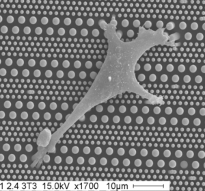
A L’ÉCHELLE DE LA CELLULES UNIQUE

Nous analysons la mécanosensation et la mécanotransduction des adhésions cellule-matrice extracellulaire associées au intégrines, et cellule-cellule associées au cadhérines. Pour répondre à ces questions, nous avons développé des modèles de cellules uniques et de doublets de cellules permettant un contrôle de la formation des adhésions dans un microenvironnement physique et mécanique défini. Couplées à des approches de microscopie avancée, au développement de capteurs de force et à la biologie cellulaire classique, ces approches nous permettent d’étudier les mécanismes moléculaires qui contrôlent le remodelage des adhésions et du cytosquelette, de la forme et de la migration des cellules et leur adaptation à la rigidité de l’environnement ainsi qu’aux propriétés viscoélastiques du cytosquelette et à la tension interne générée par les moteurs à myosine.


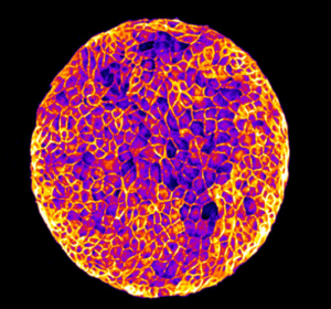
A L’ÉCHELLE MULTICELLULAIRE

Nous étudions le comportement collectif des cellules dans les feuillets épithéliaux, dans le contexte de l’homéostasie tissulaire et de la cicatrisation. Pour répondre à ces questions, nous développons des outils microfabriqués et des outils biophysiques pour mesurer et contrôler les propriétés mécaniques et la topologie du microenvironnement des cellules. Ces outils sont combinés à des approches moléculaires, des techniques avancées de microscopie optique, d’analyse d’images et de modélisation pour étudier l’influence des propriétés physiques de l’environnement sur l’organisation des couches épithéliales, la migration collective des cellules, la polarisation des cellules individuelles et collectives, la division cellulaire et l’extrusion des cellules. Nous caractérisons comment les contraintes physiques peuvent conduire à des propriétés dynamiques et mécaniques émergentes de divers tissus épithéliaux.


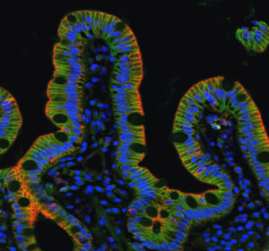
AU NIVEAU DES TISSUS ET DES ORGANOÏDES

Nous étudions comment des tissus épithéliaux plus complexes, formés de populations de cellules différentes (cellules normales/déficientes pour l’adhésion, normales/cancéreuses, différenciées/cellules souches) confrontées à des substrats homogènes ou hétérogènes (chimie de la matrice extracellulaire, rigidité, géométrie et topographie), régulent l’homéostasie, se séparent et/ou s’auto-assemblent. Nos objectifs sont de déterminer i) comment les contraintes physiques du microenvironnement modulent les propriétés mécaniques des cellules et des tissus épithéliaux, ii) comment elles dirigent une variété de comportements cellulaires incluant la prolifération des cellules souches, l’extrusion ou la délamination de cellules, la migration cellulaire, la différenciation et la polarité, et iii) comment elles ont un impact sur la morphogenèse des tissus épithéliaux normaux, ainsi que sur le développement pathologique des maladies rares de l’intestin. Les substrats biomimétiques couplés à l’imagerie à haute résolution et à la biochimie sont essentiels pour atteindre ces objectifs.

Responsables :

Benoît LADOUX
Téléphone : +33 (0)157278071 / Email : benoit.ladoux (at) ijm.fr

René-Marc MÈGE
Téléphone : +33 (0)157278067 / Email : rene-marc.mege (at) ijm.fr


Membres de l’équipe :

ANGER Lucas Ingénieur en biologie
D’ALESSANDRO Joseph Chercheur
DE BECO Simon Enseignant-chercheur
DUBEY Sushil Postdoctorant
FARDIN Marc-Antoine Chercheur
DANG Tien Ingénieure en biologie
PENETI Sudheer Kumar Doctorant
ROSSE Carine chercheur invitée
SCHONIT Andreas Doctorant
SHEN Yuan Postdoctorant
WODRASCKA Fanny Doctorante
WU Huiqiong Postdoctorante
XI Wang Chercheur
CHILUPURI Ranjith Kumar Doctorant
JIANG Pan Postdoctorant
MARTINS Joana Doctorante, visiteur
VIGNES Hélène Postdoctorante
GRUDTSYNA Valeriia Doctorante, visiteur
THIANT Clémence Doctorante
ARKOWITZ Grégory Doctorant
DAI Wufei Doctorant
KAILASAM MANI Satish Postdoctorant
RACHIDI Joud Master 2

Balasubramaniam L, Doostmohammadi A, Saw TB, Narayana GHNS, Mueller R, Dang T, Thomas M, Gupta S, Sonam S, Yap AS, Toyama Y, Mège RM, Yeomans JM, Ladoux B. Investigating the nature of active forces in tissues reveals how contractile cells can form extensile monolayers. Nat Mater. 2021 Aug;20(8):1156-1166. doi: 10.1038/s41563-021-00919-2. Epub 2021 Feb 18. Erratum in: Nat Mater. 2021 Mar 9;: PMID: 33603188; PMCID: PMC7611436.

Gaston C, De Beco S, Doss B, Pan M, Gauquelin E, D’Alessandro J, Lim CT, Ladoux B, Delacour D. EpCAM promotes endosomal modulation of the cortical RhoA zone for epithelial organization. Nat Commun. 2021 Apr 13;12(1):2226. doi: 10.1038/s41467-021-22482-9. PMID: 33850145; PMCID: PMC8044225.

Jain S, Cachoux VML, Narayana GHNS, de Beco S, D’Alessandro J, Cellerin V, Chen T, Heuzé ML, Marcq P, Mège RM, Kabla AJ, Lim CT, Ladoux B. The role of single cell mechanical behavior and polarity in driving collective cell migration. Nat Phys. 2020 Jul;16(7):802-809. doi: 10.1038/s41567-020-0875-z. Epub 2020 May 4. PMID: 32641972; PMCID: PMC7343533.

Le AP, Rupprecht JF, Mège RM, Toyama Y, Lim CT, Ladoux B. Adhesion-mediated heterogeneous actin organization governs apoptotic cell extrusion. Nat Commun. 2021 Jan 15;12(1):397. doi: 10.1038/s41467-020-20563-9. PMID: 33452264; PMCID: PMC7810754.

Doss BL, Pan M, Gupta M, Grenci G, Mège RM, Lim CT, Sheetz MP, Voituriez R, Ladoux B. Cell response to substrate rigidity is regulated by active and passive cytoskeletal stress. Proc Natl Acad Sci U S A. 2020 Jun 9;117(23):12817-12825.
doi: 10.1073/pnas.1917555117. Epub 2020 May 22. PMID: 32444491; PMCID: PMC7293595.

Heuzé ML, Sankara Narayana GHN, D’Alessandro J, Cellerin V, Dang T, Williams DS, Van Hest JC, Marcq P, Mège RM, Ladoux B. Myosin II isoforms play distinct roles in <i>adherens</i> junction biogenesis. Elife. 2019 Sep 5;8:e46599. doi: 10.7554/eLife.46599. PMID: 31486768; PMCID: PMC6756789.

Chen T, Callan-Jones A, Fedorov E, Ravasio A, Brugués A, Ong HT, Toyama Y, Low BC, Trepat X, Shemesh T, Voituriez R, Ladoux B. Large-scale curvature sensing by directional actin flow drives cellular migration mode switching. Nat Phys. 2019 Apr;15:393-402. doi: 10.1038/s41567-018-0383-6. Epub 2019 Jan 21. PMID: 30984281; PMCID: PMC6456019.

Seddiki R, Narayana GHNS, Strale PO, Balcioglu HE, Peyret G, Yao M, Le AP, Teck Lim C, Yan J, Ladoux B, Mège RM. Force-dependent binding of vinculin to α-catenin regulates cell-cell contact stability and collective cell behavior.
Mol Biol Cell. 2018 Feb 15;29(4):380-388. doi: 10.1091/mbc.E17-04-0231. Epub 2017 Dec 27. PMID: 29282282; PMCID: PMC6014167.

Saw TB, Doostmohammadi A, Nier V, Kocgozlu L, Thampi S, Toyama Y, Marcq P, Lim CT, Yeomans JM, Ladoux B. Topological defects in epithelia govern cell death and extrusion. Nature. 2017 Apr 12;544(7649):212-216.
Abstract

Salomon J, Gaston C, Magescas J, Duvauchelle B, Canioni D, Sengmanivong L, Mayeux A, Michaux G, Campeotto F, Lemale J, Viala J, Poirier F, Minc N, Schmitz J, Brousse N, Ladoux B, Goulet O, Delacour D. Contractile forces at tricellular contacts modulate epithelial organization and monolayer integrity. Nat Commun. 2017 Jan 13;8:13998.
Abstract

Publications

2913254 9RMNZZGV items 1 0 date desc 8991 https://www.ijm.fr/wp-content/plugins/zotpress/
%7B%22status%22%3A%22success%22%2C%22updateneeded%22%3Afalse%2C%22instance%22%3A%22zotpress-515e6bffab9a89492378ebdd8b890ec5%22%2C%22meta%22%3A%7B%22request_last%22%3A50%2C%22request_next%22%3A50%2C%22used_cache%22%3Atrue%7D%2C%22data%22%3A%5B%7B%22key%22%3A%22RKEUT5SU%22%2C%22library%22%3A%7B%22id%22%3A2913254%7D%2C%22meta%22%3A%7B%22creatorSummary%22%3A%22Bertrand%20et%20al.%22%2C%22parsedDate%22%3A%222024-04-04%22%2C%22numChildren%22%3A2%7D%2C%22bib%22%3A%22%3Cdiv%20class%3D%5C%22csl-bib-body%5C%22%20style%3D%5C%22line-height%3A%202%3B%20padding-left%3A%201em%3B%20text-indent%3A-1em%3B%5C%22%3E%5Cn%20%20%3Cdiv%20class%3D%5C%22csl-entry%5C%22%3EBertrand%2C%20T.%2C%20d%26%23x2019%3BAlessandro%2C%20J.%2C%20Maitra%2C%20A.%2C%20Jain%2C%20S.%2C%20Mercier%2C%20B.%2C%20M%26%23xE8%3Bge%2C%20R.-M.%2C%20Ladoux%2C%20B.%2C%20%26amp%3B%20Voituriez%2C%20R.%20%282024%29.%20Clustering%20and%20ordering%20in%20cell%20assemblies%20with%20generic%20asymmetric%20aligning%20interactions.%20%3Ci%3EPhysical%20Review%20Research%3C%5C%2Fi%3E%2C%20%3Ci%3E6%3C%5C%2Fi%3E%282%29%2C%20023022.%20%3Ca%20href%3D%27https%3A%5C%2F%5C%2Fdoi.org%5C%2F10.1103%5C%2FPhysRevResearch.6.023022%27%3Ehttps%3A%5C%2F%5C%2Fdoi.org%5C%2F10.1103%5C%2FPhysRevResearch.6.023022%3C%5C%2Fa%3E%3C%5C%2Fdiv%3E%5Cn%3C%5C%2Fdiv%3E%22%2C%22data%22%3A%7B%22itemType%22%3A%22journalArticle%22%2C%22title%22%3A%22Clustering%20and%20ordering%20in%20cell%20assemblies%20with%20generic%20asymmetric%20aligning%20interactions%22%2C%22creators%22%3A%5B%7B%22creatorType%22%3A%22author%22%2C%22firstName%22%3A%22Thibault%22%2C%22lastName%22%3A%22Bertrand%22%7D%2C%7B%22creatorType%22%3A%22author%22%2C%22firstName%22%3A%22Joseph%22%2C%22lastName%22%3A%22d%27Alessandro%22%7D%2C%7B%22creatorType%22%3A%22author%22%2C%22firstName%22%3A%22Ananyo%22%2C%22lastName%22%3A%22Maitra%22%7D%2C%7B%22creatorType%22%3A%22author%22%2C%22firstName%22%3A%22Shreyansh%22%2C%22lastName%22%3A%22Jain%22%7D%2C%7B%22creatorType%22%3A%22author%22%2C%22firstName%22%3A%22Barbara%22%2C%22lastName%22%3A%22Mercier%22%7D%2C%7B%22creatorType%22%3A%22author%22%2C%22firstName%22%3A%22Ren%5Cu00e9-Marc%22%2C%22lastName%22%3A%22M%5Cu00e8ge%22%7D%2C%7B%22creatorType%22%3A%22author%22%2C%22firstName%22%3A%22Benoit%22%2C%22lastName%22%3A%22Ladoux%22%7D%2C%7B%22creatorType%22%3A%22author%22%2C%22firstName%22%3A%22Rapha%5Cu00ebl%22%2C%22lastName%22%3A%22Voituriez%22%7D%5D%2C%22abstractNote%22%3A%22Collective%20cell%20migration%20plays%20an%20essential%20role%20in%20various%20biological%20processes%2C%20such%20as%20development%20or%20cancer%20proliferation.%20While%20cell-cell%20interactions%20are%20clearly%20key%20determinants%20of%20collective%20cell%20migration%2C%20the%20physical%20mechanisms%20that%20control%20the%20emergence%20of%20cell%20clustering%20and%20collective%20cell%20migration%20are%20still%20poorly%20understood.%20In%20particular%2C%20observations%20have%20shown%20that%20binary%20cell-cell%20collisions%20generally%20lead%20to%20antialignment%20of%20cell%20polarities%20and%20separation%20of%20pairs%5Cu2014a%20process%20called%20contact%20inhibition%20of%20locomotion%20%28CIL%29%2C%20which%20is%20expected%20to%20disfavor%20the%20formation%20of%20large-scale%20cell%20clusters%20with%20coherent%20motion%20even%20though%20the%20latter%20is%20often%20observed%20in%20tissues.%20To%20solve%20this%20puzzle%2C%20we%20adopt%20a%20joint%20experimental%20and%20theoretical%20approach%20to%20determine%20the%20large-scale%20dynamics%20of%20cell%20assemblies%20from%20elementary%20pairwise%20cell-cell%20interaction%20rules.%20We%20quantify%20experimentally%20binary%20cell-cell%20interactions%20and%20show%20that%20they%20can%20be%20captured%20by%20a%20minimal%20equilibriumlike%20pairwise%20asymmetric%20aligning%20interaction%20potential%20that%20reproduces%20the%20CIL%20phenomenology.%20We%20identify%20its%20symmetry%20class%2C%20build%20the%20corresponding%20active%20hydrodynamic%20theory%2C%20and%20show%20on%20general%20grounds%20that%20such%20asymmetric%20aligning%20interaction%20destroys%20large-scale%20clustering%20and%20ordering%2C%20leading%20instead%20to%20a%20liquidlike%20microphase%20of%20cell%20clusters%20of%20finite%20size%20and%20short%20lived%20polarity%20or%20to%20a%20fully%20dispersed%20isotropic%20phase.%20Finally%2C%20this%20shows%20that%20CIL-like%20asymmetric%20interactions%20in%20cellular%20systems%5Cu2014or%20general%20active%20systems%5Cu2014control%20cluster%20sizes%20and%20polarity%2C%20and%20can%20prevent%20large-scale%20coarsening%20and%20long-range%20polarity%2C%20except%20in%20the%20singular%20regime%20of%20dense%20confluent%20systems.%22%2C%22date%22%3A%222024-04-04%22%2C%22language%22%3A%22%22%2C%22DOI%22%3A%2210.1103%5C%2FPhysRevResearch.6.023022%22%2C%22ISSN%22%3A%22%22%2C%22url%22%3A%22https%3A%5C%2F%5C%2Flink.aps.org%5C%2Fdoi%5C%2F10.1103%5C%2FPhysRevResearch.6.023022%22%2C%22collections%22%3A%5B%229RMNZZGV%22%5D%2C%22dateModified%22%3A%222024-04-05T13%3A07%3A50Z%22%7D%7D%2C%7B%22key%22%3A%22CXRR97VC%22%2C%22library%22%3A%7B%22id%22%3A2913254%7D%2C%22meta%22%3A%7B%22creatorSummary%22%3A%22Sadhu%20et%20al.%22%2C%22parsedDate%22%3A%222024-03-19%22%2C%22numChildren%22%3A1%7D%2C%22bib%22%3A%22%3Cdiv%20class%3D%5C%22csl-bib-body%5C%22%20style%3D%5C%22line-height%3A%202%3B%20padding-left%3A%201em%3B%20text-indent%3A-1em%3B%5C%22%3E%5Cn%20%20%3Cdiv%20class%3D%5C%22csl-entry%5C%22%3ESadhu%2C%20R.%20K.%2C%20Luciano%2C%20M.%2C%20Xi%2C%20W.%2C%20Martinez-Torres%2C%20C.%2C%20Schr%26%23xF6%3Bder%2C%20M.%2C%20Blum%2C%20C.%2C%20Tarantola%2C%20M.%2C%20Villa%2C%20S.%2C%20Peni%26%23x10D%3B%2C%20S.%2C%20Igli%26%23x10D%3B%2C%20A.%2C%20Beta%2C%20C.%2C%20Steinbock%2C%20O.%2C%20Bodenschatz%2C%20E.%2C%20Ladoux%2C%20B.%2C%20Gabriele%2C%20S.%2C%20%26amp%3B%20Gov%2C%20N.%20S.%20%282024%29.%20A%20minimal%20physical%20model%20for%20curvotaxis%20driven%20by%20curved%20protein%20complexes%20at%20the%20cell%26%23x2019%3Bs%20leading%20edge.%20%3Ci%3EProceedings%20of%20the%20National%20Academy%20of%20Sciences%3C%5C%2Fi%3E%2C%20%3Ci%3E121%3C%5C%2Fi%3E%2812%29%2C%20e2306818121.%20%3Ca%20href%3D%27https%3A%5C%2F%5C%2Fdoi.org%5C%2F10.1073%5C%2Fpnas.2306818121%27%3Ehttps%3A%5C%2F%5C%2Fdoi.org%5C%2F10.1073%5C%2Fpnas.2306818121%3C%5C%2Fa%3E%3C%5C%2Fdiv%3E%5Cn%3C%5C%2Fdiv%3E%22%2C%22data%22%3A%7B%22itemType%22%3A%22journalArticle%22%2C%22title%22%3A%22A%20minimal%20physical%20model%20for%20curvotaxis%20driven%20by%20curved%20protein%20complexes%20at%20the%20cell%5Cu2019s%20leading%20edge%22%2C%22creators%22%3A%5B%7B%22creatorType%22%3A%22author%22%2C%22firstName%22%3A%22Raj%20Kumar%22%2C%22lastName%22%3A%22Sadhu%22%7D%2C%7B%22creatorType%22%3A%22author%22%2C%22firstName%22%3A%22Marine%22%2C%22lastName%22%3A%22Luciano%22%7D%2C%7B%22creatorType%22%3A%22author%22%2C%22firstName%22%3A%22Wang%22%2C%22lastName%22%3A%22Xi%22%7D%2C%7B%22creatorType%22%3A%22author%22%2C%22firstName%22%3A%22Cristina%22%2C%22lastName%22%3A%22Martinez-Torres%22%7D%2C%7B%22creatorType%22%3A%22author%22%2C%22firstName%22%3A%22Marcel%22%2C%22lastName%22%3A%22Schr%5Cu00f6der%22%7D%2C%7B%22creatorType%22%3A%22author%22%2C%22firstName%22%3A%22Christoph%22%2C%22lastName%22%3A%22Blum%22%7D%2C%7B%22creatorType%22%3A%22author%22%2C%22firstName%22%3A%22Marco%22%2C%22lastName%22%3A%22Tarantola%22%7D%2C%7B%22creatorType%22%3A%22author%22%2C%22firstName%22%3A%22Stefano%22%2C%22lastName%22%3A%22Villa%22%7D%2C%7B%22creatorType%22%3A%22author%22%2C%22firstName%22%3A%22Samo%22%2C%22lastName%22%3A%22Peni%5Cu010d%22%7D%2C%7B%22creatorType%22%3A%22author%22%2C%22firstName%22%3A%22Ale%5Cu0161%22%2C%22lastName%22%3A%22Igli%5Cu010d%22%7D%2C%7B%22creatorType%22%3A%22author%22%2C%22firstName%22%3A%22Carsten%22%2C%22lastName%22%3A%22Beta%22%7D%2C%7B%22creatorType%22%3A%22author%22%2C%22firstName%22%3A%22Oliver%22%2C%22lastName%22%3A%22Steinbock%22%7D%2C%7B%22creatorType%22%3A%22author%22%2C%22firstName%22%3A%22Eberhard%22%2C%22lastName%22%3A%22Bodenschatz%22%7D%2C%7B%22creatorType%22%3A%22author%22%2C%22firstName%22%3A%22Beno%5Cu00eet%22%2C%22lastName%22%3A%22Ladoux%22%7D%2C%7B%22creatorType%22%3A%22author%22%2C%22firstName%22%3A%22Sylvain%22%2C%22lastName%22%3A%22Gabriele%22%7D%2C%7B%22creatorType%22%3A%22author%22%2C%22firstName%22%3A%22Nir%20S.%22%2C%22lastName%22%3A%22Gov%22%7D%5D%2C%22abstractNote%22%3A%22Cells%20often%20migrate%20on%20curved%20surfaces%20inside%20the%20body%2C%20such%20as%20curved%20tissues%2C%20blood%20vessels%2C%20or%20highly%20curved%20protrusions%20of%20other%20cells.%20Recent%20in%20vitro%20experiments%20provide%20clear%20evidence%20that%20motile%20cells%20are%20affected%20by%20the%20curvature%20of%20the%20substrate%20on%20which%20they%20migrate%2C%20preferring%20certain%20curvatures%20to%20others%2C%20termed%20%5Cu201ccurvotaxis.%5Cu201d%20The%20origin%20and%20underlying%20mechanism%20that%20gives%20rise%20to%20this%20curvature%20sensitivity%20are%20not%20well%20understood.%20Here%2C%20we%20employ%20a%20%5Cu201cminimal%20cell%5Cu201d%20model%20which%20is%20composed%20of%20a%20vesicle%20that%20contains%20curved%20membrane%20protein%20complexes%2C%20that%20exert%20protrusive%20forces%20on%20the%20membrane%20%28representing%20the%20pressure%20due%20to%20actin%20polymerization%29.%20This%20minimal-cell%20model%20gives%20rise%20to%20spontaneous%20emergence%20of%20a%20motile%20phenotype%2C%20driven%20by%20a%20lamellipodia-like%20leading%20edge.%20By%20systematically%20screening%20the%20behavior%20of%20this%20model%20on%20different%20types%20of%20curved%20substrates%20%28sinusoidal%2C%20cylinder%2C%20and%20tube%29%2C%20we%20show%20that%20minimal%20ingredients%20and%20energy%20terms%20capture%20the%20experimental%20data.%20The%20model%20recovers%20the%20observed%20migration%20on%20the%20sinusoidal%20substrate%2C%20where%20cells%20move%20along%20the%20grooves%20%28minima%29%2C%20while%20avoiding%20motion%20along%20the%20ridges.%20In%20addition%2C%20the%20model%20predicts%20the%20tendency%20of%20cells%20to%20migrate%20circumferentially%20on%20convex%20substrates%20and%20axially%20on%20concave%20ones.%20Both%20of%20these%20predictions%20are%20verified%20experimentally%2C%20on%20several%20cell%20types.%20Altogether%2C%20our%20results%20identify%20the%20minimization%20of%20membrane-substrate%20adhesion%20energy%20and%20binding%20energy%20between%20the%20membrane%20protein%20complexes%20as%20key%20players%20of%20curvotaxis%20in%20cell%20migration.%22%2C%22date%22%3A%222024-03-19%22%2C%22language%22%3A%22%22%2C%22DOI%22%3A%2210.1073%5C%2Fpnas.2306818121%22%2C%22ISSN%22%3A%22%22%2C%22url%22%3A%22https%3A%5C%2F%5C%2Fwww.pnas.org%5C%2Fdoi%5C%2F10.1073%5C%2Fpnas.2306818121%22%2C%22collections%22%3A%5B%229RMNZZGV%22%5D%2C%22dateModified%22%3A%222024-04-05T09%3A24%3A28Z%22%7D%7D%2C%7B%22key%22%3A%22MF5RGAL9%22%2C%22library%22%3A%7B%22id%22%3A2913254%7D%2C%22meta%22%3A%7B%22creatorSummary%22%3A%22Monteiro%20et%20al.%22%2C%22parsedDate%22%3A%222023-10-30%22%2C%22numChildren%22%3A1%7D%2C%22bib%22%3A%22%3Cdiv%20class%3D%5C%22csl-bib-body%5C%22%20style%3D%5C%22line-height%3A%202%3B%20padding-left%3A%201em%3B%20text-indent%3A-1em%3B%5C%22%3E%5Cn%20%20%3Cdiv%20class%3D%5C%22csl-entry%5C%22%3EMonteiro%2C%20P.%2C%20Remy%2C%20D.%2C%20Lemerle%2C%20E.%2C%20Routet%2C%20F.%2C%20Mac%26%23xE9%3B%2C%20A.-S.%2C%20Guedj%2C%20C.%2C%20Ladoux%2C%20B.%2C%20Vassilopoulos%2C%20S.%2C%20Lamaze%2C%20C.%2C%20%26amp%3B%20Chavrier%2C%20P.%20%282023%29.%20A%20mechanosensitive%20caveolae%26%23x2013%3Binvadosome%20interplay%20drives%20matrix%20remodelling%20for%20cancer%20cell%20invasion.%20%3Ci%3ENature%20Cell%20Biology%3C%5C%2Fi%3E%2C%201%26%23x2013%3B17.%20%3Ca%20href%3D%27https%3A%5C%2F%5C%2Fdoi.org%5C%2F10.1038%5C%2Fs41556-023-01272-z%27%3Ehttps%3A%5C%2F%5C%2Fdoi.org%5C%2F10.1038%5C%2Fs41556-023-01272-z%3C%5C%2Fa%3E%3C%5C%2Fdiv%3E%5Cn%3C%5C%2Fdiv%3E%22%2C%22data%22%3A%7B%22itemType%22%3A%22journalArticle%22%2C%22title%22%3A%22A%20mechanosensitive%20caveolae%5Cu2013invadosome%20interplay%20drives%20matrix%20remodelling%20for%20cancer%20cell%20invasion%22%2C%22creators%22%3A%5B%7B%22creatorType%22%3A%22author%22%2C%22firstName%22%3A%22Pedro%22%2C%22lastName%22%3A%22Monteiro%22%7D%2C%7B%22creatorType%22%3A%22author%22%2C%22firstName%22%3A%22David%22%2C%22lastName%22%3A%22Remy%22%7D%2C%7B%22creatorType%22%3A%22author%22%2C%22firstName%22%3A%22Eline%22%2C%22lastName%22%3A%22Lemerle%22%7D%2C%7B%22creatorType%22%3A%22author%22%2C%22firstName%22%3A%22Fiona%22%2C%22lastName%22%3A%22Routet%22%7D%2C%7B%22creatorType%22%3A%22author%22%2C%22firstName%22%3A%22Anne-Sophie%22%2C%22lastName%22%3A%22Mac%5Cu00e9%22%7D%2C%7B%22creatorType%22%3A%22author%22%2C%22firstName%22%3A%22Chlo%5Cu00e9%22%2C%22lastName%22%3A%22Guedj%22%7D%2C%7B%22creatorType%22%3A%22author%22%2C%22firstName%22%3A%22Benoit%22%2C%22lastName%22%3A%22Ladoux%22%7D%2C%7B%22creatorType%22%3A%22author%22%2C%22firstName%22%3A%22St%5Cu00e9phane%22%2C%22lastName%22%3A%22Vassilopoulos%22%7D%2C%7B%22creatorType%22%3A%22author%22%2C%22firstName%22%3A%22Christophe%22%2C%22lastName%22%3A%22Lamaze%22%7D%2C%7B%22creatorType%22%3A%22author%22%2C%22firstName%22%3A%22Philippe%22%2C%22lastName%22%3A%22Chavrier%22%7D%5D%2C%22abstractNote%22%3A%22Invadosomes%20and%20caveolae%20are%20mechanosensitive%20structures%20that%20are%20implicated%20in%20metastasis.%20Here%2C%20we%20describe%20a%20unique%20juxtaposition%20of%20caveola%20clusters%20and%20matrix%20degradative%20invadosomes%20at%20contact%20sites%20between%20the%20plasma%20membrane%20of%20cancer%20cells%20and%20constricting%20fibrils%20both%20in%202D%20and%203D%20type%20I%20collagen%20matrix%20environments.%20Preferential%20association%20between%20caveolae%20and%20straight%20segments%20of%20the%20fibrils%2C%20and%20between%20invadosomes%20and%20bent%20segments%20of%20the%20fibrils%2C%20was%20observed%20along%20with%20matrix%20remodelling.%20Caveola%20recruitment%20precedes%20and%20is%20required%20for%20invadosome%20formation%20and%20activity.%20Reciprocally%2C%20invadosome%20disruption%20results%20in%20the%20accumulation%20of%20fibril-associated%20caveolae.%20Moreover%2C%20caveolae%20and%20the%20collagen%20receptor%20%5Cu03b21%20integrin%20co-localize%20at%20contact%20sites%20with%20the%20fibrils%2C%20and%20integrins%20control%20caveola%20recruitment%20to%20fibrils.%20In%20turn%2C%20caveolae%20mediate%20the%20clearance%20of%20%5Cu03b21%20integrin%20and%20collagen%20uptake%20in%20an%20invadosome-dependent%20and%20collagen-cleavage-dependent%20mechanism.%20Our%20data%20reveal%20a%20reciprocal%20interplay%20between%20caveolae%20and%20invadosomes%20that%20coordinates%20adhesion%20to%20and%20proteolytic%20remodelling%20of%20confining%20fibrils%20to%20support%20tumour%20cell%20dissemination.%22%2C%22date%22%3A%222023-10-30%22%2C%22language%22%3A%22en%22%2C%22DOI%22%3A%2210.1038%5C%2Fs41556-023-01272-z%22%2C%22ISSN%22%3A%221476-4679%22%2C%22url%22%3A%22https%3A%5C%2F%5C%2Fwww.nature.com%5C%2Farticles%5C%2Fs41556-023-01272-z%22%2C%22collections%22%3A%5B%229RMNZZGV%22%5D%2C%22dateModified%22%3A%222023-11-03T12%3A17%3A58Z%22%7D%7D%2C%7B%22key%22%3A%227QCDYPEX%22%2C%22library%22%3A%7B%22id%22%3A2913254%7D%2C%22meta%22%3A%7B%22creatorSummary%22%3A%22Eckert%20et%20al.%22%2C%22parsedDate%22%3A%222023-09-16%22%2C%22numChildren%22%3A1%7D%2C%22bib%22%3A%22%3Cdiv%20class%3D%5C%22csl-bib-body%5C%22%20style%3D%5C%22line-height%3A%202%3B%20padding-left%3A%201em%3B%20text-indent%3A-1em%3B%5C%22%3E%5Cn%20%20%3Cdiv%20class%3D%5C%22csl-entry%5C%22%3EEckert%2C%20J.%2C%20Ladoux%2C%20B.%2C%20M%26%23xE8%3Bge%2C%20R.-M.%2C%20Giomi%2C%20L.%2C%20%26amp%3B%20Schmidt%2C%20T.%20%282023%29.%20Hexanematic%20crossover%20in%20epithelial%20monolayers%20depends%20on%20cell%20adhesion%20and%20cell%20density.%20%3Ci%3ENature%20Communications%3C%5C%2Fi%3E%2C%20%3Ci%3E14%3C%5C%2Fi%3E%281%29%2C%205762.%20%3Ca%20href%3D%27https%3A%5C%2F%5C%2Fdoi.org%5C%2F10.1038%5C%2Fs41467-023-41449-6%27%3Ehttps%3A%5C%2F%5C%2Fdoi.org%5C%2F10.1038%5C%2Fs41467-023-41449-6%3C%5C%2Fa%3E%3C%5C%2Fdiv%3E%5Cn%3C%5C%2Fdiv%3E%22%2C%22data%22%3A%7B%22itemType%22%3A%22journalArticle%22%2C%22title%22%3A%22Hexanematic%20crossover%20in%20epithelial%20monolayers%20depends%20on%20cell%20adhesion%20and%20cell%20density%22%2C%22creators%22%3A%5B%7B%22creatorType%22%3A%22author%22%2C%22firstName%22%3A%22Julia%22%2C%22lastName%22%3A%22Eckert%22%7D%2C%7B%22creatorType%22%3A%22author%22%2C%22firstName%22%3A%22Beno%5Cu00eet%22%2C%22lastName%22%3A%22Ladoux%22%7D%2C%7B%22creatorType%22%3A%22author%22%2C%22firstName%22%3A%22Ren%5Cu00e9-Marc%22%2C%22lastName%22%3A%22M%5Cu00e8ge%22%7D%2C%7B%22creatorType%22%3A%22author%22%2C%22firstName%22%3A%22Luca%22%2C%22lastName%22%3A%22Giomi%22%7D%2C%7B%22creatorType%22%3A%22author%22%2C%22firstName%22%3A%22Thomas%22%2C%22lastName%22%3A%22Schmidt%22%7D%5D%2C%22abstractNote%22%3A%22Changes%20in%20tissue%20geometry%20during%20developmental%20processes%20are%20associated%20with%20collective%20migration%20of%20cells.%20Recent%20experimental%20and%20numerical%20results%20suggest%20that%20these%20changes%20could%20leverage%20on%20the%20coexistence%20of%20nematic%20and%20hexatic%20orientational%20order%20at%20different%20length%20scales.%20How%20this%20multiscale%20organization%20is%20affected%20by%20the%20material%20properties%20of%20the%20cells%20and%20their%20substrate%20is%20presently%20unknown.%20In%20this%20study%2C%20we%20address%20these%20questions%20in%20monolayers%20of%20Madin-Darby%20canine%20kidney%20cells%20having%20various%20cell%20densities%20and%20molecular%20repertoires.%20At%20small%20length%20scales%2C%20confluent%20monolayers%20are%20characterized%20by%20a%20prominent%20hexatic%20order%2C%20independent%20of%20the%20presence%20of%20E-cadherin%2C%20monolayer%20density%2C%20and%20underlying%20substrate%20stiffness.%20However%2C%20all%20three%20properties%20affect%20the%20meso-scale%20tissue%20organization.%20The%20length%20scale%20at%20which%20hexatic%20order%20transits%20to%20nematic%20order%2C%20the%20%5Cu201chexanematic%5Cu201d%20crossover%20scale%2C%20strongly%20depends%20on%20cell-cell%20adhesions%20and%20correlates%20with%20monolayer%20density.%20Our%20study%20demonstrates%20how%20epithelial%20organization%20is%20affected%20by%20mechanical%20properties%2C%20and%20provides%20a%20robust%20description%20of%20tissue%20organization%20during%20developmental%20processes.%22%2C%22date%22%3A%222023-09-16%22%2C%22language%22%3A%22en%22%2C%22DOI%22%3A%2210.1038%5C%2Fs41467-023-41449-6%22%2C%22ISSN%22%3A%222041-1723%22%2C%22url%22%3A%22https%3A%5C%2F%5C%2Fwww.nature.com%5C%2Farticles%5C%2Fs41467-023-41449-6%22%2C%22collections%22%3A%5B%229RMNZZGV%22%5D%2C%22dateModified%22%3A%222023-09-22T10%3A27%3A38Z%22%7D%7D%2C%7B%22key%22%3A%22FF4ZDWYJ%22%2C%22library%22%3A%7B%22id%22%3A2913254%7D%2C%22meta%22%3A%7B%22creatorSummary%22%3A%22Lignieres%20et%20al.%22%2C%22parsedDate%22%3A%222023-03-03%22%2C%22numChildren%22%3A1%7D%2C%22bib%22%3A%22%3Cdiv%20class%3D%5C%22csl-bib-body%5C%22%20style%3D%5C%22line-height%3A%202%3B%20padding-left%3A%201em%3B%20text-indent%3A-1em%3B%5C%22%3E%5Cn%20%20%3Cdiv%20class%3D%5C%22csl-entry%5C%22%3ELignieres%2C%20L.%2C%20S%26%23xE9%3Bn%26%23xE9%3Bcaut%2C%20N.%2C%20Dang%2C%20T.%2C%20Bellutti%2C%20L.%2C%20Hamon%2C%20M.%2C%20Terrier%2C%20S.%2C%20Legros%2C%20V.%2C%20Chevreux%2C%20G.%2C%20Lelandais%2C%20G.%2C%20M%26%23xE8%3Bge%2C%20R.-M.%2C%20Dumont%2C%20J.%2C%20%26amp%3B%20Camadro%2C%20J.-M.%20%282023%29.%20Extending%20the%20Range%20of%20SLIM-Labeling%20Applications%3A%20From%20Human%20Cell%20Lines%20in%20Culture%20to%20Caenorhabditis%20elegans%20Whole-Organism%20Labeling.%20%3Ci%3EJournal%20of%20Proteome%20Research%3C%5C%2Fi%3E%2C%20%3Ci%3E22%3C%5C%2Fi%3E%283%29%2C%20996%26%23x2013%3B1002.%20%3Ca%20href%3D%27https%3A%5C%2F%5C%2Fdoi.org%5C%2F10.1021%5C%2Facs.jproteome.2c00699%27%3Ehttps%3A%5C%2F%5C%2Fdoi.org%5C%2F10.1021%5C%2Facs.jproteome.2c00699%3C%5C%2Fa%3E%3C%5C%2Fdiv%3E%5Cn%3C%5C%2Fdiv%3E%22%2C%22data%22%3A%7B%22itemType%22%3A%22journalArticle%22%2C%22title%22%3A%22Extending%20the%20Range%20of%20SLIM-Labeling%20Applications%3A%20From%20Human%20Cell%20Lines%20in%20Culture%20to%20Caenorhabditis%20elegans%20Whole-Organism%20Labeling%22%2C%22creators%22%3A%5B%7B%22creatorType%22%3A%22author%22%2C%22firstName%22%3A%22Laurent%22%2C%22lastName%22%3A%22Lignieres%22%7D%2C%7B%22creatorType%22%3A%22author%22%2C%22firstName%22%3A%22Nicolas%22%2C%22lastName%22%3A%22S%5Cu00e9n%5Cu00e9caut%22%7D%2C%7B%22creatorType%22%3A%22author%22%2C%22firstName%22%3A%22Tien%22%2C%22lastName%22%3A%22Dang%22%7D%2C%7B%22creatorType%22%3A%22author%22%2C%22firstName%22%3A%22Laura%22%2C%22lastName%22%3A%22Bellutti%22%7D%2C%7B%22creatorType%22%3A%22author%22%2C%22firstName%22%3A%22Marion%22%2C%22lastName%22%3A%22Hamon%22%7D%2C%7B%22creatorType%22%3A%22author%22%2C%22firstName%22%3A%22Samuel%22%2C%22lastName%22%3A%22Terrier%22%7D%2C%7B%22creatorType%22%3A%22author%22%2C%22firstName%22%3A%22V%5Cu00e9ronique%22%2C%22lastName%22%3A%22Legros%22%7D%2C%7B%22creatorType%22%3A%22author%22%2C%22firstName%22%3A%22Guillaume%22%2C%22lastName%22%3A%22Chevreux%22%7D%2C%7B%22creatorType%22%3A%22author%22%2C%22firstName%22%3A%22Ga%5Cu00eblle%22%2C%22lastName%22%3A%22Lelandais%22%7D%2C%7B%22creatorType%22%3A%22author%22%2C%22firstName%22%3A%22Ren%5Cu00e9-Marc%22%2C%22lastName%22%3A%22M%5Cu00e8ge%22%7D%2C%7B%22creatorType%22%3A%22author%22%2C%22firstName%22%3A%22Julien%22%2C%22lastName%22%3A%22Dumont%22%7D%2C%7B%22creatorType%22%3A%22author%22%2C%22firstName%22%3A%22Jean-Michel%22%2C%22lastName%22%3A%22Camadro%22%7D%5D%2C%22abstractNote%22%3A%22The%20simple%20light%20isotope%20metabolic-labeling%20technique%20relies%20on%20the%20in%20vivo%20biosynthesis%20of%20amino%20acids%20from%20U-%5B12C%5D-labeled%20molecules%20provided%20as%20the%20sole%20carbon%20source.%20The%20incorporation%20of%20the%20resulting%20U-%5B12C%5D-amino%20acids%20into%20proteins%20presents%20several%20key%20advantages%20for%20mass-spectrometry-based%20proteomics%20analysis%2C%20as%20it%20results%20in%20more%20intense%20monoisotopic%20ions%2C%20with%20a%20better%20signal-to-noise%20ratio%20in%20bottom-up%20analysis.%20In%20our%20initial%20studies%2C%20we%20developed%20the%20simple%20light%20isotope%20metabolic%20%28SLIM%29-labeling%20strategy%20using%20prototrophic%20eukaryotic%20microorganisms%2C%20the%20yeasts%20Candida%20albicans%20and%20Saccharomyces%20cerevisiae%2C%20as%20well%20as%20strains%20with%20genetic%20markers%20that%20lead%20to%20amino-acid%20auxotrophy.%20To%20extend%20the%20range%20of%20SLIM-labeling%20applications%2C%20we%20evaluated%20%28i%29%20the%20incorporation%20of%20U-%5B12C%5D-glucose%20into%20proteins%20of%20human%20cells%20grown%20in%20a%20complex%20RPMI-based%20medium%20containing%20the%20labeled%20molecule%2C%20considering%20that%20human%20cell%20lines%20require%20a%20large%20number%20of%20essential%20amino-acids%20to%20support%20their%20growth%2C%20and%20%28ii%29%20an%20indirect%20labeling%20strategy%20in%20which%20the%20nematode%20Caenorhabditis%20elegans%20grown%20on%20plates%20was%20fed%20U-%5B12C%5D-labeled%20bacteria%20%28Escherichia%20coli%29%20and%20the%20worm%20proteome%20analyzed%20for%2012C%20incorporation%20into%20proteins.%20In%20both%20cases%2C%20we%20were%20able%20to%20demonstrate%20efficient%20incorporation%20of%2012C%20into%20the%20newly%20synthesized%20proteins%2C%20opening%20the%20way%20for%20original%20approaches%20in%20quantitative%20proteomics.%22%2C%22date%22%3A%222023-03-03%22%2C%22language%22%3A%22eng%22%2C%22DOI%22%3A%2210.1021%5C%2Facs.jproteome.2c00699%22%2C%22ISSN%22%3A%221535-3907%22%2C%22url%22%3A%22%22%2C%22collections%22%3A%5B%229RMNZZGV%22%5D%2C%22dateModified%22%3A%222023-05-03T13%3A25%3A39Z%22%7D%7D%2C%7B%22key%22%3A%22IU44CHP3%22%2C%22library%22%3A%7B%22id%22%3A2913254%7D%2C%22meta%22%3A%7B%22creatorSummary%22%3A%22Kawaue%20et%20al.%22%2C%22parsedDate%22%3A%222023-02-27%22%2C%22numChildren%22%3A1%7D%2C%22bib%22%3A%22%3Cdiv%20class%3D%5C%22csl-bib-body%5C%22%20style%3D%5C%22line-height%3A%202%3B%20padding-left%3A%201em%3B%20text-indent%3A-1em%3B%5C%22%3E%5Cn%20%20%3Cdiv%20class%3D%5C%22csl-entry%5C%22%3EKawaue%2C%20T.%2C%20Yow%2C%20I.%2C%20Pan%2C%20Y.%2C%20Le%2C%20A.%20P.%2C%20Lou%2C%20Y.%2C%20Loberas%2C%20M.%2C%20Shagirov%2C%20M.%2C%20Teng%2C%20X.%2C%20Prost%2C%20J.%2C%20Hiraiwa%2C%20T.%2C%20Ladoux%2C%20B.%2C%20%26amp%3B%20Toyama%2C%20Y.%20%282023%29.%20Inhomogeneous%20mechanotransduction%20defines%20the%20spatial%20pattern%20of%20apoptosis-induced%20compensatory%20proliferation.%20%3Ci%3EDevelopmental%20Cell%3C%5C%2Fi%3E%2C%20%3Ci%3E58%3C%5C%2Fi%3E%284%29%2C%20267-277.e5.%20%3Ca%20href%3D%27https%3A%5C%2F%5C%2Fdoi.org%5C%2F10.1016%5C%2Fj.devcel.2023.01.005%27%3Ehttps%3A%5C%2F%5C%2Fdoi.org%5C%2F10.1016%5C%2Fj.devcel.2023.01.005%3C%5C%2Fa%3E%3C%5C%2Fdiv%3E%5Cn%3C%5C%2Fdiv%3E%22%2C%22data%22%3A%7B%22itemType%22%3A%22journalArticle%22%2C%22title%22%3A%22Inhomogeneous%20mechanotransduction%20defines%20the%20spatial%20pattern%20of%20apoptosis-induced%20compensatory%20proliferation%22%2C%22creators%22%3A%5B%7B%22creatorType%22%3A%22author%22%2C%22firstName%22%3A%22Takumi%22%2C%22lastName%22%3A%22Kawaue%22%7D%2C%7B%22creatorType%22%3A%22author%22%2C%22firstName%22%3A%22Ivan%22%2C%22lastName%22%3A%22Yow%22%7D%2C%7B%22creatorType%22%3A%22author%22%2C%22firstName%22%3A%22Yuping%22%2C%22lastName%22%3A%22Pan%22%7D%2C%7B%22creatorType%22%3A%22author%22%2C%22firstName%22%3A%22Anh%20Phuong%22%2C%22lastName%22%3A%22Le%22%7D%2C%7B%22creatorType%22%3A%22author%22%2C%22firstName%22%3A%22Yuting%22%2C%22lastName%22%3A%22Lou%22%7D%2C%7B%22creatorType%22%3A%22author%22%2C%22firstName%22%3A%22Mavis%22%2C%22lastName%22%3A%22Loberas%22%7D%2C%7B%22creatorType%22%3A%22author%22%2C%22firstName%22%3A%22Murat%22%2C%22lastName%22%3A%22Shagirov%22%7D%2C%7B%22creatorType%22%3A%22author%22%2C%22firstName%22%3A%22Xiang%22%2C%22lastName%22%3A%22Teng%22%7D%2C%7B%22creatorType%22%3A%22author%22%2C%22firstName%22%3A%22Jacques%22%2C%22lastName%22%3A%22Prost%22%7D%2C%7B%22creatorType%22%3A%22author%22%2C%22firstName%22%3A%22Tetsuya%22%2C%22lastName%22%3A%22Hiraiwa%22%7D%2C%7B%22creatorType%22%3A%22author%22%2C%22firstName%22%3A%22Benoit%22%2C%22lastName%22%3A%22Ladoux%22%7D%2C%7B%22creatorType%22%3A%22author%22%2C%22firstName%22%3A%22Yusuke%22%2C%22lastName%22%3A%22Toyama%22%7D%5D%2C%22abstractNote%22%3A%22The%20number%20of%20cells%20in%20tissues%20is%20controlled%20by%20cell%20division%20and%20cell%20death%2C%20and%20its%20misregulation%20could%20lead%20to%20pathological%20conditions%20such%20as%20cancer.%20To%20maintain%20the%20cell%20numbers%2C%20a%20cell-elimination%20process%20called%20apoptosis%20also%20stimulates%20the%20proliferation%20of%20neighboring%20cells.%20This%20mechanism%2C%20apoptosis-induced%20compensatory%20proliferation%2C%20was%20originally%20described%20more%20than%2040%20years%20ago.%20Although%20only%20a%20limited%20number%20of%20the%20neighboring%20cells%20need%20to%20divide%20to%20compensate%20for%20the%20apoptotic%20cell%20loss%2C%20the%20mechanisms%20that%20select%20cells%20to%20divide%20have%20remained%20elusive.%20Here%2C%20we%20found%20that%20spatial%20inhomogeneity%20in%20Yes-associated%20protein%20%28YAP%29-mediated%20mechanotransduction%20in%20neighboring%20tissues%20determines%20the%20inhomogeneity%20of%20compensatory%20proliferation%20in%20Madin-Darby%20canine%20kidney%20%28MDCK%29%20cells.%20Such%20inhomogeneity%20arises%20from%20the%20non-uniform%20distribution%20of%20nuclear%20size%20and%20the%20non-uniform%20pattern%20of%20mechanical%20force%20applied%20to%20neighboring%20cells.%20Our%20findings%20from%20a%20mechanical%20perspective%20provide%20additional%20insight%20into%20how%20tissues%20precisely%20maintain%20homeostasis.%22%2C%22date%22%3A%222023-02-27%22%2C%22language%22%3A%22eng%22%2C%22DOI%22%3A%2210.1016%5C%2Fj.devcel.2023.01.005%22%2C%22ISSN%22%3A%221878-1551%22%2C%22url%22%3A%22%22%2C%22collections%22%3A%5B%229RMNZZGV%22%5D%2C%22dateModified%22%3A%222023-05-03T12%3A57%3A20Z%22%7D%7D%2C%7B%22key%22%3A%22UKRW8XEM%22%2C%22library%22%3A%7B%22id%22%3A2913254%7D%2C%22meta%22%3A%7B%22creatorSummary%22%3A%22Rose%20et%20al.%22%2C%22parsedDate%22%3A%222023-02%22%2C%22numChildren%22%3A2%7D%2C%22bib%22%3A%22%3Cdiv%20class%3D%5C%22csl-bib-body%5C%22%20style%3D%5C%22line-height%3A%202%3B%20padding-left%3A%201em%3B%20text-indent%3A-1em%3B%5C%22%3E%5Cn%20%20%3Cdiv%20class%3D%5C%22csl-entry%5C%22%3ERose%2C%20N.%2C%20Estrada%20Chavez%2C%20B.%2C%20Sonam%2C%20S.%2C%20Nguyen%2C%20T.%2C%20Grenci%2C%20G.%2C%20Bigot%2C%20A.%2C%20Muchir%2C%20A.%2C%20Ladoux%2C%20B.%2C%20Cadot%2C%20B.%2C%20Le%20Grand%2C%20F.%2C%20%26amp%3B%20Trichet%2C%20L.%20%282023%29.%20Bioengineering%20a%20miniaturized%20in%20vitro%203D%20myotube%20contraction%20monitoring%20chip%20to%20model%20muscular%20dystrophies.%20%3Ci%3EBiomaterials%3C%5C%2Fi%3E%2C%20%3Ci%3E293%3C%5C%2Fi%3E%2C%20121935.%20%3Ca%20href%3D%27https%3A%5C%2F%5C%2Fdoi.org%5C%2F10.1016%5C%2Fj.biomaterials.2022.121935%27%3Ehttps%3A%5C%2F%5C%2Fdoi.org%5C%2F10.1016%5C%2Fj.biomaterials.2022.121935%3C%5C%2Fa%3E%3C%5C%2Fdiv%3E%5Cn%3C%5C%2Fdiv%3E%22%2C%22data%22%3A%7B%22itemType%22%3A%22journalArticle%22%2C%22title%22%3A%22Bioengineering%20a%20miniaturized%20in%20vitro%203D%20myotube%20contraction%20monitoring%20chip%20to%20model%20muscular%20dystrophies%22%2C%22creators%22%3A%5B%7B%22creatorType%22%3A%22author%22%2C%22firstName%22%3A%22Nicolas%22%2C%22lastName%22%3A%22Rose%22%7D%2C%7B%22creatorType%22%3A%22author%22%2C%22firstName%22%3A%22Berenice%22%2C%22lastName%22%3A%22Estrada%20Chavez%22%7D%2C%7B%22creatorType%22%3A%22author%22%2C%22firstName%22%3A%22Surabhi%22%2C%22lastName%22%3A%22Sonam%22%7D%2C%7B%22creatorType%22%3A%22author%22%2C%22firstName%22%3A%22Thao%22%2C%22lastName%22%3A%22Nguyen%22%7D%2C%7B%22creatorType%22%3A%22author%22%2C%22firstName%22%3A%22Gianluca%22%2C%22lastName%22%3A%22Grenci%22%7D%2C%7B%22creatorType%22%3A%22author%22%2C%22firstName%22%3A%22Anne%22%2C%22lastName%22%3A%22Bigot%22%7D%2C%7B%22creatorType%22%3A%22author%22%2C%22firstName%22%3A%22Antoine%22%2C%22lastName%22%3A%22Muchir%22%7D%2C%7B%22creatorType%22%3A%22author%22%2C%22firstName%22%3A%22Beno%5Cu00eet%22%2C%22lastName%22%3A%22Ladoux%22%7D%2C%7B%22creatorType%22%3A%22author%22%2C%22firstName%22%3A%22Bruno%22%2C%22lastName%22%3A%22Cadot%22%7D%2C%7B%22creatorType%22%3A%22author%22%2C%22firstName%22%3A%22Fabien%22%2C%22lastName%22%3A%22Le%20Grand%22%7D%2C%7B%22creatorType%22%3A%22author%22%2C%22firstName%22%3A%22L%5Cu00e9a%22%2C%22lastName%22%3A%22Trichet%22%7D%5D%2C%22abstractNote%22%3A%22Quantification%20of%20skeletal%20muscle%20functional%20contraction%20is%20essential%20to%20assess%20the%20outcomes%20of%20therapeutic%20procedures%20for%20neuromuscular%20disorders.%20Muscle%20three-dimensional%20%5C%22Organ-on-chip%5C%22%20models%20usually%20require%20a%20substantial%20amount%20of%20biological%20material%2C%20which%20rarely%20can%20be%20obtained%20from%20patient%20biopsies.%20Here%2C%20we%20developed%20a%20miniaturized%203D%20myotube%20culture%20chip%20with%20contraction%20monitoring%20capacity%20at%20the%20single%20cell%20level.%20Optimized%20micropatterned%20substrate%20design%20enabled%20to%20obtain%20high%20culture%20yields%20in%20tightly%20controlled%20microenvironments%2C%20with%20myotubes%20derived%20from%20primary%20human%20myoblasts%20displaying%20spontaneous%20contractions.%20Analysis%20of%20nuclear%20morphology%20confirmed%20similar%20myonuclei%20structure%20between%20obtained%20myotubes%20and%20in%20vivo%20myofibers%2C%20as%20compared%20to%202D%20monolayers.%20LMNA-related%20Congenital%20Muscular%20Dystrophy%20%28L-CMD%29%20was%20modeled%20with%20successful%20development%20of%20diseased%203D%20myotubes%20displaying%20reduced%20contraction.%20The%20miniaturized%20myotube%20technology%20can%20thus%20be%20used%20to%20study%20contraction%20characteristics%20and%20evaluate%20how%20diseases%20affect%20muscle%20organization%20and%20force%20generation.%20Importantly%2C%20it%20requires%20significantly%20fewer%20starting%20materials%20than%20current%20systems%2C%20which%20should%20substantially%20improve%20drug%20screening%20capability.%22%2C%22date%22%3A%222023-02%22%2C%22language%22%3A%22eng%22%2C%22DOI%22%3A%2210.1016%5C%2Fj.biomaterials.2022.121935%22%2C%22ISSN%22%3A%221878-5905%22%2C%22url%22%3A%22%22%2C%22collections%22%3A%5B%229RMNZZGV%22%5D%2C%22dateModified%22%3A%222023-05-03T12%3A50%3A46Z%22%7D%7D%2C%7B%22key%22%3A%22XPAPJ8D3%22%2C%22library%22%3A%7B%22id%22%3A2913254%7D%2C%22meta%22%3A%7B%22creatorSummary%22%3A%22Sonam%20et%20al.%22%2C%22parsedDate%22%3A%222023-01%22%2C%22numChildren%22%3A3%7D%2C%22bib%22%3A%22%3Cdiv%20class%3D%5C%22csl-bib-body%5C%22%20style%3D%5C%22line-height%3A%202%3B%20padding-left%3A%201em%3B%20text-indent%3A-1em%3B%5C%22%3E%5Cn%20%20%3Cdiv%20class%3D%5C%22csl-entry%5C%22%3ESonam%2C%20S.%2C%20Balasubramaniam%2C%20L.%2C%20Lin%2C%20S.-Z.%2C%20Ivan%2C%20Y.%20M.%20Y.%2C%20Jaum%26%23xE0%3B%2C%20I.%20P.%2C%20Jebane%2C%20C.%2C%20Karnat%2C%20M.%2C%20Toyama%2C%20Y.%2C%20Marcq%2C%20P.%2C%20Prost%2C%20J.%2C%20M%26%23xE8%3Bge%2C%20R.-M.%2C%20Rupprecht%2C%20J.-F.%2C%20%26amp%3B%20Ladoux%2C%20B.%20%282023%29.%20Mechanical%20stress%20driven%20by%20rigidity%20sensing%20governs%20epithelial%20stability.%20%3Ci%3ENature%20Physics%3C%5C%2Fi%3E%2C%20%3Ci%3E19%3C%5C%2Fi%3E%2C%20132%26%23x2013%3B141.%20%3Ca%20href%3D%27https%3A%5C%2F%5C%2Fdoi.org%5C%2F10.1038%5C%2Fs41567-022-01826-2%27%3Ehttps%3A%5C%2F%5C%2Fdoi.org%5C%2F10.1038%5C%2Fs41567-022-01826-2%3C%5C%2Fa%3E%3C%5C%2Fdiv%3E%5Cn%3C%5C%2Fdiv%3E%22%2C%22data%22%3A%7B%22itemType%22%3A%22journalArticle%22%2C%22title%22%3A%22Mechanical%20stress%20driven%20by%20rigidity%20sensing%20governs%20epithelial%20stability%22%2C%22creators%22%3A%5B%7B%22creatorType%22%3A%22author%22%2C%22firstName%22%3A%22Surabhi%22%2C%22lastName%22%3A%22Sonam%22%7D%2C%7B%22creatorType%22%3A%22author%22%2C%22firstName%22%3A%22Lakshmi%22%2C%22lastName%22%3A%22Balasubramaniam%22%7D%2C%7B%22creatorType%22%3A%22author%22%2C%22firstName%22%3A%22Shao-Zhen%22%2C%22lastName%22%3A%22Lin%22%7D%2C%7B%22creatorType%22%3A%22author%22%2C%22firstName%22%3A%22Ying%20Ming%20Yow%22%2C%22lastName%22%3A%22Ivan%22%7D%2C%7B%22creatorType%22%3A%22author%22%2C%22firstName%22%3A%22Irina%20Pi%22%2C%22lastName%22%3A%22Jaum%5Cu00e0%22%7D%2C%7B%22creatorType%22%3A%22author%22%2C%22firstName%22%3A%22Cecile%22%2C%22lastName%22%3A%22Jebane%22%7D%2C%7B%22creatorType%22%3A%22author%22%2C%22firstName%22%3A%22Marc%22%2C%22lastName%22%3A%22Karnat%22%7D%2C%7B%22creatorType%22%3A%22author%22%2C%22firstName%22%3A%22Yusuke%22%2C%22lastName%22%3A%22Toyama%22%7D%2C%7B%22creatorType%22%3A%22author%22%2C%22firstName%22%3A%22Philippe%22%2C%22lastName%22%3A%22Marcq%22%7D%2C%7B%22creatorType%22%3A%22author%22%2C%22firstName%22%3A%22Jacques%22%2C%22lastName%22%3A%22Prost%22%7D%2C%7B%22creatorType%22%3A%22author%22%2C%22firstName%22%3A%22Ren%5Cu00e9-Marc%22%2C%22lastName%22%3A%22M%5Cu00e8ge%22%7D%2C%7B%22creatorType%22%3A%22author%22%2C%22firstName%22%3A%22Jean-Fran%5Cu00e7ois%22%2C%22lastName%22%3A%22Rupprecht%22%7D%2C%7B%22creatorType%22%3A%22author%22%2C%22firstName%22%3A%22Beno%5Cu00eet%22%2C%22lastName%22%3A%22Ladoux%22%7D%5D%2C%22abstractNote%22%3A%22Epithelia%20act%20as%20a%20barrier%20against%20environmental%20stress%20and%20abrasion%20and%20in%20vivo%20they%20are%20continuously%20exposed%20to%20environments%20of%20various%20mechanical%20properties.%20The%20impact%20of%20this%20environment%20on%20epithelial%20integrity%20remains%20elusive.%20By%20culturing%20epithelial%20cells%20on%202D%20hydrogels%2C%20we%20observe%20a%20loss%20of%20epithelial%20monolayer%20integrity%20through%20spontaneous%20hole%20formation%20when%20grown%20on%20soft%20substrates.%20Substrate%20stiffness%20triggers%20an%20unanticipated%20mechanical%20switch%20of%20epithelial%20monolayers%20from%20tensile%20on%20soft%20to%20compressive%20on%20stiff%20substrates.%20Through%20active%20nematic%20modelling%2C%20we%20find%20that%20spontaneous%20half-integer%20defect%20formation%20underpinning%20large%20isotropic%20stress%20fluctuations%20initiate%20hole%20opening%20events.%20Our%20data%20show%20that%20monolayer%20rupture%20due%20to%20high%20tensile%20stress%20is%20promoted%20by%20the%20weakening%20of%20cell-cell%20junctions%20that%20could%20be%20induced%20by%20cell%20division%20events%20or%20local%20cellular%20stretching.%20Our%20results%20show%20that%20substrate%20stiffness%20provides%20feedback%20on%20monolayer%20mechanical%20state%20and%20that%20topological%20defects%20can%20trigger%20stochastic%20mechanical%20failure%2C%20with%20potential%20application%20towards%20a%20mechanistic%20understanding%20of%20compromised%20epithelial%20integrity%20during%20immune%20response%20and%20morphogenesis.%22%2C%22date%22%3A%222023-01%22%2C%22language%22%3A%22eng%22%2C%22DOI%22%3A%2210.1038%5C%2Fs41567-022-01826-2%22%2C%22ISSN%22%3A%221745-2473%22%2C%22url%22%3A%22%22%2C%22collections%22%3A%5B%229RMNZZGV%22%5D%2C%22dateModified%22%3A%222023-05-03T12%3A50%3A45Z%22%7D%7D%2C%7B%22key%22%3A%22MNB69NEF%22%2C%22library%22%3A%7B%22id%22%3A2913254%7D%2C%22meta%22%3A%7B%22creatorSummary%22%3A%22Gautier%20et%20al.%22%2C%22parsedDate%22%3A%222022-12-08%22%2C%22numChildren%22%3A3%7D%2C%22bib%22%3A%22%3Cdiv%20class%3D%5C%22csl-bib-body%5C%22%20style%3D%5C%22line-height%3A%202%3B%20padding-left%3A%201em%3B%20text-indent%3A-1em%3B%5C%22%3E%5Cn%20%20%3Cdiv%20class%3D%5C%22csl-entry%5C%22%3EGautier%2C%20B.%2C%20Meneux%2C%20L.%2C%20Feret%2C%20N.%2C%20Audrain%2C%20C.%2C%20Hudecek%2C%20L.%2C%20Kuony%2C%20A.%2C%20Bourdon%2C%20A.%2C%20Le%20Guiner%2C%20C.%2C%20Blouin%2C%20V.%2C%20Delettre%2C%20C.%2C%20%26amp%3B%20Michon%2C%20F.%20%282022%29.%20AAV2%5C%2F9-mediated%20gene%20transfer%20into%20murine%20lacrimal%20gland%20leads%20to%20a%20long-term%20targeted%20tear%20film%20modification.%20%3Ci%3EMolecular%20Therapy.%20Methods%20%26amp%3B%20Clinical%20Development%3C%5C%2Fi%3E%2C%20%3Ci%3E27%3C%5C%2Fi%3E%2C%201%26%23x2013%3B16.%20%3Ca%20href%3D%27https%3A%5C%2F%5C%2Fdoi.org%5C%2F10.1016%5C%2Fj.omtm.2022.08.006%27%3Ehttps%3A%5C%2F%5C%2Fdoi.org%5C%2F10.1016%5C%2Fj.omtm.2022.08.006%3C%5C%2Fa%3E%3C%5C%2Fdiv%3E%5Cn%3C%5C%2Fdiv%3E%22%2C%22data%22%3A%7B%22itemType%22%3A%22journalArticle%22%2C%22title%22%3A%22AAV2%5C%2F9-mediated%20gene%20transfer%20into%20murine%20lacrimal%20gland%20leads%20to%20a%20long-term%20targeted%20tear%20film%20modification%22%2C%22creators%22%3A%5B%7B%22creatorType%22%3A%22author%22%2C%22firstName%22%3A%22Benoit%22%2C%22lastName%22%3A%22Gautier%22%7D%2C%7B%22creatorType%22%3A%22author%22%2C%22firstName%22%3A%22L%5Cu00e9na%22%2C%22lastName%22%3A%22Meneux%22%7D%2C%7B%22creatorType%22%3A%22author%22%2C%22firstName%22%3A%22Nad%5Cu00e8ge%22%2C%22lastName%22%3A%22Feret%22%7D%2C%7B%22creatorType%22%3A%22author%22%2C%22firstName%22%3A%22Christine%22%2C%22lastName%22%3A%22Audrain%22%7D%2C%7B%22creatorType%22%3A%22author%22%2C%22firstName%22%3A%22Laetitia%22%2C%22lastName%22%3A%22Hudecek%22%7D%2C%7B%22creatorType%22%3A%22author%22%2C%22firstName%22%3A%22Alison%22%2C%22lastName%22%3A%22Kuony%22%7D%2C%7B%22creatorType%22%3A%22author%22%2C%22firstName%22%3A%22Audrey%22%2C%22lastName%22%3A%22Bourdon%22%7D%2C%7B%22creatorType%22%3A%22author%22%2C%22firstName%22%3A%22Caroline%22%2C%22lastName%22%3A%22Le%20Guiner%22%7D%2C%7B%22creatorType%22%3A%22author%22%2C%22firstName%22%3A%22V%5Cu00e9ronique%22%2C%22lastName%22%3A%22Blouin%22%7D%2C%7B%22creatorType%22%3A%22author%22%2C%22firstName%22%3A%22C%5Cu00e9cile%22%2C%22lastName%22%3A%22Delettre%22%7D%2C%7B%22creatorType%22%3A%22author%22%2C%22firstName%22%3A%22Fr%5Cu00e9d%5Cu00e9ric%22%2C%22lastName%22%3A%22Michon%22%7D%5D%2C%22abstractNote%22%3A%22Corneal%20blindness%20is%20the%20fourth%20leading%20cause%20of%20blindness%20worldwide.%20Since%20corneal%20epithelium%20is%20constantly%20renewed%2C%20non-integrative%20gene%20transfer%20cannot%20be%20used%20to%20treat%20corneal%20diseases.%20In%20many%20of%20these%20diseases%2C%20the%20tear%20film%20is%20defective.%20Tears%20are%20a%20complex%20biological%20fluid%20secreted%20by%20the%20lacrimal%20apparatus.%20Their%20composition%20is%20modulated%20according%20to%20the%20context.%20After%20a%20corneal%20wound%2C%20the%20lacrimal%20gland%20secretes%20reflex%20tears%2C%20which%20contain%20growth%20factors%20supporting%20the%20wound%20healing%20process.%20In%20various%20pathological%20contexts%2C%20the%20tear%20composition%20can%20support%20neither%20corneal%20homeostasis%20nor%20wound%20healing.%20Here%2C%20we%20propose%20to%20use%20the%20lacrimal%20gland%20as%20bioreactor%20to%20produce%20and%20secrete%20specific%20factors%20supporting%20corneal%20physiology.%20In%20this%20study%2C%20we%20use%20an%20AAV2%5C%2F9-mediated%20gene%20transfer%20to%20supplement%20the%20tear%20film.%20First%2C%20we%20demonstrate%20that%20a%20single%20injection%20of%20AAV2%5C%2F9%20is%20sufficient%20to%20transduce%20all%20epithelial%20cell%20types%20of%20the%20lacrimal%20gland%20efficiently%20and%20widely.%20Second%2C%20we%20detect%20no%20adverse%20effect%20after%20AAV2%5C%2F9-mediated%20nerve%20growth%20factor%20expression%20in%20the%20lacrimal%20gland.%20Only%20a%20transitory%20increase%20in%20tear%20flow%20is%20measured.%20Remarkably%2C%20AAV2%5C%2F9%20induces%20an%20important%20and%20long-lasting%20secretion%20of%20this%20growth%20factor%20in%20the%20tear%20film.%20Altogether%2C%20our%20findings%20provide%20a%20new%20clinically%20applicable%20approach%20to%20tackle%20corneal%20blindness.%22%2C%22date%22%3A%222022-12-08%22%2C%22language%22%3A%22eng%22%2C%22DOI%22%3A%2210.1016%5C%2Fj.omtm.2022.08.006%22%2C%22ISSN%22%3A%222329-0501%22%2C%22url%22%3A%22%22%2C%22collections%22%3A%5B%229RMNZZGV%22%5D%2C%22dateModified%22%3A%222023-05-03T12%3A57%3A20Z%22%7D%7D%2C%7B%22key%22%3A%22KVQX2G5J%22%2C%22library%22%3A%7B%22id%22%3A2913254%7D%2C%22meta%22%3A%7B%22creatorSummary%22%3A%22Kabla%20et%20al.%22%2C%22parsedDate%22%3A%222022-11-09%22%2C%22numChildren%22%3A3%7D%2C%22bib%22%3A%22%3Cdiv%20class%3D%5C%22csl-bib-body%5C%22%20style%3D%5C%22line-height%3A%202%3B%20padding-left%3A%201em%3B%20text-indent%3A-1em%3B%5C%22%3E%5Cn%20%20%3Cdiv%20class%3D%5C%22csl-entry%5C%22%3EKabla%2C%20A.%2C%20Ladoux%2C%20B.%2C%20%26amp%3B%20Di%20Meglio%2C%20J.-M.%20%282022%29.%20Tissue%20mechanics.%20%3Ci%3EThe%20European%20Physical%20Journal.%20E%2C%20Soft%20Matter%3C%5C%2Fi%3E%2C%20%3Ci%3E45%3C%5C%2Fi%3E%2811%29%2C%2090.%20%3Ca%20href%3D%27https%3A%5C%2F%5C%2Fdoi.org%5C%2F10.1140%5C%2Fepje%5C%2Fs10189-022-00240-z%27%3Ehttps%3A%5C%2F%5C%2Fdoi.org%5C%2F10.1140%5C%2Fepje%5C%2Fs10189-022-00240-z%3C%5C%2Fa%3E%3C%5C%2Fdiv%3E%5Cn%3C%5C%2Fdiv%3E%22%2C%22data%22%3A%7B%22itemType%22%3A%22journalArticle%22%2C%22title%22%3A%22Tissue%20mechanics%22%2C%22creators%22%3A%5B%7B%22creatorType%22%3A%22author%22%2C%22firstName%22%3A%22Alexandre%22%2C%22lastName%22%3A%22Kabla%22%7D%2C%7B%22creatorType%22%3A%22author%22%2C%22firstName%22%3A%22Beno%5Cu00eet%22%2C%22lastName%22%3A%22Ladoux%22%7D%2C%7B%22creatorType%22%3A%22author%22%2C%22firstName%22%3A%22Jean-Marc%22%2C%22lastName%22%3A%22Di%20Meglio%22%7D%5D%2C%22abstractNote%22%3A%22%22%2C%22date%22%3A%222022-11-09%22%2C%22language%22%3A%22eng%22%2C%22DOI%22%3A%2210.1140%5C%2Fepje%5C%2Fs10189-022-00240-z%22%2C%22ISSN%22%3A%221292-895X%22%2C%22url%22%3A%22%22%2C%22collections%22%3A%5B%229RMNZZGV%22%5D%2C%22dateModified%22%3A%222023-05-03T12%3A57%3A20Z%22%7D%7D%2C%7B%22key%22%3A%22FTJVCPLQ%22%2C%22library%22%3A%7B%22id%22%3A2913254%7D%2C%22meta%22%3A%7B%22creatorSummary%22%3A%22d%27Alessandro%20et%20al.%22%2C%22parsedDate%22%3A%222022-11%22%2C%22numChildren%22%3A3%7D%2C%22bib%22%3A%22%3Cdiv%20class%3D%5C%22csl-bib-body%5C%22%20style%3D%5C%22line-height%3A%202%3B%20padding-left%3A%201em%3B%20text-indent%3A-1em%3B%5C%22%3E%5Cn%20%20%3Cdiv%20class%3D%5C%22csl-entry%5C%22%3Ed%26%23x2019%3BAlessandro%2C%20J.%2C%20Barbier-Chebbah%2C%20A.%2C%20Voituriez%2C%20R.%2C%20%26amp%3B%20Ladoux%2C%20B.%20%282022%29.%20%5BCells%20as%20%26%23xAB%3B%26%23xA0%3BTom%20Thumbs%26%23xA0%3B%26%23xBB%3B%20of%20living%20tissues%5D.%20%3Ci%3EMedecine%20Sciences%3A%20M%5C%2FS%3C%5C%2Fi%3E%2C%20%3Ci%3E38%3C%5C%2Fi%3E%2811%29%2C%20861%26%23x2013%3B863.%20%3Ca%20href%3D%27https%3A%5C%2F%5C%2Fdoi.org%5C%2F10.1051%5C%2Fmedsci%5C%2F2022138%27%3Ehttps%3A%5C%2F%5C%2Fdoi.org%5C%2F10.1051%5C%2Fmedsci%5C%2F2022138%3C%5C%2Fa%3E%3C%5C%2Fdiv%3E%5Cn%3C%5C%2Fdiv%3E%22%2C%22data%22%3A%7B%22itemType%22%3A%22journalArticle%22%2C%22title%22%3A%22%5BCells%20as%20%5Cu00ab%5Cu00a0Tom%20Thumbs%5Cu00a0%5Cu00bb%20of%20living%20tissues%5D%22%2C%22creators%22%3A%5B%7B%22creatorType%22%3A%22author%22%2C%22firstName%22%3A%22Joseph%22%2C%22lastName%22%3A%22d%27Alessandro%22%7D%2C%7B%22creatorType%22%3A%22author%22%2C%22firstName%22%3A%22Alex%22%2C%22lastName%22%3A%22Barbier-Chebbah%22%7D%2C%7B%22creatorType%22%3A%22author%22%2C%22firstName%22%3A%22Rapha%5Cu00ebl%22%2C%22lastName%22%3A%22Voituriez%22%7D%2C%7B%22creatorType%22%3A%22author%22%2C%22firstName%22%3A%22Beno%5Cu00eet%22%2C%22lastName%22%3A%22Ladoux%22%7D%5D%2C%22abstractNote%22%3A%22%22%2C%22date%22%3A%222022-11%22%2C%22language%22%3A%22fre%22%2C%22DOI%22%3A%2210.1051%5C%2Fmedsci%5C%2F2022138%22%2C%22ISSN%22%3A%221958-5381%22%2C%22url%22%3A%22%22%2C%22collections%22%3A%5B%229RMNZZGV%22%5D%2C%22dateModified%22%3A%222023-05-03T12%3A50%3A46Z%22%7D%7D%2C%7B%22key%22%3A%224RF8EG6I%22%2C%22library%22%3A%7B%22id%22%3A2913254%7D%2C%22meta%22%3A%7B%22creatorSummary%22%3A%22Petracchini%20et%20al.%22%2C%22parsedDate%22%3A%222022-10-13%22%2C%22numChildren%22%3A3%7D%2C%22bib%22%3A%22%3Cdiv%20class%3D%5C%22csl-bib-body%5C%22%20style%3D%5C%22line-height%3A%202%3B%20padding-left%3A%201em%3B%20text-indent%3A-1em%3B%5C%22%3E%5Cn%20%20%3Cdiv%20class%3D%5C%22csl-entry%5C%22%3EPetracchini%2C%20S.%2C%20Hamaoui%2C%20D.%2C%20Doye%2C%20A.%2C%20Asnacios%2C%20A.%2C%20Fage%2C%20F.%2C%20Vitiello%2C%20E.%2C%20Balland%2C%20M.%2C%20Janel%2C%20S.%2C%20Lafont%2C%20F.%2C%20Gupta%2C%20M.%2C%20Ladoux%2C%20B.%2C%20Gilleron%2C%20J.%2C%20Maia%2C%20T.%20M.%2C%20Impens%2C%20F.%2C%20Gagnoux-Palacios%2C%20L.%2C%20Daugaard%2C%20M.%2C%20Sorensen%2C%20P.%20H.%2C%20Lemichez%2C%20E.%2C%20%26amp%3B%20Mettouchi%2C%20A.%20%282022%29.%20Optineurin%20links%20Hace1-dependent%20Rac%20ubiquitylation%20to%20integrin-mediated%20mechanotransduction%20to%20control%20bacterial%20invasion%20and%20cell%20division.%20%3Ci%3ENature%20Communications%3C%5C%2Fi%3E%2C%20%3Ci%3E13%3C%5C%2Fi%3E%281%29%2C%206059.%20%3Ca%20href%3D%27https%3A%5C%2F%5C%2Fdoi.org%5C%2F10.1038%5C%2Fs41467-022-33803-x%27%3Ehttps%3A%5C%2F%5C%2Fdoi.org%5C%2F10.1038%5C%2Fs41467-022-33803-x%3C%5C%2Fa%3E%3C%5C%2Fdiv%3E%5Cn%3C%5C%2Fdiv%3E%22%2C%22data%22%3A%7B%22itemType%22%3A%22journalArticle%22%2C%22title%22%3A%22Optineurin%20links%20Hace1-dependent%20Rac%20ubiquitylation%20to%20integrin-mediated%20mechanotransduction%20to%20control%20bacterial%20invasion%20and%20cell%20division%22%2C%22creators%22%3A%5B%7B%22creatorType%22%3A%22author%22%2C%22firstName%22%3A%22Serena%22%2C%22lastName%22%3A%22Petracchini%22%7D%2C%7B%22creatorType%22%3A%22author%22%2C%22firstName%22%3A%22Daniel%22%2C%22lastName%22%3A%22Hamaoui%22%7D%2C%7B%22creatorType%22%3A%22author%22%2C%22firstName%22%3A%22Anne%22%2C%22lastName%22%3A%22Doye%22%7D%2C%7B%22creatorType%22%3A%22author%22%2C%22firstName%22%3A%22Atef%22%2C%22lastName%22%3A%22Asnacios%22%7D%2C%7B%22creatorType%22%3A%22author%22%2C%22firstName%22%3A%22Florian%22%2C%22lastName%22%3A%22Fage%22%7D%2C%7B%22creatorType%22%3A%22author%22%2C%22firstName%22%3A%22Elisa%22%2C%22lastName%22%3A%22Vitiello%22%7D%2C%7B%22creatorType%22%3A%22author%22%2C%22firstName%22%3A%22Martial%22%2C%22lastName%22%3A%22Balland%22%7D%2C%7B%22creatorType%22%3A%22author%22%2C%22firstName%22%3A%22Sebastien%22%2C%22lastName%22%3A%22Janel%22%7D%2C%7B%22creatorType%22%3A%22author%22%2C%22firstName%22%3A%22Frank%22%2C%22lastName%22%3A%22Lafont%22%7D%2C%7B%22creatorType%22%3A%22author%22%2C%22firstName%22%3A%22Mukund%22%2C%22lastName%22%3A%22Gupta%22%7D%2C%7B%22creatorType%22%3A%22author%22%2C%22firstName%22%3A%22Benoit%22%2C%22lastName%22%3A%22Ladoux%22%7D%2C%7B%22creatorType%22%3A%22author%22%2C%22firstName%22%3A%22Jer%5Cu00f4me%22%2C%22lastName%22%3A%22Gilleron%22%7D%2C%7B%22creatorType%22%3A%22author%22%2C%22firstName%22%3A%22Teresa%20M.%22%2C%22lastName%22%3A%22Maia%22%7D%2C%7B%22creatorType%22%3A%22author%22%2C%22firstName%22%3A%22Francis%22%2C%22lastName%22%3A%22Impens%22%7D%2C%7B%22creatorType%22%3A%22author%22%2C%22firstName%22%3A%22Laurent%22%2C%22lastName%22%3A%22Gagnoux-Palacios%22%7D%2C%7B%22creatorType%22%3A%22author%22%2C%22firstName%22%3A%22Mads%22%2C%22lastName%22%3A%22Daugaard%22%7D%2C%7B%22creatorType%22%3A%22author%22%2C%22firstName%22%3A%22Poul%20H.%22%2C%22lastName%22%3A%22Sorensen%22%7D%2C%7B%22creatorType%22%3A%22author%22%2C%22firstName%22%3A%22Emmanuel%22%2C%22lastName%22%3A%22Lemichez%22%7D%2C%7B%22creatorType%22%3A%22author%22%2C%22firstName%22%3A%22Amel%22%2C%22lastName%22%3A%22Mettouchi%22%7D%5D%2C%22abstractNote%22%3A%22Extracellular%20matrix%20%28ECM%29%20elasticity%20is%20perceived%20by%20cells%20via%20focal%20adhesion%20structures%2C%20which%20transduce%20mechanical%20cues%20into%20chemical%20signalling%20to%20conform%20cell%20behavior.%20Although%20the%20contribution%20of%20ECM%20compliance%20to%20the%20control%20of%20cell%20migration%20or%20division%20is%20extensively%20studied%2C%20little%20is%20reported%20regarding%20infectious%20processes.%20We%20study%20this%20phenomenon%20with%20the%20extraintestinal%20Escherichia%20coli%20pathogen%20UTI89.%20We%20show%20that%20UTI89%20takes%20advantage%2C%20via%20its%20CNF1%20toxin%2C%20of%20integrin%20mechanoactivation%20to%20trigger%20its%20invasion%20into%20cells.%20We%20identify%20the%20HACE1%20E3%20ligase-interacting%20protein%20Optineurin%20%28OPTN%29%20as%20a%20protein%20regulated%20by%20ECM%20stiffness.%20Functional%20analysis%20establishes%20a%20role%20of%20OPTN%20in%20bacterial%20invasion%20and%20integrin%20mechanical%20coupling%20and%20for%20stimulation%20of%20HACE1%20E3%20ligase%20activity%20towards%20the%20Rac1%20GTPase.%20Consistent%20with%20a%20role%20of%20OPTN%20in%20cell%20mechanics%2C%20OPTN%20knockdown%20cells%20display%20defective%20integrin-mediated%20traction%20force%20buildup%2C%20associated%20with%20limited%20cellular%20invasion%20by%20UTI89.%20Nevertheless%2C%20OPTN%20knockdown%20cells%20display%20strong%20mechanochemical%20adhesion%20signalling%2C%20enhanced%20Rac1%20activation%20and%20increased%20cyclin%20D1%20translation%2C%20together%20with%20enhanced%20cell%20proliferation%20independent%20of%20ECM%20stiffness.%20Together%2C%20our%20data%20ascribe%20a%20new%20function%20to%20OPTN%20in%20mechanobiology.%22%2C%22date%22%3A%222022-10-13%22%2C%22language%22%3A%22eng%22%2C%22DOI%22%3A%2210.1038%5C%2Fs41467-022-33803-x%22%2C%22ISSN%22%3A%222041-1723%22%2C%22url%22%3A%22%22%2C%22collections%22%3A%5B%229RMNZZGV%22%5D%2C%22dateModified%22%3A%222023-05-03T12%3A50%3A49Z%22%7D%7D%2C%7B%22key%22%3A%22MDNCIFAB%22%2C%22library%22%3A%7B%22id%22%3A2913254%7D%2C%22meta%22%3A%7B%22creatorSummary%22%3A%22Glentis%20et%20al.%22%2C%22parsedDate%22%3A%222022-09-16%22%2C%22numChildren%22%3A3%7D%2C%22bib%22%3A%22%3Cdiv%20class%3D%5C%22csl-bib-body%5C%22%20style%3D%5C%22line-height%3A%202%3B%20padding-left%3A%201em%3B%20text-indent%3A-1em%3B%5C%22%3E%5Cn%20%20%3Cdiv%20class%3D%5C%22csl-entry%5C%22%3EGlentis%2C%20A.%2C%20Blanch-Mercader%2C%20C.%2C%20Balasubramaniam%2C%20L.%2C%20Saw%2C%20T.%20B.%2C%20d%26%23x2019%3BAlessandro%2C%20J.%2C%20Janel%2C%20S.%2C%20Douanier%2C%20A.%2C%20Delaval%2C%20B.%2C%20Lafont%2C%20F.%2C%20Lim%2C%20C.%20T.%2C%20Delacour%2C%20D.%2C%20Prost%2C%20J.%2C%20Xi%2C%20W.%2C%20%26amp%3B%20Ladoux%2C%20B.%20%282022%29.%20The%20emergence%20of%20spontaneous%20coordinated%20epithelial%20rotation%20on%20cylindrical%20curved%20surfaces.%20%3Ci%3EScience%20Advances%3C%5C%2Fi%3E%2C%20%3Ci%3E8%3C%5C%2Fi%3E%2837%29%2C%20eabn5406.%20%3Ca%20href%3D%27https%3A%5C%2F%5C%2Fdoi.org%5C%2F10.1126%5C%2Fsciadv.abn5406%27%3Ehttps%3A%5C%2F%5C%2Fdoi.org%5C%2F10.1126%5C%2Fsciadv.abn5406%3C%5C%2Fa%3E%3C%5C%2Fdiv%3E%5Cn%3C%5C%2Fdiv%3E%22%2C%22data%22%3A%7B%22itemType%22%3A%22journalArticle%22%2C%22title%22%3A%22The%20emergence%20of%20spontaneous%20coordinated%20epithelial%20rotation%20on%20cylindrical%20curved%20surfaces%22%2C%22creators%22%3A%5B%7B%22creatorType%22%3A%22author%22%2C%22firstName%22%3A%22Alexandros%22%2C%22lastName%22%3A%22Glentis%22%7D%2C%7B%22creatorType%22%3A%22author%22%2C%22firstName%22%3A%22Carles%22%2C%22lastName%22%3A%22Blanch-Mercader%22%7D%2C%7B%22creatorType%22%3A%22author%22%2C%22firstName%22%3A%22Lakshmi%22%2C%22lastName%22%3A%22Balasubramaniam%22%7D%2C%7B%22creatorType%22%3A%22author%22%2C%22firstName%22%3A%22Thuan%20Beng%22%2C%22lastName%22%3A%22Saw%22%7D%2C%7B%22creatorType%22%3A%22author%22%2C%22firstName%22%3A%22Joseph%22%2C%22lastName%22%3A%22d%27Alessandro%22%7D%2C%7B%22creatorType%22%3A%22author%22%2C%22firstName%22%3A%22Sebastien%22%2C%22lastName%22%3A%22Janel%22%7D%2C%7B%22creatorType%22%3A%22author%22%2C%22firstName%22%3A%22Audrey%22%2C%22lastName%22%3A%22Douanier%22%7D%2C%7B%22creatorType%22%3A%22author%22%2C%22firstName%22%3A%22Benedicte%22%2C%22lastName%22%3A%22Delaval%22%7D%2C%7B%22creatorType%22%3A%22author%22%2C%22firstName%22%3A%22Frank%22%2C%22lastName%22%3A%22Lafont%22%7D%2C%7B%22creatorType%22%3A%22author%22%2C%22firstName%22%3A%22Chwee%20Teck%22%2C%22lastName%22%3A%22Lim%22%7D%2C%7B%22creatorType%22%3A%22author%22%2C%22firstName%22%3A%22Delphine%22%2C%22lastName%22%3A%22Delacour%22%7D%2C%7B%22creatorType%22%3A%22author%22%2C%22firstName%22%3A%22Jacques%22%2C%22lastName%22%3A%22Prost%22%7D%2C%7B%22creatorType%22%3A%22author%22%2C%22firstName%22%3A%22Wang%22%2C%22lastName%22%3A%22Xi%22%7D%2C%7B%22creatorType%22%3A%22author%22%2C%22firstName%22%3A%22Benoit%22%2C%22lastName%22%3A%22Ladoux%22%7D%5D%2C%22abstractNote%22%3A%22Three-dimensional%20collective%20epithelial%20rotation%20around%20a%20given%20axis%20represents%20a%20coordinated%20cellular%20movement%20driving%20tissue%20morphogenesis%20and%20transformation.%20Questions%20regarding%20these%20behaviors%20and%20their%20relationship%20with%20substrate%20curvatures%20are%20intimately%20linked%20to%20spontaneous%20active%20matter%20processes%20and%20to%20vital%20morphogenetic%20and%20embryonic%20processes.%20Here%2C%20using%20interdisciplinary%20approaches%2C%20we%20study%20the%20dynamics%20of%20epithelial%20layers%20lining%20different%20cylindrical%20surfaces.%20We%20observe%20large-scale%2C%20persistent%2C%20and%20circumferential%20rotation%20in%20both%20concavely%20and%20convexly%20curved%20cylindrical%20tissues.%20While%20epithelia%20of%20inverse%20curvature%20show%20an%20orthogonal%20switch%20in%20actomyosin%20network%20orientation%20and%20opposite%20apicobasal%20polarities%2C%20their%20rotational%20movements%20emerge%20and%20vary%20similarly%20within%20a%20common%20curvature%20window.%20We%20further%20reveal%20that%20this%20persisting%20rotation%20requires%20stable%20cell-cell%20adhesion%20and%20Rac-1-dependent%20cell%20polarity.%20Using%20an%20active%20polar%20gel%20model%2C%20we%20unveil%20the%20different%20relationships%20of%20collective%20cell%20polarity%20and%20actin%20alignment%20with%20curvatures%2C%20which%20lead%20to%20coordinated%20rotational%20behavior%20despite%20the%20inverted%20curvature%20and%20cytoskeleton%20order.%22%2C%22date%22%3A%222022-09-16%22%2C%22language%22%3A%22eng%22%2C%22DOI%22%3A%2210.1126%5C%2Fsciadv.abn5406%22%2C%22ISSN%22%3A%222375-2548%22%2C%22url%22%3A%22%22%2C%22collections%22%3A%5B%229RMNZZGV%22%5D%2C%22dateModified%22%3A%222023-05-03T12%3A56%3A51Z%22%7D%7D%2C%7B%22key%22%3A%22VI7K2AEL%22%2C%22library%22%3A%7B%22id%22%3A2913254%7D%2C%22meta%22%3A%7B%22lastModifiedByUser%22%3A%7B%22id%22%3A11274337%2C%22username%22%3A%22Charlotte_Brancaz%22%2C%22name%22%3A%22%22%2C%22links%22%3A%7B%22alternate%22%3A%7B%22href%22%3A%22https%3A%5C%2F%5C%2Fwww.zotero.org%5C%2Fcharlotte_brancaz%22%2C%22type%22%3A%22text%5C%2Fhtml%22%7D%7D%7D%2C%22creatorSummary%22%3A%22Willems%20et%20al.%22%2C%22parsedDate%22%3A%222022-08-02%22%2C%22numChildren%22%3A3%7D%2C%22bib%22%3A%22%3Cdiv%20class%3D%5C%22csl-bib-body%5C%22%20style%3D%5C%22line-height%3A%202%3B%20padding-left%3A%201em%3B%20text-indent%3A-1em%3B%5C%22%3E%5Cn%20%20%3Cdiv%20class%3D%5C%22csl-entry%5C%22%3EWillems%2C%20M.%2C%20Wells%2C%20C.%20F.%2C%20Coubes%2C%20C.%2C%20Pequignot%2C%20M.%2C%20Kuony%2C%20A.%2C%20%26amp%3B%20Michon%2C%20F.%20%282022%29.%20Hypolacrimia%20and%20Alacrimia%20as%20Diagnostic%20Features%20for%20Genetic%20or%20Congenital%20Conditions.%20%3Ci%3EInvestigative%20Ophthalmology%20%26amp%3B%20Visual%20Science%3C%5C%2Fi%3E%2C%20%3Ci%3E63%3C%5C%2Fi%3E%289%29%2C%203.%20%3Ca%20href%3D%27https%3A%5C%2F%5C%2Fdoi.org%5C%2F10.1167%5C%2Fiovs.63.9.3%27%3Ehttps%3A%5C%2F%5C%2Fdoi.org%5C%2F10.1167%5C%2Fiovs.63.9.3%3C%5C%2Fa%3E%3C%5C%2Fdiv%3E%5Cn%3C%5C%2Fdiv%3E%22%2C%22data%22%3A%7B%22itemType%22%3A%22journalArticle%22%2C%22title%22%3A%22Hypolacrimia%20and%20Alacrimia%20as%20Diagnostic%20Features%20for%20Genetic%20or%20Congenital%20Conditions%22%2C%22creators%22%3A%5B%7B%22creatorType%22%3A%22author%22%2C%22firstName%22%3A%22Marjolaine%22%2C%22lastName%22%3A%22Willems%22%7D%2C%7B%22creatorType%22%3A%22author%22%2C%22firstName%22%3A%22Constance%20F.%22%2C%22lastName%22%3A%22Wells%22%7D%2C%7B%22creatorType%22%3A%22author%22%2C%22firstName%22%3A%22Christine%22%2C%22lastName%22%3A%22Coubes%22%7D%2C%7B%22creatorType%22%3A%22author%22%2C%22firstName%22%3A%22Marie%22%2C%22lastName%22%3A%22Pequignot%22%7D%2C%7B%22creatorType%22%3A%22author%22%2C%22firstName%22%3A%22Alison%22%2C%22lastName%22%3A%22Kuony%22%7D%2C%7B%22creatorType%22%3A%22author%22%2C%22firstName%22%3A%22Frederic%22%2C%22lastName%22%3A%22Michon%22%7D%5D%2C%22abstractNote%22%3A%22As%20part%20of%20the%20lacrimal%20apparatus%2C%20the%20lacrimal%20gland%20participates%20in%20the%20maintenance%20of%20a%20healthy%20eye%20surface%20by%20producing%20the%20aqueous%20part%20of%20the%20tear%20film.%20Alacrimia%20and%20hypolacrimia%2C%20which%20are%20relatively%20rare%20during%20childhood%20or%20young%20adulthood%2C%20have%20their%20origin%20in%20a%20number%20of%20mechanisms%20which%20include%20agenesia%2C%20aplasia%2C%20hypoplasia%2C%20or%20incorrect%20maturation%20of%20the%20gland.%20Moreover%2C%20impaired%20innervation%20of%20the%20gland%20and%5C%2For%20the%20cornea%20and%20alterations%20of%20protein%20secretion%20pathways%20can%20lead%20to%20a%20defective%20tear%20film.%20In%20most%20conditions%20leading%20to%20alacrimia%20or%20hypolacrimia%2C%20however%2C%20the%20altered%20tear%20film%20is%20only%20one%20of%20numerous%20defects%20that%20arise%20and%20therefore%20is%20commonly%20disregarded.%20Here%2C%20we%20have%20systematically%20reviewed%20all%20of%20those%20genetic%20conditions%20or%20congenital%20disorders%20that%20have%20alacrimia%20or%20hypolacrimia%20as%20a%20feature.%20Where%20it%20is%20known%2C%20we%20describe%20the%20mechanism%20of%20the%20defect%20in%20question.%20It%20has%20been%20possible%20to%20clearly%20establish%20the%20physiopathology%20of%20only%20a%20minority%20of%20these%20conditions.%20As%20hypolacrimia%20and%20alacrimia%20are%20rare%20features%2C%20this%20review%20could%20be%20used%20as%20a%20tool%20in%20clinical%20genetics%20to%20perform%20a%20quick%20diagnosis%2C%20necessary%20for%20appropriate%20care%20and%20counseling.%22%2C%22date%22%3A%222022-08-02%22%2C%22language%22%3A%22eng%22%2C%22DOI%22%3A%2210.1167%5C%2Fiovs.63.9.3%22%2C%22ISSN%22%3A%221552-5783%22%2C%22url%22%3A%22%22%2C%22collections%22%3A%5B%229RMNZZGV%22%5D%2C%22dateModified%22%3A%222022-09-05T13%3A25%3A09Z%22%7D%7D%2C%7B%22key%22%3A%22FQULBGYN%22%2C%22library%22%3A%7B%22id%22%3A2913254%7D%2C%22meta%22%3A%7B%22lastModifiedByUser%22%3A%7B%22id%22%3A11274337%2C%22username%22%3A%22Charlotte_Brancaz%22%2C%22name%22%3A%22%22%2C%22links%22%3A%7B%22alternate%22%3A%7B%22href%22%3A%22https%3A%5C%2F%5C%2Fwww.zotero.org%5C%2Fcharlotte_brancaz%22%2C%22type%22%3A%22text%5C%2Fhtml%22%7D%7D%7D%2C%22creatorSummary%22%3A%22Fardin%20et%20al.%22%2C%22parsedDate%22%3A%222022-05-04%22%2C%22numChildren%22%3A3%7D%2C%22bib%22%3A%22%3Cdiv%20class%3D%5C%22csl-bib-body%5C%22%20style%3D%5C%22line-height%3A%202%3B%20padding-left%3A%201em%3B%20text-indent%3A-1em%3B%5C%22%3E%5Cn%20%20%3Cdiv%20class%3D%5C%22csl-entry%5C%22%3EFardin%2C%20M.%20A.%2C%20Hautefeuille%2C%20M.%2C%20%26amp%3B%20Sharma%2C%20V.%20%282022%29.%20Spreading%2C%20pinching%2C%20and%20coalescence%3A%20the%20Ohnesorge%20units.%20%3Ci%3ESoft%20Matter%3C%5C%2Fi%3E%2C%20%3Ci%3E18%3C%5C%2Fi%3E%2817%29%2C%203291%26%23x2013%3B3303.%20%3Ca%20href%3D%27https%3A%5C%2F%5C%2Fdoi.org%5C%2F10.1039%5C%2Fd2sm00069e%27%3Ehttps%3A%5C%2F%5C%2Fdoi.org%5C%2F10.1039%5C%2Fd2sm00069e%3C%5C%2Fa%3E%3C%5C%2Fdiv%3E%5Cn%3C%5C%2Fdiv%3E%22%2C%22data%22%3A%7B%22itemType%22%3A%22journalArticle%22%2C%22title%22%3A%22Spreading%2C%20pinching%2C%20and%20coalescence%3A%20the%20Ohnesorge%20units%22%2C%22creators%22%3A%5B%7B%22creatorType%22%3A%22author%22%2C%22firstName%22%3A%22Marc%20A.%22%2C%22lastName%22%3A%22Fardin%22%7D%2C%7B%22creatorType%22%3A%22author%22%2C%22firstName%22%3A%22Mathieu%22%2C%22lastName%22%3A%22Hautefeuille%22%7D%2C%7B%22creatorType%22%3A%22author%22%2C%22firstName%22%3A%22Vivek%22%2C%22lastName%22%3A%22Sharma%22%7D%5D%2C%22abstractNote%22%3A%22Understanding%20the%20kinematics%20and%20dynamics%20of%20spreading%2C%20pinching%2C%20and%20coalescence%20of%20drops%20is%20critically%20important%20for%20a%20diverse%20range%20of%20applications%20involving%20spraying%2C%20printing%2C%20coating%2C%20dispensing%2C%20emulsification%2C%20and%20atomization.%20Hence%20experimental%20studies%20visualize%20and%20characterize%20the%20increase%20in%20size%20over%20time%20for%20drops%20spreading%20over%20substrates%2C%20or%20liquid%20bridges%20between%20coalescing%20drops%2C%20or%20the%20decrease%20in%20the%20radius%20of%20pinching%20necks%20during%20drop%20formation.%20Even%20for%20Newtonian%20fluids%2C%20the%20interplay%20of%20inertial%2C%20viscous%2C%20and%20capillary%20stresses%20can%20lead%20to%20a%20number%20of%20scaling%20laws%2C%20with%20three%20limiting%20self-similar%20cases%3A%20visco-inertial%20%28VI%29%2C%20visco-capillary%20%28VC%29%20and%20inertio-capillary%20%28IC%29.%20Though%20experiments%20are%20presented%20as%20examples%20of%20the%20methods%20of%20dimensional%20analysis%2C%20the%20lack%20of%20precise%20values%20or%20estimates%20for%20pre-factors%2C%20transitions%2C%20and%20scaling%20exponents%20presents%20difficulties%20for%20quantitative%20analysis%20and%20material%20characterization.%20In%20this%20tutorial%20review%2C%20we%20reanalyze%20and%20summarize%20an%20elaborate%20set%20of%20landmark%20published%20experimental%20studies%20on%20a%20wide%20range%20of%20Newtonian%20fluids.%20We%20show%20that%20moving%20beyond%20VI%2C%20VC%2C%20and%20IC%20units%20in%20favor%20of%20intrinsic%20timescale%20and%20lengthscale%20determined%20by%20all%20three%20material%20properties%20%28viscosity%2C%20surface%20tension%20and%20density%29%2C%20creates%20a%20complementary%20system%20that%20we%20call%20the%20Ohnesorge%20units.%20We%20find%20that%20in%20spite%20of%20large%20differences%20in%20topological%20features%2C%20timescales%2C%20and%20material%20properties%2C%20the%20analysis%20of%20spreading%2C%20pinching%20and%20coalescing%20drops%20in%20the%20Ohnesorge%20units%20results%20in%20a%20remarkable%20collapse%20of%20the%20experimental%20datasets%2C%20highlighting%20the%20shared%20and%20universal%20features%20displayed%20in%20such%20flows.%22%2C%22date%22%3A%222022-05-04%22%2C%22language%22%3A%22eng%22%2C%22DOI%22%3A%2210.1039%5C%2Fd2sm00069e%22%2C%22ISSN%22%3A%221744-6848%22%2C%22url%22%3A%22%22%2C%22collections%22%3A%5B%229RMNZZGV%22%5D%2C%22dateModified%22%3A%222022-09-05T13%3A25%3A09Z%22%7D%7D%2C%7B%22key%22%3A%22A5RKTRI4%22%2C%22library%22%3A%7B%22id%22%3A2913254%7D%2C%22meta%22%3A%7B%22creatorSummary%22%3A%22Liu%20et%20al.%22%2C%22parsedDate%22%3A%222022-04-26%22%2C%22numChildren%22%3A0%7D%2C%22bib%22%3A%22%3Cdiv%20class%3D%5C%22csl-bib-body%5C%22%20style%3D%5C%22line-height%3A%202%3B%20padding-left%3A%201em%3B%20text-indent%3A-1em%3B%5C%22%3E%5Cn%20%20%3Cdiv%20class%3D%5C%22csl-entry%5C%22%3ELiu%2C%20X.%2C%20Tsubota%2C%20K.%2C%20Yu%2C%20Y.%2C%20Xi%2C%20W.%2C%20%26amp%3B%20Gong%2C%20X.%20%282022%29.%20A%20numerical%20method%20to%20predict%20the%20membrane%20tension%20distribution%20of%20spreading%20cells%20based%20on%20the%20reconstruction%20of%20focal%20adhesions.%20%3Ci%3EScience%20China%20Physics%2C%20Mechanics%20%26amp%3B%20Astronomy%3C%5C%2Fi%3E%2C%20%3Ci%3E65%3C%5C%2Fi%3E%286%29%2C%20264612.%20%3Ca%20href%3D%27https%3A%5C%2F%5C%2Fdoi.org%5C%2F10.1007%5C%2Fs11433-021-1873-1%27%3Ehttps%3A%5C%2F%5C%2Fdoi.org%5C%2F10.1007%5C%2Fs11433-021-1873-1%3C%5C%2Fa%3E%3C%5C%2Fdiv%3E%5Cn%3C%5C%2Fdiv%3E%22%2C%22data%22%3A%7B%22itemType%22%3A%22journalArticle%22%2C%22title%22%3A%22A%20numerical%20method%20to%20predict%20the%20membrane%20tension%20distribution%20of%20spreading%20cells%20based%20on%20the%20reconstruction%20of%20focal%20adhesions%22%2C%22creators%22%3A%5B%7B%22creatorType%22%3A%22author%22%2C%22firstName%22%3A%22XinYue%22%2C%22lastName%22%3A%22Liu%22%7D%2C%7B%22creatorType%22%3A%22author%22%2C%22firstName%22%3A%22Keni-chi%22%2C%22lastName%22%3A%22Tsubota%22%7D%2C%7B%22creatorType%22%3A%22author%22%2C%22firstName%22%3A%22Yi%22%2C%22lastName%22%3A%22Yu%22%7D%2C%7B%22creatorType%22%3A%22author%22%2C%22firstName%22%3A%22Wang%22%2C%22lastName%22%3A%22Xi%22%7D%2C%7B%22creatorType%22%3A%22author%22%2C%22firstName%22%3A%22XiaoBo%22%2C%22lastName%22%3A%22Gong%22%7D%5D%2C%22abstractNote%22%3A%22Changes%20in%20membrane%20tension%20significantly%20affect%20the%20physiological%20functions%20of%20cells%20in%20various%20ways.%20However%2C%20directly%20measuring%20the%20spatial%20distribution%20of%20membrane%20tension%20remains%20an%20ongoing%20issue.%20In%20this%20study%2C%20an%20algorithm%20is%20proposed%20to%20determine%20the%20membrane%20tension%20inversely%20by%20executing%20a%20particle-based%20method%20and%20searching%20for%20the%20minimum%20deformation%20energy%20based%20on%20the%20cell%20images%20and%20focal%20adhesions.%20A%20standard%20spreading%20cell%20model%20is%20established%20using%203D%20reconstructions%20with%20images%20from%20structured%20illumination%20microscopy%20as%20the%20reference%20cell%20shape.%20The%20membrane%20tension%20distribution%2C%20forces%20across%20focal%20adhesions%2C%20and%20profile%20of%20the%20spread%20cell%20are%20obtained%20using%20this%20method%2C%20until%20the%20cell%20deformation%20energy%20function%20optimization%20converges.%20Qualitative%20and%20quantitative%20comparisons%20with%20previous%20experimental%20results%20validated%20the%20reliability%20of%20this%20method.%20The%20results%20show%20that%20in%20the%20standard%20spreading%20cell%20model%2C%20the%20membrane%20tension%20decreases%20from%20the%20bottom%20to%20the%20top%20of%20the%20membrane.%20This%20method%20can%20be%20applied%20to%20predict%20the%20membrane%20tension%20distribution%20of%20cells%20freely%20spreading%20into%20different%20shapes%2C%20which%20could%20improve%20the%20quantitative%20analysis%20of%20cellular%20membrane%20tension%20in%20various%20studies%20for%20cell%20mechanics.%22%2C%22date%22%3A%222022-04-26%22%2C%22language%22%3A%22en%22%2C%22DOI%22%3A%2210.1007%5C%2Fs11433-021-1873-1%22%2C%22ISSN%22%3A%221869-1927%22%2C%22url%22%3A%22https%3A%5C%2F%5C%2Fdoi.org%5C%2F10.1007%5C%2Fs11433-021-1873-1%22%2C%22collections%22%3A%5B%229RMNZZGV%22%5D%2C%22dateModified%22%3A%222023-06-06T12%3A33%3A58Z%22%7D%7D%2C%7B%22key%22%3A%226FCPQVRI%22%2C%22library%22%3A%7B%22id%22%3A2913254%7D%2C%22meta%22%3A%7B%22lastModifiedByUser%22%3A%7B%22id%22%3A11274337%2C%22username%22%3A%22Charlotte_Brancaz%22%2C%22name%22%3A%22%22%2C%22links%22%3A%7B%22alternate%22%3A%7B%22href%22%3A%22https%3A%5C%2F%5C%2Fwww.zotero.org%5C%2Fcharlotte_brancaz%22%2C%22type%22%3A%22text%5C%2Fhtml%22%7D%7D%7D%2C%22creatorSummary%22%3A%22Xi%20et%20al.%22%2C%22parsedDate%22%3A%222022-03%22%2C%22numChildren%22%3A3%7D%2C%22bib%22%3A%22%3Cdiv%20class%3D%5C%22csl-bib-body%5C%22%20style%3D%5C%22line-height%3A%202%3B%20padding-left%3A%201em%3B%20text-indent%3A-1em%3B%5C%22%3E%5Cn%20%20%3Cdiv%20class%3D%5C%22csl-entry%5C%22%3EXi%2C%20W.%2C%20Saleh%2C%20J.%2C%20Yamada%2C%20A.%2C%20Tomba%2C%20C.%2C%20Mercier%2C%20B.%2C%20Janel%2C%20S.%2C%20Dang%2C%20T.%2C%20Soleilhac%2C%20M.%2C%20Djemat%2C%20A.%2C%20Wu%2C%20H.%2C%20Romagnolo%2C%20B.%2C%20Lafont%2C%20F.%2C%20M%26%23xE8%3Bge%2C%20R.-M.%2C%20Chen%2C%20Y.%2C%20%26amp%3B%20Delacour%2C%20D.%20%282022%29.%20Modulation%20of%20designer%20biomimetic%20matrices%20for%20optimized%20differentiated%20intestinal%20epithelial%20cultures.%20%3Ci%3EBiomaterials%3C%5C%2Fi%3E%2C%20%3Ci%3E282%3C%5C%2Fi%3E%2C%20121380.%20%3Ca%20href%3D%27https%3A%5C%2F%5C%2Fdoi.org%5C%2F10.1016%5C%2Fj.biomaterials.2022.121380%27%3Ehttps%3A%5C%2F%5C%2Fdoi.org%5C%2F10.1016%5C%2Fj.biomaterials.2022.121380%3C%5C%2Fa%3E%3C%5C%2Fdiv%3E%5Cn%3C%5C%2Fdiv%3E%22%2C%22data%22%3A%7B%22itemType%22%3A%22journalArticle%22%2C%22title%22%3A%22Modulation%20of%20designer%20biomimetic%20matrices%20for%20optimized%20differentiated%20intestinal%20epithelial%20cultures%22%2C%22creators%22%3A%5B%7B%22creatorType%22%3A%22author%22%2C%22firstName%22%3A%22Wang%22%2C%22lastName%22%3A%22Xi%22%7D%2C%7B%22creatorType%22%3A%22author%22%2C%22firstName%22%3A%22Jad%22%2C%22lastName%22%3A%22Saleh%22%7D%2C%7B%22creatorType%22%3A%22author%22%2C%22firstName%22%3A%22Ayako%22%2C%22lastName%22%3A%22Yamada%22%7D%2C%7B%22creatorType%22%3A%22author%22%2C%22firstName%22%3A%22Caterina%22%2C%22lastName%22%3A%22Tomba%22%7D%2C%7B%22creatorType%22%3A%22author%22%2C%22firstName%22%3A%22Barbara%22%2C%22lastName%22%3A%22Mercier%22%7D%2C%7B%22creatorType%22%3A%22author%22%2C%22firstName%22%3A%22S%5Cu00e9bastien%22%2C%22lastName%22%3A%22Janel%22%7D%2C%7B%22creatorType%22%3A%22author%22%2C%22firstName%22%3A%22Tien%22%2C%22lastName%22%3A%22Dang%22%7D%2C%7B%22creatorType%22%3A%22author%22%2C%22firstName%22%3A%22Matis%22%2C%22lastName%22%3A%22Soleilhac%22%7D%2C%7B%22creatorType%22%3A%22author%22%2C%22firstName%22%3A%22Aur%5Cu00e9lie%22%2C%22lastName%22%3A%22Djemat%22%7D%2C%7B%22creatorType%22%3A%22author%22%2C%22firstName%22%3A%22Huiqiong%22%2C%22lastName%22%3A%22Wu%22%7D%2C%7B%22creatorType%22%3A%22author%22%2C%22firstName%22%3A%22B%5Cu00e9atrice%22%2C%22lastName%22%3A%22Romagnolo%22%7D%2C%7B%22creatorType%22%3A%22author%22%2C%22firstName%22%3A%22Frank%22%2C%22lastName%22%3A%22Lafont%22%7D%2C%7B%22creatorType%22%3A%22author%22%2C%22firstName%22%3A%22Ren%5Cu00e9-Marc%22%2C%22lastName%22%3A%22M%5Cu00e8ge%22%7D%2C%7B%22creatorType%22%3A%22author%22%2C%22firstName%22%3A%22Yong%22%2C%22lastName%22%3A%22Chen%22%7D%2C%7B%22creatorType%22%3A%22author%22%2C%22firstName%22%3A%22Delphine%22%2C%22lastName%22%3A%22Delacour%22%7D%5D%2C%22abstractNote%22%3A%22The%20field%20of%20intestinal%20biology%20is%20thirstily%20searching%20for%20different%20culture%20methods%20that%20complement%20the%20limitations%20of%20organoids%2C%20particularly%20the%20lack%20of%20a%20differentiated%20intestinal%20compartment.%20While%20being%20recognized%20as%20an%20important%20milestone%20for%20basic%20and%20translational%20biological%20studies%2C%20many%20primary%20cultures%20of%20intestinal%20epithelium%20%28IE%29%20rely%20on%20empirical%20trials%20using%20hydrogels%20of%20various%20stiffness%2C%20whose%20mechanical%20impact%20on%20epithelial%20organization%20remains%20vague%20until%20now.%20Here%2C%20we%20report%20the%20development%20of%20hydrogel%20scaffolds%20with%20a%20range%20of%20elasticities%20and%20their%20influence%20on%20IE%20expansion%2C%20organization%2C%20and%20differentiation.%20On%20stiff%20substrates%20%28%3E5%5Cu00a0kPa%29%2C%20mouse%20IE%20cells%20adopt%20a%20flat%20cell%20shape%20and%20detach%20in%20the%20short-term.%20In%20contrast%2C%20on%20soft%20substrates%20%2880-500%5Cu00a0Pa%29%2C%20they%20sustain%20for%20a%20long-term%2C%20pack%20into%20high%20density%2C%20develop%20columnar%20shape%20with%20improved%20apical-basal%20polarity%20and%20differentiation%20marker%20expression%2C%20a%20phenotype%20reminiscent%20of%20features%20in%20vivo%20mouse%20IE.%20We%20then%20developed%20a%20soft%20gel%20molding%20process%20to%20produce%203D%20Matrigel%20scaffolds%20of%20close-to-nature%20stiffness%2C%20which%20support%20and%20maintain%20a%20culture%20of%20mouse%20IE%20into%20crypt-villus%20architecture.%20Thus%2C%20the%20present%20work%20is%20up-to-date%20informative%20for%20the%20design%20of%20biomaterials%20for%20ex%20vivo%20intestinal%20models%2C%20offering%20self-renewal%20in%20vitro%20culture%20that%20emulates%20the%20mouse%20IE.%22%2C%22date%22%3A%222022-03%22%2C%22language%22%3A%22eng%22%2C%22DOI%22%3A%2210.1016%5C%2Fj.biomaterials.2022.121380%22%2C%22ISSN%22%3A%221878-5905%22%2C%22url%22%3A%22%22%2C%22collections%22%3A%5B%229RMNZZGV%22%5D%2C%22dateModified%22%3A%222022-09-05T13%3A26%3A09Z%22%7D%7D%2C%7B%22key%22%3A%225MQDPYTG%22%2C%22library%22%3A%7B%22id%22%3A2913254%7D%2C%22meta%22%3A%7B%22lastModifiedByUser%22%3A%7B%22id%22%3A11274337%2C%22username%22%3A%22Charlotte_Brancaz%22%2C%22name%22%3A%22%22%2C%22links%22%3A%7B%22alternate%22%3A%7B%22href%22%3A%22https%3A%5C%2F%5C%2Fwww.zotero.org%5C%2Fcharlotte_brancaz%22%2C%22type%22%3A%22text%5C%2Fhtml%22%7D%7D%7D%2C%22creatorSummary%22%3A%22Yang%20et%20al.%22%2C%22parsedDate%22%3A%222022-01-28%22%2C%22numChildren%22%3A3%7D%2C%22bib%22%3A%22%3Cdiv%20class%3D%5C%22csl-bib-body%5C%22%20style%3D%5C%22line-height%3A%202%3B%20padding-left%3A%201em%3B%20text-indent%3A-1em%3B%5C%22%3E%5Cn%20%20%3Cdiv%20class%3D%5C%22csl-entry%5C%22%3EYang%2C%20Y.-A.%2C%20Nguyen%2C%20E.%2C%20Sankara%20Narayana%2C%20G.%20H.%20N.%2C%20Heuz%26%23xE9%3B%2C%20M.%2C%20Fu%2C%20C.%2C%20Yu%2C%20H.%2C%20M%26%23xE8%3Bge%2C%20R.-M.%2C%20Ladoux%2C%20B.%2C%20%26amp%3B%20Sheetz%2C%20M.%20P.%20%282022%29.%20Local%20contractions%20regulate%20E-cadherin%20rigidity%20sensing.%20%3Ci%3EScience%20Advances%3C%5C%2Fi%3E%2C%20%3Ci%3E8%3C%5C%2Fi%3E%284%29%2C%20eabk0387.%20%3Ca%20href%3D%27https%3A%5C%2F%5C%2Fdoi.org%5C%2F10.1126%5C%2Fsciadv.abk0387%27%3Ehttps%3A%5C%2F%5C%2Fdoi.org%5C%2F10.1126%5C%2Fsciadv.abk0387%3C%5C%2Fa%3E%3C%5C%2Fdiv%3E%5Cn%3C%5C%2Fdiv%3E%22%2C%22data%22%3A%7B%22itemType%22%3A%22journalArticle%22%2C%22title%22%3A%22Local%20contractions%20regulate%20E-cadherin%20rigidity%20sensing%22%2C%22creators%22%3A%5B%7B%22creatorType%22%3A%22author%22%2C%22firstName%22%3A%22Yi-An%22%2C%22lastName%22%3A%22Yang%22%7D%2C%7B%22creatorType%22%3A%22author%22%2C%22firstName%22%3A%22Emmanuelle%22%2C%22lastName%22%3A%22Nguyen%22%7D%2C%7B%22creatorType%22%3A%22author%22%2C%22firstName%22%3A%22Gautham%20Hari%20Narayana%22%2C%22lastName%22%3A%22Sankara%20Narayana%22%7D%2C%7B%22creatorType%22%3A%22author%22%2C%22firstName%22%3A%22Melina%22%2C%22lastName%22%3A%22Heuz%5Cu00e9%22%7D%2C%7B%22creatorType%22%3A%22author%22%2C%22firstName%22%3A%22Chaoyu%22%2C%22lastName%22%3A%22Fu%22%7D%2C%7B%22creatorType%22%3A%22author%22%2C%22firstName%22%3A%22Hanry%22%2C%22lastName%22%3A%22Yu%22%7D%2C%7B%22creatorType%22%3A%22author%22%2C%22firstName%22%3A%22Ren%5Cu00e9-Marc%22%2C%22lastName%22%3A%22M%5Cu00e8ge%22%7D%2C%7B%22creatorType%22%3A%22author%22%2C%22firstName%22%3A%22Benoit%22%2C%22lastName%22%3A%22Ladoux%22%7D%2C%7B%22creatorType%22%3A%22author%22%2C%22firstName%22%3A%22Michael%20P.%22%2C%22lastName%22%3A%22Sheetz%22%7D%5D%2C%22abstractNote%22%3A%22E-cadherin%20is%20a%20major%20cell-cell%20adhesion%20molecule%20involved%20in%20mechanotransduction%20at%20cell-cell%20contacts%20in%20tissues.%20Because%20epithelial%20cells%20respond%20to%20rigidity%20and%20tension%20in%20tissue%20through%20E-cadherin%2C%20there%20must%20be%20active%20processes%20that%20test%20and%20respond%20to%20the%20mechanical%20properties%20of%20these%20adhesive%20contacts.%20Using%20submicrometer%2C%20E-cadherin-coated%20polydimethylsiloxane%20pillars%2C%20we%20find%20that%20cells%20generate%20local%20contractions%20between%20E-cadherin%20adhesions%20and%20pull%20to%20a%20constant%20distance%20for%20a%20constant%20duration%2C%20irrespective%20of%20pillar%20rigidity.%20These%20cadherin%20contractions%20require%20nonmuscle%20myosin%20IIB%2C%20tropomyosin%202.1%2C%20%5Cu03b1-catenin%2C%20and%20binding%20of%20vinculin%20to%20%5Cu03b1-catenin.%20Cells%20spread%20to%20different%20areas%20on%20soft%20and%20rigid%20surfaces%20with%20contractions%2C%20but%20spread%20equally%20on%20soft%20and%20rigid%20without.%20We%20further%20observe%20that%20cadherin%20contractions%20enable%20cells%20to%20test%20myosin%20IIA-mediated%20tension%20of%20neighboring%20cells%20and%20sort%20out%20myosin%20IIA-depleted%20cells.%20Thus%2C%20we%20suggest%20that%20epithelial%20cells%20test%20and%20respond%20to%20the%20mechanical%20characteristics%20of%20neighboring%20cells%20through%20cadherin%20contractions.%22%2C%22date%22%3A%222022-01-28%22%2C%22language%22%3A%22eng%22%2C%22DOI%22%3A%2210.1126%5C%2Fsciadv.abk0387%22%2C%22ISSN%22%3A%222375-2548%22%2C%22url%22%3A%22%22%2C%22collections%22%3A%5B%229RMNZZGV%22%5D%2C%22dateModified%22%3A%222022-09-05T13%3A26%3A09Z%22%7D%7D%2C%7B%22key%22%3A%22FW3FLGRY%22%2C%22library%22%3A%7B%22id%22%3A2913254%7D%2C%22meta%22%3A%7B%22lastModifiedByUser%22%3A%7B%22id%22%3A11274337%2C%22username%22%3A%22Charlotte_Brancaz%22%2C%22name%22%3A%22%22%2C%22links%22%3A%7B%22alternate%22%3A%7B%22href%22%3A%22https%3A%5C%2F%5C%2Fwww.zotero.org%5C%2Fcharlotte_brancaz%22%2C%22type%22%3A%22text%5C%2Fhtml%22%7D%7D%7D%2C%22creatorSummary%22%3A%22Zisis%20et%20al.%22%2C%22parsedDate%22%3A%222022-01-04%22%2C%22numChildren%22%3A3%7D%2C%22bib%22%3A%22%3Cdiv%20class%3D%5C%22csl-bib-body%5C%22%20style%3D%5C%22line-height%3A%202%3B%20padding-left%3A%201em%3B%20text-indent%3A-1em%3B%5C%22%3E%5Cn%20%20%3Cdiv%20class%3D%5C%22csl-entry%5C%22%3EZisis%2C%20T.%2C%20Br%26%23xFC%3Bckner%2C%20D.%20B.%2C%20Brandst%26%23xE4%3Btter%2C%20T.%2C%20Siow%2C%20W.%20X.%2C%20d%26%23x2019%3BAlessandro%2C%20J.%2C%20Vollmar%2C%20A.%20M.%2C%20Broedersz%2C%20C.%20P.%2C%20%26amp%3B%20Zahler%2C%20S.%20%282022%29.%20Disentangling%20cadherin-mediated%20cell-cell%20interactions%20in%20collective%20cancer%20cell%20migration.%20%3Ci%3EBiophysical%20Journal%3C%5C%2Fi%3E%2C%20%3Ci%3E121%3C%5C%2Fi%3E%281%29%2C%2044%26%23x2013%3B60.%20%3Ca%20href%3D%27https%3A%5C%2F%5C%2Fdoi.org%5C%2F10.1016%5C%2Fj.bpj.2021.12.006%27%3Ehttps%3A%5C%2F%5C%2Fdoi.org%5C%2F10.1016%5C%2Fj.bpj.2021.12.006%3C%5C%2Fa%3E%3C%5C%2Fdiv%3E%5Cn%3C%5C%2Fdiv%3E%22%2C%22data%22%3A%7B%22itemType%22%3A%22journalArticle%22%2C%22title%22%3A%22Disentangling%20cadherin-mediated%20cell-cell%20interactions%20in%20collective%20cancer%20cell%20migration%22%2C%22creators%22%3A%5B%7B%22creatorType%22%3A%22author%22%2C%22firstName%22%3A%22Themistoklis%22%2C%22lastName%22%3A%22Zisis%22%7D%2C%7B%22creatorType%22%3A%22author%22%2C%22firstName%22%3A%22David%20B.%22%2C%22lastName%22%3A%22Br%5Cu00fcckner%22%7D%2C%7B%22creatorType%22%3A%22author%22%2C%22firstName%22%3A%22Tom%22%2C%22lastName%22%3A%22Brandst%5Cu00e4tter%22%7D%2C%7B%22creatorType%22%3A%22author%22%2C%22firstName%22%3A%22Wei%20Xiong%22%2C%22lastName%22%3A%22Siow%22%7D%2C%7B%22creatorType%22%3A%22author%22%2C%22firstName%22%3A%22Joseph%22%2C%22lastName%22%3A%22d%27Alessandro%22%7D%2C%7B%22creatorType%22%3A%22author%22%2C%22firstName%22%3A%22Angelika%20M.%22%2C%22lastName%22%3A%22Vollmar%22%7D%2C%7B%22creatorType%22%3A%22author%22%2C%22firstName%22%3A%22Chase%20P.%22%2C%22lastName%22%3A%22Broedersz%22%7D%2C%7B%22creatorType%22%3A%22author%22%2C%22firstName%22%3A%22Stefan%22%2C%22lastName%22%3A%22Zahler%22%7D%5D%2C%22abstractNote%22%3A%22Cell%20dispersion%20from%20a%20confined%20area%20is%20fundamental%20in%20a%20number%20of%20biological%20processes%2C%20including%20cancer%20metastasis.%20To%20date%2C%20a%20quantitative%20understanding%20of%20the%20interplay%20of%20single-cell%20motility%2C%20cell%20proliferation%2C%20and%20intercellular%20contacts%20remains%20elusive.%20In%20particular%2C%20the%20role%20of%20E-%20and%20N-cadherin%20junctions%2C%20central%20components%20of%20intercellular%20contacts%2C%20is%20still%20controversial.%20Combining%20theoretical%20modeling%20with%20in%5Cu00a0vitro%20observations%2C%20we%20investigate%20the%20collective%20spreading%20behavior%20of%20colonies%20of%20human%20cancer%20cells%20%28T24%29.%20The%20spreading%20of%20these%20colonies%20is%20driven%20by%20stochastic%20single-cell%20migration%20with%20frequent%20transient%20cell-cell%20contacts.%20We%20find%20that%20inhibition%20of%20E-%20and%20N-cadherin%20junctions%20decreases%20colony%20spreading%20and%20average%20spreading%20velocities%2C%20without%20affecting%20the%20strength%20of%20correlations%20in%20spreading%20velocities%20of%20neighboring%20cells.%20Based%20on%20a%20biophysical%20simulation%20model%20for%20cell%20migration%2C%20we%20show%20that%20the%20behavioral%20changes%20upon%20disruption%20of%20these%20junctions%20can%20be%20explained%20by%20reduced%20repulsive%20excluded%20volume%20interactions%20between%20cells.%20This%20suggests%20that%20in%20cancer%20cell%20migration%2C%20cadherin-based%20intercellular%20contacts%20sharpen%20cell%20boundaries%20leading%20to%20repulsive%20rather%20than%20cohesive%20interactions%20between%20cells%2C%20thereby%20promoting%20efficient%20cell%20spreading%20during%20collective%20migration.%22%2C%22date%22%3A%222022-01-04%22%2C%22language%22%3A%22eng%22%2C%22DOI%22%3A%2210.1016%5C%2Fj.bpj.2021.12.006%22%2C%22ISSN%22%3A%221542-0086%22%2C%22url%22%3A%22%22%2C%22collections%22%3A%5B%229RMNZZGV%22%5D%2C%22dateModified%22%3A%222022-09-05T13%3A26%3A09Z%22%7D%7D%2C%7B%22key%22%3A%22BQ2GUNXI%22%2C%22library%22%3A%7B%22id%22%3A2913254%7D%2C%22meta%22%3A%7B%22lastModifiedByUser%22%3A%7B%22id%22%3A11274337%2C%22username%22%3A%22Charlotte_Brancaz%22%2C%22name%22%3A%22%22%2C%22links%22%3A%7B%22alternate%22%3A%7B%22href%22%3A%22https%3A%5C%2F%5C%2Fwww.zotero.org%5C%2Fcharlotte_brancaz%22%2C%22type%22%3A%22text%5C%2Fhtml%22%7D%7D%7D%2C%22creatorSummary%22%3A%22Sonam%20et%20al.%22%2C%22parsedDate%22%3A%222021-11%22%2C%22numChildren%22%3A4%7D%2C%22bib%22%3A%22%3Cdiv%20class%3D%5C%22csl-bib-body%5C%22%20style%3D%5C%22line-height%3A%202%3B%20padding-left%3A%201em%3B%20text-indent%3A-1em%3B%5C%22%3E%5Cn%20%20%3Cdiv%20class%3D%5C%22csl-entry%5C%22%3ESonam%2C%20S.%2C%20Vigouroux%2C%20C.%2C%20J%26%23xE9%3Bgou%2C%20A.%2C%20Romet-Lemonne%2C%20G.%2C%20Le%20Clainche%2C%20C.%2C%20Ladoux%2C%20B.%2C%20%26amp%3B%20M%26%23xE8%3Bge%2C%20R.%20M.%20%282021%29.%20Direct%20measurement%20of%20near-nano-Newton%20forces%20developed%20by%20self-organizing%20actomyosin%20fibers%20bound%20%26%23x3B1%3B-catenin.%20%3Ci%3EBiology%20of%20the%20Cell%3C%5C%2Fi%3E%2C%20%3Ci%3E113%3C%5C%2Fi%3E%2811%29%2C%20441%26%23x2013%3B449.%20%3Ca%20href%3D%27https%3A%5C%2F%5C%2Fdoi.org%5C%2F10.1111%5C%2Fboc.202100014%27%3Ehttps%3A%5C%2F%5C%2Fdoi.org%5C%2F10.1111%5C%2Fboc.202100014%3C%5C%2Fa%3E%3C%5C%2Fdiv%3E%5Cn%3C%5C%2Fdiv%3E%22%2C%22data%22%3A%7B%22itemType%22%3A%22journalArticle%22%2C%22title%22%3A%22Direct%20measurement%20of%20near-nano-Newton%20forces%20developed%20by%20self-organizing%20actomyosin%20fibers%20bound%20%5Cu03b1-catenin%22%2C%22creators%22%3A%5B%7B%22creatorType%22%3A%22author%22%2C%22firstName%22%3A%22Surabhi%22%2C%22lastName%22%3A%22Sonam%22%7D%2C%7B%22creatorType%22%3A%22author%22%2C%22firstName%22%3A%22Cl%5Cu00e9mence%22%2C%22lastName%22%3A%22Vigouroux%22%7D%2C%7B%22creatorType%22%3A%22author%22%2C%22firstName%22%3A%22Antoine%22%2C%22lastName%22%3A%22J%5Cu00e9gou%22%7D%2C%7B%22creatorType%22%3A%22author%22%2C%22firstName%22%3A%22Guillaume%22%2C%22lastName%22%3A%22Romet-Lemonne%22%7D%2C%7B%22creatorType%22%3A%22author%22%2C%22firstName%22%3A%22Christophe%22%2C%22lastName%22%3A%22Le%20Clainche%22%7D%2C%7B%22creatorType%22%3A%22author%22%2C%22firstName%22%3A%22Benoit%22%2C%22lastName%22%3A%22Ladoux%22%7D%2C%7B%22creatorType%22%3A%22author%22%2C%22firstName%22%3A%22Ren%5Cu00e9%20Marc%22%2C%22lastName%22%3A%22M%5Cu00e8ge%22%7D%5D%2C%22abstractNote%22%3A%22BACKGROUND%20INFORMATION%3A%20Actin%20cytoskeleton%20contractility%20plays%20a%20critical%20role%20in%20morphogenetic%20processes%20by%20generating%20forces%20that%20are%20then%20transmitted%20to%20cell-cell%20and%20cell-ECM%20adhesion%20complexes.%20In%20turn%2C%20mechanical%20properties%20of%20the%20environment%20are%20sensed%20and%20transmitted%20to%20the%20cytoskeleton%20at%20cell%20adhesion%20sites%2C%20influencing%20cellular%20processes%20such%20as%20cell%20migration%2C%20differentiation%20and%20survival.%20Anchoring%20of%20the%20actomyosin%20cytoskeleton%20to%20adhesion%20sites%20is%20mediated%20by%20adaptor%20proteins%20such%20as%20talin%20or%20%5Cu03b1-catenin%20that%20link%20F-actin%20to%20transmembrane%20cell%20adhesion%20receptors%2C%20thereby%20allowing%20mechanical%20coupling%20between%20the%20intracellular%20and%20extracellular%20compartments.%20Thus%2C%20a%20key%20issue%20is%20to%20be%20able%20to%20measure%20the%20forces%20generated%20by%20actomyosin%20and%20transmitted%20to%20the%20adhesion%20complexes.%20Approaches%20developed%20in%20cells%20and%20those%20probing%20single%20molecule%20mechanical%20properties%20of%20%5Cu03b1-catenin%20molecules%20allowed%20to%20identify%20%5Cu03b1-catenin%2C%20an%20F-actin%20binding%20protein%20which%20binds%20to%20the%20cadherin%20complexes%20as%20a%20major%20player%20in%20cadherin-based%20mechanotransduction.%20However%2C%20it%20is%20still%20very%20difficult%20to%20bridge%20intercellular%20forces%20measured%20at%20cellular%20levels%20and%20those%20measured%20at%20the%20single-molecule%20level.%5CnRESULTS%3A%20Here%2C%20we%20applied%20an%20intermediate%20approach%20allowing%20reconstruction%20of%20the%20actomyosin-%5Cu03b1-catenin%20complex%20in%20acellular%20conditions%20to%20probe%20directly%20the%20transmitted%20forces.%20For%20this%2C%20we%20combined%20micropatterning%20of%20purified%20%5Cu03b1-catenin%20and%20spontaneous%20actomyosin%20network%20assembly%20in%20the%20presence%20of%20G-actin%20and%20Myosin%20II%20with%20microforce%20sensor%20arrays%20used%20so%20far%20to%20measure%20cell-generated%20forces.%5CnCONCLUSIONS%3A%20Using%20this%20method%2C%20we%20show%20that%20self-organizing%20actomyosin%20bundles%20bound%20to%20micrometric%20%5Cu03b1-catenin%20patches%20can%20apply%20near-nano-Newton%20forces.%5CnSIGNIFICANCE%3A%20Our%20results%20pave%20the%20way%20for%20future%20studies%20on%20molecular%5C%2Fcellular%20mechanotransduction%20and%20mechanosensing.%22%2C%22date%22%3A%222021-11%22%2C%22language%22%3A%22eng%22%2C%22DOI%22%3A%2210.1111%5C%2Fboc.202100014%22%2C%22ISSN%22%3A%221768-322X%22%2C%22url%22%3A%22%22%2C%22collections%22%3A%5B%229RMNZZGV%22%5D%2C%22dateModified%22%3A%222022-09-05T13%3A26%3A24Z%22%7D%7D%2C%7B%22key%22%3A%22WLW2WP86%22%2C%22library%22%3A%7B%22id%22%3A2913254%7D%2C%22meta%22%3A%7B%22lastModifiedByUser%22%3A%7B%22id%22%3A11274337%2C%22username%22%3A%22Charlotte_Brancaz%22%2C%22name%22%3A%22%22%2C%22links%22%3A%7B%22alternate%22%3A%7B%22href%22%3A%22https%3A%5C%2F%5C%2Fwww.zotero.org%5C%2Fcharlotte_brancaz%22%2C%22type%22%3A%22text%5C%2Fhtml%22%7D%7D%7D%2C%22creatorSummary%22%3A%22Jain%20et%20al.%22%2C%22parsedDate%22%3A%222021-10%22%2C%22numChildren%22%3A3%7D%2C%22bib%22%3A%22%3Cdiv%20class%3D%5C%22csl-bib-body%5C%22%20style%3D%5C%22line-height%3A%202%3B%20padding-left%3A%201em%3B%20text-indent%3A-1em%3B%5C%22%3E%5Cn%20%20%3Cdiv%20class%3D%5C%22csl-entry%5C%22%3EJain%2C%20S.%2C%20Ladoux%2C%20B.%2C%20%26amp%3B%20M%26%23xE8%3Bge%2C%20R.-M.%20%282021%29.%20Mechanical%20plasticity%20in%20collective%20cell%20migration.%20%3Ci%3ECurrent%20Opinion%20in%20Cell%20Biology%3C%5C%2Fi%3E%2C%20%3Ci%3E72%3C%5C%2Fi%3E%2C%2054%26%23x2013%3B62.%20%3Ca%20href%3D%27https%3A%5C%2F%5C%2Fdoi.org%5C%2F10.1016%5C%2Fj.ceb.2021.04.006%27%3Ehttps%3A%5C%2F%5C%2Fdoi.org%5C%2F10.1016%5C%2Fj.ceb.2021.04.006%3C%5C%2Fa%3E%3C%5C%2Fdiv%3E%5Cn%3C%5C%2Fdiv%3E%22%2C%22data%22%3A%7B%22itemType%22%3A%22journalArticle%22%2C%22title%22%3A%22Mechanical%20plasticity%20in%20collective%20cell%20migration%22%2C%22creators%22%3A%5B%7B%22creatorType%22%3A%22author%22%2C%22firstName%22%3A%22Shreyansh%22%2C%22lastName%22%3A%22Jain%22%7D%2C%7B%22creatorType%22%3A%22author%22%2C%22firstName%22%3A%22Benoit%22%2C%22lastName%22%3A%22Ladoux%22%7D%2C%7B%22creatorType%22%3A%22author%22%2C%22firstName%22%3A%22Ren%5Cu00e9-Marc%22%2C%22lastName%22%3A%22M%5Cu00e8ge%22%7D%5D%2C%22abstractNote%22%3A%22Collective%20cell%20migration%20is%20crucial%20to%20maintain%20epithelium%20integrity%20during%20developmental%20and%20repair%20processes.%20It%20requires%20a%20tight%20regulation%20of%20mechanical%20coordination%20between%20neighboring%20cells.%20This%20coordination%20embraces%20different%20features%20including%20mechanical%20self-propulsion%20of%20individual%20cells%20within%20cellular%20colonies%20and%20large-scale%20force%20transmission%20through%20cell-cell%20junctions.%20This%20review%20discusses%20how%20the%20plasticity%20of%20biomechanical%20interactions%20at%20cell-cell%20contacts%20could%20help%20cellular%20systems%20to%20perform%20coordinated%20motions%20and%20adapt%20to%20the%20properties%20of%20the%20external%20environment.%22%2C%22date%22%3A%222021-10%22%2C%22language%22%3A%22eng%22%2C%22DOI%22%3A%2210.1016%5C%2Fj.ceb.2021.04.006%22%2C%22ISSN%22%3A%221879-0410%22%2C%22url%22%3A%22%22%2C%22collections%22%3A%5B%229RMNZZGV%22%5D%2C%22dateModified%22%3A%222022-09-05T13%3A26%3A24Z%22%7D%7D%2C%7B%22key%22%3A%22CWEDZ2E9%22%2C%22library%22%3A%7B%22id%22%3A2913254%7D%2C%22meta%22%3A%7B%22lastModifiedByUser%22%3A%7B%22id%22%3A11274337%2C%22username%22%3A%22Charlotte_Brancaz%22%2C%22name%22%3A%22%22%2C%22links%22%3A%7B%22alternate%22%3A%7B%22href%22%3A%22https%3A%5C%2F%5C%2Fwww.zotero.org%5C%2Fcharlotte_brancaz%22%2C%22type%22%3A%22text%5C%2Fhtml%22%7D%7D%7D%2C%22creatorSummary%22%3A%22Balasubramaniam%20et%20al.%22%2C%22parsedDate%22%3A%222021-08%22%2C%22numChildren%22%3A3%7D%2C%22bib%22%3A%22%3Cdiv%20class%3D%5C%22csl-bib-body%5C%22%20style%3D%5C%22line-height%3A%202%3B%20padding-left%3A%201em%3B%20text-indent%3A-1em%3B%5C%22%3E%5Cn%20%20%3Cdiv%20class%3D%5C%22csl-entry%5C%22%3EBalasubramaniam%2C%20L.%2C%20Doostmohammadi%2C%20A.%2C%20Saw%2C%20T.%20B.%2C%20Narayana%2C%20G.%20H.%20N.%20S.%2C%20Mueller%2C%20R.%2C%20Dang%2C%20T.%2C%20Thomas%2C%20M.%2C%20Gupta%2C%20S.%2C%20Sonam%2C%20S.%2C%20Yap%2C%20A.%20S.%2C%20Toyama%2C%20Y.%2C%20M%26%23xE8%3Bge%2C%20R.-M.%2C%20Yeomans%2C%20J.%20M.%2C%20%26amp%3B%20Ladoux%2C%20B.%20%282021%29.%20Investigating%20the%20nature%20of%20active%20forces%20in%20tissues%20reveals%20how%20contractile%20cells%20can%20form%20extensile%20monolayers.%20%3Ci%3ENature%20Materials%3C%5C%2Fi%3E%2C%20%3Ci%3E20%3C%5C%2Fi%3E%288%29%2C%201156%26%23x2013%3B1166.%20%3Ca%20href%3D%27https%3A%5C%2F%5C%2Fdoi.org%5C%2F10.1038%5C%2Fs41563-021-00919-2%27%3Ehttps%3A%5C%2F%5C%2Fdoi.org%5C%2F10.1038%5C%2Fs41563-021-00919-2%3C%5C%2Fa%3E%3C%5C%2Fdiv%3E%5Cn%3C%5C%2Fdiv%3E%22%2C%22data%22%3A%7B%22itemType%22%3A%22journalArticle%22%2C%22title%22%3A%22Investigating%20the%20nature%20of%20active%20forces%20in%20tissues%20reveals%20how%20contractile%20cells%20can%20form%20extensile%20monolayers%22%2C%22creators%22%3A%5B%7B%22creatorType%22%3A%22author%22%2C%22firstName%22%3A%22Lakshmi%22%2C%22lastName%22%3A%22Balasubramaniam%22%7D%2C%7B%22creatorType%22%3A%22author%22%2C%22firstName%22%3A%22Amin%22%2C%22lastName%22%3A%22Doostmohammadi%22%7D%2C%7B%22creatorType%22%3A%22author%22%2C%22firstName%22%3A%22Thuan%20Beng%22%2C%22lastName%22%3A%22Saw%22%7D%2C%7B%22creatorType%22%3A%22author%22%2C%22firstName%22%3A%22Gautham%20Hari%20Narayana%20Sankara%22%2C%22lastName%22%3A%22Narayana%22%7D%2C%7B%22creatorType%22%3A%22author%22%2C%22firstName%22%3A%22Romain%22%2C%22lastName%22%3A%22Mueller%22%7D%2C%7B%22creatorType%22%3A%22author%22%2C%22firstName%22%3A%22Tien%22%2C%22lastName%22%3A%22Dang%22%7D%2C%7B%22creatorType%22%3A%22author%22%2C%22firstName%22%3A%22Minnah%22%2C%22lastName%22%3A%22Thomas%22%7D%2C%7B%22creatorType%22%3A%22author%22%2C%22firstName%22%3A%22Shafali%22%2C%22lastName%22%3A%22Gupta%22%7D%2C%7B%22creatorType%22%3A%22author%22%2C%22firstName%22%3A%22Surabhi%22%2C%22lastName%22%3A%22Sonam%22%7D%2C%7B%22creatorType%22%3A%22author%22%2C%22firstName%22%3A%22Alpha%20S.%22%2C%22lastName%22%3A%22Yap%22%7D%2C%7B%22creatorType%22%3A%22author%22%2C%22firstName%22%3A%22Yusuke%22%2C%22lastName%22%3A%22Toyama%22%7D%2C%7B%22creatorType%22%3A%22author%22%2C%22firstName%22%3A%22Ren%5Cu00e9-Marc%22%2C%22lastName%22%3A%22M%5Cu00e8ge%22%7D%2C%7B%22creatorType%22%3A%22author%22%2C%22firstName%22%3A%22Julia%20M.%22%2C%22lastName%22%3A%22Yeomans%22%7D%2C%7B%22creatorType%22%3A%22author%22%2C%22firstName%22%3A%22Beno%5Cu00eet%22%2C%22lastName%22%3A%22Ladoux%22%7D%5D%2C%22abstractNote%22%3A%22Actomyosin%20machinery%20endows%20cells%20with%20contractility%20at%20a%20single-cell%20level.%20However%2C%20within%20a%20monolayer%2C%20cells%20can%20be%20contractile%20or%20extensile%20based%20on%20the%20direction%20of%20pushing%20or%20pulling%20forces%20exerted%20by%20their%20neighbours%20or%20on%20the%20substrate.%20It%20has%20been%20shown%20that%20a%20monolayer%20of%20fibroblasts%20behaves%20as%20a%20contractile%20system%20while%20epithelial%20or%20neural%20progentior%20monolayers%20behave%20as%20an%20extensile%20system.%20Through%20a%20combination%20of%20cell%20culture%20experiments%20and%20in%20silico%20modelling%2C%20we%20reveal%20the%20mechanism%20behind%20this%20switch%20in%20extensile%20to%20contractile%20as%20the%20weakening%20of%20intercellular%20contacts.%20This%20switch%20promotes%20the%20build-up%20of%20tension%20at%20the%20cell-substrate%20interface%20through%20an%20increase%20in%20actin%20stress%20fibres%20and%20traction%20forces.%20This%20is%20accompanied%20by%20mechanotransductive%20changes%20in%20vinculin%20and%20YAP%20activation.%20We%20further%20show%20that%20contractile%20and%20extensile%20differences%20in%20cell%20activity%20sort%20cells%20in%20mixtures%2C%20uncovering%20a%20generic%20mechanism%20for%20pattern%20formation%20during%20cell%20competition%2C%20and%20morphogenesis.%22%2C%22date%22%3A%222021-08%22%2C%22language%22%3A%22eng%22%2C%22DOI%22%3A%2210.1038%5C%2Fs41563-021-00919-2%22%2C%22ISSN%22%3A%221476-1122%22%2C%22url%22%3A%22%22%2C%22collections%22%3A%5B%229RMNZZGV%22%5D%2C%22dateModified%22%3A%222022-09-05T13%3A26%3A24Z%22%7D%7D%2C%7B%22key%22%3A%226S9YSR5P%22%2C%22library%22%3A%7B%22id%22%3A2913254%7D%2C%22meta%22%3A%7B%22lastModifiedByUser%22%3A%7B%22id%22%3A11274337%2C%22username%22%3A%22Charlotte_Brancaz%22%2C%22name%22%3A%22%22%2C%22links%22%3A%7B%22alternate%22%3A%7B%22href%22%3A%22https%3A%5C%2F%5C%2Fwww.zotero.org%5C%2Fcharlotte_brancaz%22%2C%22type%22%3A%22text%5C%2Fhtml%22%7D%7D%7D%2C%22creatorSummary%22%3A%22Balasubramaniam%20et%20al.%22%2C%22parsedDate%22%3A%222021-08%22%2C%22numChildren%22%3A3%7D%2C%22bib%22%3A%22%3Cdiv%20class%3D%5C%22csl-bib-body%5C%22%20style%3D%5C%22line-height%3A%202%3B%20padding-left%3A%201em%3B%20text-indent%3A-1em%3B%5C%22%3E%5Cn%20%20%3Cdiv%20class%3D%5C%22csl-entry%5C%22%3EBalasubramaniam%2C%20L.%2C%20Doostmohammadi%2C%20A.%2C%20Saw%2C%20T.%20B.%2C%20Narayana%2C%20G.%20H.%20N.%20S.%2C%20Mueller%2C%20R.%2C%20Dang%2C%20T.%2C%20Thomas%2C%20M.%2C%20Gupta%2C%20S.%2C%20Sonam%2C%20S.%2C%20Yap%2C%20A.%20S.%2C%20Toyama%2C%20Y.%2C%20M%26%23xE8%3Bge%2C%20R.-M.%2C%20Yeomans%2C%20J.%20M.%2C%20%26amp%3B%20Ladoux%2C%20B.%20%282021%29.%20Author%20Correction%3A%20Investigating%20the%20nature%20of%20active%20forces%20in%20tissues%20reveals%20how%20contractile%20cells%20can%20form%20extensile%20monolayers.%20%3Ci%3ENature%20Materials%3C%5C%2Fi%3E%2C%20%3Ci%3E20%3C%5C%2Fi%3E%288%29%2C%201167.%20%3Ca%20href%3D%27https%3A%5C%2F%5C%2Fdoi.org%5C%2F10.1038%5C%2Fs41563-021-00974-9%27%3Ehttps%3A%5C%2F%5C%2Fdoi.org%5C%2F10.1038%5C%2Fs41563-021-00974-9%3C%5C%2Fa%3E%3C%5C%2Fdiv%3E%5Cn%3C%5C%2Fdiv%3E%22%2C%22data%22%3A%7B%22itemType%22%3A%22journalArticle%22%2C%22title%22%3A%22Author%20Correction%3A%20Investigating%20the%20nature%20of%20active%20forces%20in%20tissues%20reveals%20how%20contractile%20cells%20can%20form%20extensile%20monolayers%22%2C%22creators%22%3A%5B%7B%22creatorType%22%3A%22author%22%2C%22firstName%22%3A%22Lakshmi%22%2C%22lastName%22%3A%22Balasubramaniam%22%7D%2C%7B%22creatorType%22%3A%22author%22%2C%22firstName%22%3A%22Amin%22%2C%22lastName%22%3A%22Doostmohammadi%22%7D%2C%7B%22creatorType%22%3A%22author%22%2C%22firstName%22%3A%22Thuan%20Beng%22%2C%22lastName%22%3A%22Saw%22%7D%2C%7B%22creatorType%22%3A%22author%22%2C%22firstName%22%3A%22Gautham%20Hari%20Narayana%20Sankara%22%2C%22lastName%22%3A%22Narayana%22%7D%2C%7B%22creatorType%22%3A%22author%22%2C%22firstName%22%3A%22Romain%22%2C%22lastName%22%3A%22Mueller%22%7D%2C%7B%22creatorType%22%3A%22author%22%2C%22firstName%22%3A%22Tien%22%2C%22lastName%22%3A%22Dang%22%7D%2C%7B%22creatorType%22%3A%22author%22%2C%22firstName%22%3A%22Minnah%22%2C%22lastName%22%3A%22Thomas%22%7D%2C%7B%22creatorType%22%3A%22author%22%2C%22firstName%22%3A%22Shafali%22%2C%22lastName%22%3A%22Gupta%22%7D%2C%7B%22creatorType%22%3A%22author%22%2C%22firstName%22%3A%22Surabhi%22%2C%22lastName%22%3A%22Sonam%22%7D%2C%7B%22creatorType%22%3A%22author%22%2C%22firstName%22%3A%22Alpha%20S.%22%2C%22lastName%22%3A%22Yap%22%7D%2C%7B%22creatorType%22%3A%22author%22%2C%22firstName%22%3A%22Yusuke%22%2C%22lastName%22%3A%22Toyama%22%7D%2C%7B%22creatorType%22%3A%22author%22%2C%22firstName%22%3A%22Ren%5Cu00e9-Marc%22%2C%22lastName%22%3A%22M%5Cu00e8ge%22%7D%2C%7B%22creatorType%22%3A%22author%22%2C%22firstName%22%3A%22Julia%20M.%22%2C%22lastName%22%3A%22Yeomans%22%7D%2C%7B%22creatorType%22%3A%22author%22%2C%22firstName%22%3A%22Beno%5Cu00eet%22%2C%22lastName%22%3A%22Ladoux%22%7D%5D%2C%22abstractNote%22%3A%22%22%2C%22date%22%3A%222021-08%22%2C%22language%22%3A%22eng%22%2C%22DOI%22%3A%2210.1038%5C%2Fs41563-021-00974-9%22%2C%22ISSN%22%3A%221476-1122%22%2C%22url%22%3A%22%22%2C%22collections%22%3A%5B%229RMNZZGV%22%5D%2C%22dateModified%22%3A%222022-09-05T13%3A26%3A24Z%22%7D%7D%2C%7B%22key%22%3A%22SP96UJYR%22%2C%22library%22%3A%7B%22id%22%3A2913254%7D%2C%22meta%22%3A%7B%22lastModifiedByUser%22%3A%7B%22id%22%3A11274337%2C%22username%22%3A%22Charlotte_Brancaz%22%2C%22name%22%3A%22%22%2C%22links%22%3A%7B%22alternate%22%3A%7B%22href%22%3A%22https%3A%5C%2F%5C%2Fwww.zotero.org%5C%2Fcharlotte_brancaz%22%2C%22type%22%3A%22text%5C%2Fhtml%22%7D%7D%7D%2C%22creatorSummary%22%3A%22d%27Alessandro%20et%20al.%22%2C%22parsedDate%22%3A%222021-07-05%22%2C%22numChildren%22%3A3%7D%2C%22bib%22%3A%22%3Cdiv%20class%3D%5C%22csl-bib-body%5C%22%20style%3D%5C%22line-height%3A%202%3B%20padding-left%3A%201em%3B%20text-indent%3A-1em%3B%5C%22%3E%5Cn%20%20%3Cdiv%20class%3D%5C%22csl-entry%5C%22%3Ed%26%23x2019%3BAlessandro%2C%20J.%2C%20Barbier-Chebbah%2C%20A.%2C%20Cellerin%2C%20V.%2C%20Benichou%2C%20O.%2C%20M%26%23xE8%3Bge%2C%20R.%20M.%2C%20Voituriez%2C%20R.%2C%20%26amp%3B%20Ladoux%2C%20B.%20%282021%29.%20Cell%20migration%20guided%20by%20long-lived%20spatial%20memory.%20%3Ci%3ENature%20Communications%3C%5C%2Fi%3E%2C%20%3Ci%3E12%3C%5C%2Fi%3E%281%29%2C%204118.%20%3Ca%20href%3D%27https%3A%5C%2F%5C%2Fdoi.org%5C%2F10.1038%5C%2Fs41467-021-24249-8%27%3Ehttps%3A%5C%2F%5C%2Fdoi.org%5C%2F10.1038%5C%2Fs41467-021-24249-8%3C%5C%2Fa%3E%3C%5C%2Fdiv%3E%5Cn%3C%5C%2Fdiv%3E%22%2C%22data%22%3A%7B%22itemType%22%3A%22journalArticle%22%2C%22title%22%3A%22Cell%20migration%20guided%20by%20long-lived%20spatial%20memory%22%2C%22creators%22%3A%5B%7B%22creatorType%22%3A%22author%22%2C%22firstName%22%3A%22Joseph%22%2C%22lastName%22%3A%22d%27Alessandro%22%7D%2C%7B%22creatorType%22%3A%22author%22%2C%22firstName%22%3A%22Alex%22%2C%22lastName%22%3A%22Barbier-Chebbah%22%7D%2C%7B%22creatorType%22%3A%22author%22%2C%22firstName%22%3A%22Victor%22%2C%22lastName%22%3A%22Cellerin%22%7D%2C%7B%22creatorType%22%3A%22author%22%2C%22firstName%22%3A%22Olivier%22%2C%22lastName%22%3A%22Benichou%22%7D%2C%7B%22creatorType%22%3A%22author%22%2C%22firstName%22%3A%22Ren%5Cu00e9%20Marc%22%2C%22lastName%22%3A%22M%5Cu00e8ge%22%7D%2C%7B%22creatorType%22%3A%22author%22%2C%22firstName%22%3A%22Rapha%5Cu00ebl%22%2C%22lastName%22%3A%22Voituriez%22%7D%2C%7B%22creatorType%22%3A%22author%22%2C%22firstName%22%3A%22Beno%5Cu00eet%22%2C%22lastName%22%3A%22Ladoux%22%7D%5D%2C%22abstractNote%22%3A%22Living%20cells%20actively%20migrate%20in%20their%20environment%20to%20perform%20key%20biological%20functions-from%20unicellular%20organisms%20looking%20for%20food%20to%20single%20cells%20such%20as%20fibroblasts%2C%20leukocytes%20or%20cancer%20cells%20that%20can%20shape%2C%20patrol%20or%20invade%20tissues.%20Cell%20migration%20results%20from%20complex%20intracellular%20processes%20that%20enable%20cell%20self-propulsion%2C%20and%20has%20been%20shown%20to%20also%20integrate%20various%20chemical%20or%20physical%20extracellular%20signals.%20While%20it%20is%20established%20that%20cells%20can%20modify%20their%20environment%20by%20depositing%20biochemical%20signals%20or%20mechanically%20remodelling%20the%20extracellular%20matrix%2C%20the%20impact%20of%20such%20self-induced%20environmental%20perturbations%20on%20cell%20trajectories%20at%20various%20scales%20remains%20unexplored.%20Here%2C%20we%20show%20that%20cells%20can%20retrieve%20their%20path%3A%20by%20confining%20motile%20cells%20on%201D%20and%202D%20micropatterned%20surfaces%2C%20we%20demonstrate%20that%20they%20leave%20long-lived%20physicochemical%20footprints%20along%20their%20way%2C%20which%20determine%20their%20future%20path.%20On%20this%20basis%2C%20we%20argue%20that%20cell%20trajectories%20belong%20to%20the%20general%20class%20of%20self-interacting%20random%20walks%2C%20and%20show%20that%20self-interactions%20can%20rule%20large%20scale%20exploration%20by%20inducing%20long-lived%20ageing%2C%20subdiffusion%20and%20anomalous%20first-passage%20statistics.%20Altogether%2C%20our%20joint%20experimental%20and%20theoretical%20approach%20points%20to%20a%20generic%20coupling%20between%20motile%20cells%20and%20their%20environment%2C%20which%20endows%20cells%20with%20a%20spatial%20memory%20of%20their%20path%20and%20can%20dramatically%20change%20their%20space%20exploration.%22%2C%22date%22%3A%222021-07-05%22%2C%22language%22%3A%22eng%22%2C%22DOI%22%3A%2210.1038%5C%2Fs41467-021-24249-8%22%2C%22ISSN%22%3A%222041-1723%22%2C%22url%22%3A%22%22%2C%22collections%22%3A%5B%229RMNZZGV%22%5D%2C%22dateModified%22%3A%222022-09-05T13%3A26%3A24Z%22%7D%7D%2C%7B%22key%22%3A%22DGGPWWWK%22%2C%22library%22%3A%7B%22id%22%3A2913254%7D%2C%22meta%22%3A%7B%22lastModifiedByUser%22%3A%7B%22id%22%3A11274337%2C%22username%22%3A%22Charlotte_Brancaz%22%2C%22name%22%3A%22%22%2C%22links%22%3A%7B%22alternate%22%3A%7B%22href%22%3A%22https%3A%5C%2F%5C%2Fwww.zotero.org%5C%2Fcharlotte_brancaz%22%2C%22type%22%3A%22text%5C%2Fhtml%22%7D%7D%7D%2C%22creatorSummary%22%3A%22Salvi%20et%20al.%22%2C%22parsedDate%22%3A%222021-05%22%2C%22numChildren%22%3A3%7D%2C%22bib%22%3A%22%3Cdiv%20class%3D%5C%22csl-bib-body%5C%22%20style%3D%5C%22line-height%3A%202%3B%20padding-left%3A%201em%3B%20text-indent%3A-1em%3B%5C%22%3E%5Cn%20%20%3Cdiv%20class%3D%5C%22csl-entry%5C%22%3ESalvi%2C%20A.%20M.%2C%20Bays%2C%20J.%20L.%2C%20Mackin%2C%20S.%20R.%2C%20Mege%2C%20R.-M.%2C%20%26amp%3B%20DeMali%2C%20K.%20A.%20%282021%29.%20Ankyrin%20G%20organizes%20membrane%20components%20to%20promote%20coupling%20of%20cell%20mechanics%20and%20glucose%20uptake.%20%3Ci%3ENature%20Cell%20Biology%3C%5C%2Fi%3E%2C%20%3Ci%3E23%3C%5C%2Fi%3E%285%29%2C%20457%26%23x2013%3B466.%20%3Ca%20href%3D%27https%3A%5C%2F%5C%2Fdoi.org%5C%2F10.1038%5C%2Fs41556-021-00677-y%27%3Ehttps%3A%5C%2F%5C%2Fdoi.org%5C%2F10.1038%5C%2Fs41556-021-00677-y%3C%5C%2Fa%3E%3C%5C%2Fdiv%3E%5Cn%3C%5C%2Fdiv%3E%22%2C%22data%22%3A%7B%22itemType%22%3A%22journalArticle%22%2C%22title%22%3A%22Ankyrin%20G%20organizes%20membrane%20components%20to%20promote%20coupling%20of%20cell%20mechanics%20and%20glucose%20uptake%22%2C%22creators%22%3A%5B%7B%22creatorType%22%3A%22author%22%2C%22firstName%22%3A%22Alicia%20M.%22%2C%22lastName%22%3A%22Salvi%22%7D%2C%7B%22creatorType%22%3A%22author%22%2C%22firstName%22%3A%22Jennifer%20L.%22%2C%22lastName%22%3A%22Bays%22%7D%2C%7B%22creatorType%22%3A%22author%22%2C%22firstName%22%3A%22Samantha%20R.%22%2C%22lastName%22%3A%22Mackin%22%7D%2C%7B%22creatorType%22%3A%22author%22%2C%22firstName%22%3A%22Ren%5Cu00e9-Marc%22%2C%22lastName%22%3A%22Mege%22%7D%2C%7B%22creatorType%22%3A%22author%22%2C%22firstName%22%3A%22Kris%20A.%22%2C%22lastName%22%3A%22DeMali%22%7D%5D%2C%22abstractNote%22%3A%22The%20response%20of%20cells%20to%20forces%20is%20critical%20for%20their%20function%20and%20occurs%20via%20rearrangement%20of%20the%20actin%20cytoskeleton1.%20Cytoskeletal%20remodelling%20is%20energetically%20costly2%2C3%2C%20yet%20how%20cells%20signal%20for%20nutrient%20uptake%20remains%20undefined.%20Here%20we%20present%20evidence%20that%20force%20transmission%20increases%20glucose%20uptake%20by%20stimulating%20glucose%20transporter%201%20%28GLUT1%29.%20GLUT1%20recruitment%20to%20and%20retention%20at%20sites%20of%20force%20transmission%20requires%20non-muscle%20myosin%20IIA-mediated%20contractility%20and%20ankyrin%20G.%20Ankyrin%20G%20forms%20a%20bridge%20between%20the%20force-transducing%20receptors%20and%20GLUT1.%20This%20bridge%20is%20critical%20for%20enabling%20cells%20under%20tension%20to%20tune%20glucose%20uptake%20to%20support%20remodelling%20of%20the%20actin%20cytoskeleton%20and%20formation%20of%20an%20epithelial%20barrier.%20Collectively%2C%20these%20data%20reveal%20an%20unexpected%20mechanism%20for%20how%20cells%20under%20tension%20take%20up%20nutrients%20and%20provide%20insight%20into%20how%20defects%20in%20glucose%20transport%20and%20mechanics%20might%20be%20linked.%22%2C%22date%22%3A%222021-05%22%2C%22language%22%3A%22eng%22%2C%22DOI%22%3A%2210.1038%5C%2Fs41556-021-00677-y%22%2C%22ISSN%22%3A%221476-4679%22%2C%22url%22%3A%22%22%2C%22collections%22%3A%5B%229RMNZZGV%22%5D%2C%22dateModified%22%3A%222022-09-05T13%3A26%3A24Z%22%7D%7D%2C%7B%22key%22%3A%22SG889B5V%22%2C%22library%22%3A%7B%22id%22%3A2913254%7D%2C%22meta%22%3A%7B%22lastModifiedByUser%22%3A%7B%22id%22%3A11274337%2C%22username%22%3A%22Charlotte_Brancaz%22%2C%22name%22%3A%22%22%2C%22links%22%3A%7B%22alternate%22%3A%7B%22href%22%3A%22https%3A%5C%2F%5C%2Fwww.zotero.org%5C%2Fcharlotte_brancaz%22%2C%22type%22%3A%22text%5C%2Fhtml%22%7D%7D%7D%2C%22creatorSummary%22%3A%22Gaston%20et%20al.%22%2C%22parsedDate%22%3A%222021-04-13%22%2C%22numChildren%22%3A3%7D%2C%22bib%22%3A%22%3Cdiv%20class%3D%5C%22csl-bib-body%5C%22%20style%3D%5C%22line-height%3A%202%3B%20padding-left%3A%201em%3B%20text-indent%3A-1em%3B%5C%22%3E%5Cn%20%20%3Cdiv%20class%3D%5C%22csl-entry%5C%22%3EGaston%2C%20C.%2C%20De%20Beco%2C%20S.%2C%20Doss%2C%20B.%2C%20Pan%2C%20M.%2C%20Gauquelin%2C%20E.%2C%20D%26%23x2019%3BAlessandro%2C%20J.%2C%20Lim%2C%20C.%20T.%2C%20Ladoux%2C%20B.%2C%20%26amp%3B%20Delacour%2C%20D.%20%282021%29.%20EpCAM%20promotes%20endosomal%20modulation%20of%20the%20cortical%20RhoA%20zone%20for%20epithelial%20organization.%20%3Ci%3ENature%20Communications%3C%5C%2Fi%3E%2C%20%3Ci%3E12%3C%5C%2Fi%3E%281%29%2C%202226.%20%3Ca%20href%3D%27https%3A%5C%2F%5C%2Fdoi.org%5C%2F10.1038%5C%2Fs41467-021-22482-9%27%3Ehttps%3A%5C%2F%5C%2Fdoi.org%5C%2F10.1038%5C%2Fs41467-021-22482-9%3C%5C%2Fa%3E%3C%5C%2Fdiv%3E%5Cn%3C%5C%2Fdiv%3E%22%2C%22data%22%3A%7B%22itemType%22%3A%22journalArticle%22%2C%22title%22%3A%22EpCAM%20promotes%20endosomal%20modulation%20of%20the%20cortical%20RhoA%20zone%20for%20epithelial%20organization%22%2C%22creators%22%3A%5B%7B%22creatorType%22%3A%22author%22%2C%22firstName%22%3A%22C%5Cu00e9cile%22%2C%22lastName%22%3A%22Gaston%22%7D%2C%7B%22creatorType%22%3A%22author%22%2C%22firstName%22%3A%22Simon%22%2C%22lastName%22%3A%22De%20Beco%22%7D%2C%7B%22creatorType%22%3A%22author%22%2C%22firstName%22%3A%22Bryant%22%2C%22lastName%22%3A%22Doss%22%7D%2C%7B%22creatorType%22%3A%22author%22%2C%22firstName%22%3A%22Meng%22%2C%22lastName%22%3A%22Pan%22%7D%2C%7B%22creatorType%22%3A%22author%22%2C%22firstName%22%3A%22Estelle%22%2C%22lastName%22%3A%22Gauquelin%22%7D%2C%7B%22creatorType%22%3A%22author%22%2C%22firstName%22%3A%22Joseph%22%2C%22lastName%22%3A%22D%27Alessandro%22%7D%2C%7B%22creatorType%22%3A%22author%22%2C%22firstName%22%3A%22Chwee%20Teck%22%2C%22lastName%22%3A%22Lim%22%7D%2C%7B%22creatorType%22%3A%22author%22%2C%22firstName%22%3A%22Benoit%22%2C%22lastName%22%3A%22Ladoux%22%7D%2C%7B%22creatorType%22%3A%22author%22%2C%22firstName%22%3A%22Delphine%22%2C%22lastName%22%3A%22Delacour%22%7D%5D%2C%22abstractNote%22%3A%22At%20the%20basis%20of%20cell%20shape%20and%20behavior%2C%20the%20organization%20of%20actomyosin%20and%20its%20ability%20to%20generate%20forces%20are%20widely%20studied.%20However%2C%20the%20precise%20regulation%20of%20this%20contractile%20network%20in%20space%20and%20time%20is%20unclear.%20Here%2C%20we%20study%20the%20role%20of%20the%20epithelial-specific%20protein%20EpCAM%2C%20a%20contractility%20modulator%2C%20in%20cell%20shape%20and%20motility.%20We%20show%20that%20EpCAM%20is%20required%20for%20stress%20fiber%20generation%20and%20front-rear%20polarity%20acquisition%20at%20the%20single%20cell%20level.%20In%20fact%2C%20EpCAM%20participates%20in%20the%20remodeling%20of%20a%20transient%20zone%20of%20active%20RhoA%20at%20the%20cortex%20of%20spreading%20epithelial%20cells.%20EpCAM%20and%20RhoA%20route%20together%20through%20the%20Rab35%5C%2FEHD1%20fast%20recycling%20pathway.%20This%20endosomal%20pathway%20spatially%20organizes%20GTP-RhoA%20to%20fine%20tune%20the%20activity%20of%20actomyosin%20resulting%20in%20polarized%20cell%20shape%20and%20development%20of%20intracellular%5Cu00a0stiffness%20and%20traction%20forces.%20Impairment%20of%20GTP-RhoA%20endosomal%20trafficking%20either%20by%20silencing%20EpCAM%20or%20by%20expressing%20Rab35%5C%2FEHD1%20mutants%20prevents%20proper%20myosin-II%20activity%2C%20stress%20fiber%20formation%20and%20ultimately%20cell%20polarization.%20Collectively%2C%20this%20work%20shows%20that%20the%20coupling%20between%20co-trafficking%20of%20EpCAM%20and%20RhoA%2C%20and%20actomyosin%20rearrangement%20is%20pivotal%20for%20cell%20spreading%2C%20and%20advances%20our%20understanding%20of%20how%20biochemical%20and%20mechanical%20properties%20promote%20cell%20plasticity.%22%2C%22date%22%3A%222021-04-13%22%2C%22language%22%3A%22eng%22%2C%22DOI%22%3A%2210.1038%5C%2Fs41467-021-22482-9%22%2C%22ISSN%22%3A%222041-1723%22%2C%22url%22%3A%22%22%2C%22collections%22%3A%5B%229RMNZZGV%22%5D%2C%22dateModified%22%3A%222022-09-05T13%3A26%3A24Z%22%7D%7D%2C%7B%22key%22%3A%22SMZSTN95%22%2C%22library%22%3A%7B%22id%22%3A2913254%7D%2C%22meta%22%3A%7B%22lastModifiedByUser%22%3A%7B%22id%22%3A11274337%2C%22username%22%3A%22Charlotte_Brancaz%22%2C%22name%22%3A%22%22%2C%22links%22%3A%7B%22alternate%22%3A%7B%22href%22%3A%22https%3A%5C%2F%5C%2Fwww.zotero.org%5C%2Fcharlotte_brancaz%22%2C%22type%22%3A%22text%5C%2Fhtml%22%7D%7D%7D%2C%22creatorSummary%22%3A%22Latorre%20et%20al.%22%2C%22parsedDate%22%3A%222021-04%22%2C%22numChildren%22%3A3%7D%2C%22bib%22%3A%22%3Cdiv%20class%3D%5C%22csl-bib-body%5C%22%20style%3D%5C%22line-height%3A%202%3B%20padding-left%3A%201em%3B%20text-indent%3A-1em%3B%5C%22%3E%5Cn%20%20%3Cdiv%20class%3D%5C%22csl-entry%5C%22%3ELatorre%2C%20E.%2C%20Kale%2C%20S.%2C%20Casares%2C%20L.%2C%20G%26%23xF3%3Bmez-Gonz%26%23xE1%3Blez%2C%20M.%2C%20Uroz%2C%20M.%2C%20Valon%2C%20L.%2C%20Nair%2C%20R.%20V.%2C%20Garreta%2C%20E.%2C%20Montserrat%2C%20N.%2C%20Del%20Campo%2C%20A.%2C%20Ladoux%2C%20B.%2C%20Arroyo%2C%20M.%2C%20%26amp%3B%20Trepat%2C%20X.%20%282021%29.%20Addendum%3A%20Active%20superelasticity%20in%20three-dimensional%20epithelia%20of%20controlled%20shape.%20%3Ci%3ENature%3C%5C%2Fi%3E%2C%20%3Ci%3E592%3C%5C%2Fi%3E%287856%29%2C%20E30.%20%3Ca%20href%3D%27https%3A%5C%2F%5C%2Fdoi.org%5C%2F10.1038%5C%2Fs41586-021-03281-0%27%3Ehttps%3A%5C%2F%5C%2Fdoi.org%5C%2F10.1038%5C%2Fs41586-021-03281-0%3C%5C%2Fa%3E%3C%5C%2Fdiv%3E%5Cn%3C%5C%2Fdiv%3E%22%2C%22data%22%3A%7B%22itemType%22%3A%22journalArticle%22%2C%22title%22%3A%22Addendum%3A%20Active%20superelasticity%20in%20three-dimensional%20epithelia%20of%20controlled%20shape%22%2C%22creators%22%3A%5B%7B%22creatorType%22%3A%22author%22%2C%22firstName%22%3A%22Ernest%22%2C%22lastName%22%3A%22Latorre%22%7D%2C%7B%22creatorType%22%3A%22author%22%2C%22firstName%22%3A%22Sohan%22%2C%22lastName%22%3A%22Kale%22%7D%2C%7B%22creatorType%22%3A%22author%22%2C%22firstName%22%3A%22Laura%22%2C%22lastName%22%3A%22Casares%22%7D%2C%7B%22creatorType%22%3A%22author%22%2C%22firstName%22%3A%22Manuel%22%2C%22lastName%22%3A%22G%5Cu00f3mez-Gonz%5Cu00e1lez%22%7D%2C%7B%22creatorType%22%3A%22author%22%2C%22firstName%22%3A%22Marina%22%2C%22lastName%22%3A%22Uroz%22%7D%2C%7B%22creatorType%22%3A%22author%22%2C%22firstName%22%3A%22L%5Cu00e9o%22%2C%22lastName%22%3A%22Valon%22%7D%2C%7B%22creatorType%22%3A%22author%22%2C%22firstName%22%3A%22Roshna%20V.%22%2C%22lastName%22%3A%22Nair%22%7D%2C%7B%22creatorType%22%3A%22author%22%2C%22firstName%22%3A%22Elena%22%2C%22lastName%22%3A%22Garreta%22%7D%2C%7B%22creatorType%22%3A%22author%22%2C%22firstName%22%3A%22Nuria%22%2C%22lastName%22%3A%22Montserrat%22%7D%2C%7B%22creatorType%22%3A%22author%22%2C%22firstName%22%3A%22Ar%5Cu00e1nzazu%22%2C%22lastName%22%3A%22Del%20Campo%22%7D%2C%7B%22creatorType%22%3A%22author%22%2C%22firstName%22%3A%22Benoit%22%2C%22lastName%22%3A%22Ladoux%22%7D%2C%7B%22creatorType%22%3A%22author%22%2C%22firstName%22%3A%22Marino%22%2C%22lastName%22%3A%22Arroyo%22%7D%2C%7B%22creatorType%22%3A%22author%22%2C%22firstName%22%3A%22Xavier%22%2C%22lastName%22%3A%22Trepat%22%7D%5D%2C%22abstractNote%22%3A%22%22%2C%22date%22%3A%222021-04%22%2C%22language%22%3A%22eng%22%2C%22DOI%22%3A%2210.1038%5C%2Fs41586-021-03281-0%22%2C%22ISSN%22%3A%221476-4687%22%2C%22url%22%3A%22%22%2C%22collections%22%3A%5B%229RMNZZGV%22%5D%2C%22dateModified%22%3A%222022-09-05T13%3A26%3A24Z%22%7D%7D%2C%7B%22key%22%3A%22H8QJ64PT%22%2C%22library%22%3A%7B%22id%22%3A2913254%7D%2C%22meta%22%3A%7B%22lastModifiedByUser%22%3A%7B%22id%22%3A11274337%2C%22username%22%3A%22Charlotte_Brancaz%22%2C%22name%22%3A%22%22%2C%22links%22%3A%7B%22alternate%22%3A%7B%22href%22%3A%22https%3A%5C%2F%5C%2Fwww.zotero.org%5C%2Fcharlotte_brancaz%22%2C%22type%22%3A%22text%5C%2Fhtml%22%7D%7D%7D%2C%22creatorSummary%22%3A%22Le%20et%20al.%22%2C%22parsedDate%22%3A%222021-01-15%22%2C%22numChildren%22%3A3%7D%2C%22bib%22%3A%22%3Cdiv%20class%3D%5C%22csl-bib-body%5C%22%20style%3D%5C%22line-height%3A%202%3B%20padding-left%3A%201em%3B%20text-indent%3A-1em%3B%5C%22%3E%5Cn%20%20%3Cdiv%20class%3D%5C%22csl-entry%5C%22%3ELe%2C%20A.%20P.%2C%20Rupprecht%2C%20J.-F.%2C%20M%26%23xE8%3Bge%2C%20R.-M.%2C%20Toyama%2C%20Y.%2C%20Lim%2C%20C.%20T.%2C%20%26amp%3B%20Ladoux%2C%20B.%20%282021%29.%20Adhesion-mediated%20heterogeneous%20actin%20organization%20governs%20apoptotic%20cell%20extrusion.%20%3Ci%3ENature%20Communications%3C%5C%2Fi%3E%2C%20%3Ci%3E12%3C%5C%2Fi%3E%281%29%2C%20397.%20%3Ca%20href%3D%27https%3A%5C%2F%5C%2Fdoi.org%5C%2F10.1038%5C%2Fs41467-020-20563-9%27%3Ehttps%3A%5C%2F%5C%2Fdoi.org%5C%2F10.1038%5C%2Fs41467-020-20563-9%3C%5C%2Fa%3E%3C%5C%2Fdiv%3E%5Cn%3C%5C%2Fdiv%3E%22%2C%22data%22%3A%7B%22itemType%22%3A%22journalArticle%22%2C%22title%22%3A%22Adhesion-mediated%20heterogeneous%20actin%20organization%20governs%20apoptotic%20cell%20extrusion%22%2C%22creators%22%3A%5B%7B%22creatorType%22%3A%22author%22%2C%22firstName%22%3A%22Anh%20Phuong%22%2C%22lastName%22%3A%22Le%22%7D%2C%7B%22creatorType%22%3A%22author%22%2C%22firstName%22%3A%22Jean-Fran%5Cu00e7ois%22%2C%22lastName%22%3A%22Rupprecht%22%7D%2C%7B%22creatorType%22%3A%22author%22%2C%22firstName%22%3A%22Ren%5Cu00e9-Marc%22%2C%22lastName%22%3A%22M%5Cu00e8ge%22%7D%2C%7B%22creatorType%22%3A%22author%22%2C%22firstName%22%3A%22Yusuke%22%2C%22lastName%22%3A%22Toyama%22%7D%2C%7B%22creatorType%22%3A%22author%22%2C%22firstName%22%3A%22Chwee%20Teck%22%2C%22lastName%22%3A%22Lim%22%7D%2C%7B%22creatorType%22%3A%22author%22%2C%22firstName%22%3A%22Beno%5Cu00eet%22%2C%22lastName%22%3A%22Ladoux%22%7D%5D%2C%22abstractNote%22%3A%22Apoptotic%20extrusion%20is%20crucial%20in%20maintaining%20epithelial%20homeostasis.%20Current%20literature%20supports%20that%20epithelia%20respond%20to%20extrusion%20by%20forming%20a%20supracellular%20actomyosin%20purse-string%20in%20the%20neighbors.%20However%2C%20whether%20other%20actin%20structures%20could%20contribute%20to%20extrusion%20and%20how%20forces%20generated%20by%20these%20structures%20can%20be%20integrated%20are%20unknown.%20Here%2C%20we%20found%20that%20during%20extrusion%2C%20a%20heterogeneous%20actin%20network%20composed%20of%20lamellipodia%20protrusions%20and%20discontinuous%20actomyosin%20cables%2C%20was%20reorganized%20in%20the%20neighboring%20cells.%20The%20early%20presence%20of%20basal%20lamellipodia%20protrusion%20participated%20in%20both%20basal%20sealing%20of%20the%20extrusion%20site%20and%20orienting%20the%20actomyosin%20purse-string.%20The%20co-existence%20of%20these%20two%20mechanisms%20is%20determined%20by%20the%20interplay%20between%20the%20cell-cell%20and%20cell-substrate%20adhesions.%20A%20theoretical%20model%20integrates%20these%20cellular%20mechanosensitive%20components%20to%20explain%20why%20a%20dual-mode%20mechanism%2C%20which%20combines%20lamellipodia%20protrusion%20and%20purse-string%20contractility%2C%20leads%20to%20more%20efficient%20extrusion%20than%20a%20single-mode%20mechanism.%20In%20this%20work%2C%20we%20provide%20mechanistic%20insight%20into%20extrusion%2C%20an%20essential%20epithelial%20homeostasis%20process.%22%2C%22date%22%3A%222021-01-15%22%2C%22language%22%3A%22eng%22%2C%22DOI%22%3A%2210.1038%5C%2Fs41467-020-20563-9%22%2C%22ISSN%22%3A%222041-1723%22%2C%22url%22%3A%22%22%2C%22collections%22%3A%5B%229RMNZZGV%22%5D%2C%22dateModified%22%3A%222022-09-05T13%3A26%3A39Z%22%7D%7D%2C%7B%22key%22%3A%22EAJTER78%22%2C%22library%22%3A%7B%22id%22%3A2913254%7D%2C%22meta%22%3A%7B%22lastModifiedByUser%22%3A%7B%22id%22%3A11274337%2C%22username%22%3A%22Charlotte_Brancaz%22%2C%22name%22%3A%22%22%2C%22links%22%3A%7B%22alternate%22%3A%7B%22href%22%3A%22https%3A%5C%2F%5C%2Fwww.zotero.org%5C%2Fcharlotte_brancaz%22%2C%22type%22%3A%22text%5C%2Fhtml%22%7D%7D%7D%2C%22creatorSummary%22%3A%22Ghisleni%20et%20al.%22%2C%22parsedDate%22%3A%222020-10-09%22%2C%22numChildren%22%3A3%7D%2C%22bib%22%3A%22%3Cdiv%20class%3D%5C%22csl-bib-body%5C%22%20style%3D%5C%22line-height%3A%202%3B%20padding-left%3A%201em%3B%20text-indent%3A-1em%3B%5C%22%3E%5Cn%20%20%3Cdiv%20class%3D%5C%22csl-entry%5C%22%3EGhisleni%2C%20A.%2C%20Galli%2C%20C.%2C%20Monzo%2C%20P.%2C%20Ascione%2C%20F.%2C%20Fardin%2C%20M.-A.%2C%20Scita%2C%20G.%2C%20Li%2C%20Q.%2C%20Maiuri%2C%20P.%2C%20%26amp%3B%20Gauthier%2C%20N.%20C.%20%282020%29.%20Complementary%20mesoscale%20dynamics%20of%20spectrin%20and%20acto-myosin%20shape%20membrane%20territories%20during%20mechanoresponse.%20%3Ci%3ENature%20Communications%3C%5C%2Fi%3E%2C%20%3Ci%3E11%3C%5C%2Fi%3E%281%29%2C%205108.%20%3Ca%20href%3D%27https%3A%5C%2F%5C%2Fdoi.org%5C%2F10.1038%5C%2Fs41467-020-18825-7%27%3Ehttps%3A%5C%2F%5C%2Fdoi.org%5C%2F10.1038%5C%2Fs41467-020-18825-7%3C%5C%2Fa%3E%3C%5C%2Fdiv%3E%5Cn%3C%5C%2Fdiv%3E%22%2C%22data%22%3A%7B%22itemType%22%3A%22journalArticle%22%2C%22title%22%3A%22Complementary%20mesoscale%20dynamics%20of%20spectrin%20and%20acto-myosin%20shape%20membrane%20territories%20during%20mechanoresponse%22%2C%22creators%22%3A%5B%7B%22creatorType%22%3A%22author%22%2C%22firstName%22%3A%22Andrea%22%2C%22lastName%22%3A%22Ghisleni%22%7D%2C%7B%22creatorType%22%3A%22author%22%2C%22firstName%22%3A%22Camilla%22%2C%22lastName%22%3A%22Galli%22%7D%2C%7B%22creatorType%22%3A%22author%22%2C%22firstName%22%3A%22Pascale%22%2C%22lastName%22%3A%22Monzo%22%7D%2C%7B%22creatorType%22%3A%22author%22%2C%22firstName%22%3A%22Flora%22%2C%22lastName%22%3A%22Ascione%22%7D%2C%7B%22creatorType%22%3A%22author%22%2C%22firstName%22%3A%22Marc-Antoine%22%2C%22lastName%22%3A%22Fardin%22%7D%2C%7B%22creatorType%22%3A%22author%22%2C%22firstName%22%3A%22Giorgio%22%2C%22lastName%22%3A%22Scita%22%7D%2C%7B%22creatorType%22%3A%22author%22%2C%22firstName%22%3A%22Qingsen%22%2C%22lastName%22%3A%22Li%22%7D%2C%7B%22creatorType%22%3A%22author%22%2C%22firstName%22%3A%22Paolo%22%2C%22lastName%22%3A%22Maiuri%22%7D%2C%7B%22creatorType%22%3A%22author%22%2C%22firstName%22%3A%22Nils%20C.%22%2C%22lastName%22%3A%22Gauthier%22%7D%5D%2C%22abstractNote%22%3A%22The%20spectrin-based%20membrane%20skeleton%20is%20a%20major%20component%20of%20the%20cell%20cortex.%20While%20expressed%20by%20all%20metazoans%2C%20its%20dynamic%20interactions%20with%20the%20other%20cortex%20components%2C%20including%20the%20plasma%20membrane%20or%20the%20acto-myosin%20cytoskeleton%2C%20are%20poorly%20understood.%20Here%2C%20we%20investigate%20how%20spectrin%20re-organizes%20spatially%20and%20dynamically%20under%20the%20membrane%20during%20changes%20in%20cell%20mechanics.%20We%20find%20spectrin%20and%20acto-myosin%20to%20be%20spatially%20distinct%20but%20cooperating%20during%20mechanical%20challenges%2C%20such%20as%20cell%20adhesion%20and%20contraction%2C%20or%20compression%2C%20stretch%20and%20osmolarity%20fluctuations%2C%20creating%20a%20cohesive%20cortex%20supporting%20the%20plasma%20membrane.%20Actin%20territories%20control%20protrusions%20and%20contractile%20structures%20while%20spectrin%20territories%20concentrate%20in%20retractile%20zones%20and%20low-actin%20density%5C%2Finter-contractile%20regions%2C%20acting%20as%20a%20fence%20that%20organize%20membrane%20trafficking%20events.%20We%20unveil%20here%20the%20existence%20of%20a%20dynamic%20interplay%20between%20acto-myosin%20and%20spectrin%20necessary%20to%20support%20a%20mesoscale%20organization%20of%20the%20lipid%20bilayer%20into%20spatially-confined%20cortical%20territories%20during%20cell%20mechanoresponse.%22%2C%22date%22%3A%222020-10-09%22%2C%22language%22%3A%22eng%22%2C%22DOI%22%3A%2210.1038%5C%2Fs41467-020-18825-7%22%2C%22ISSN%22%3A%222041-1723%22%2C%22url%22%3A%22%22%2C%22collections%22%3A%5B%229RMNZZGV%22%5D%2C%22dateModified%22%3A%222022-09-05T13%3A26%3A39Z%22%7D%7D%2C%7B%22key%22%3A%22EFYX7T4D%22%2C%22library%22%3A%7B%22id%22%3A2913254%7D%2C%22meta%22%3A%7B%22lastModifiedByUser%22%3A%7B%22id%22%3A11274337%2C%22username%22%3A%22Charlotte_Brancaz%22%2C%22name%22%3A%22%22%2C%22links%22%3A%7B%22alternate%22%3A%7B%22href%22%3A%22https%3A%5C%2F%5C%2Fwww.zotero.org%5C%2Fcharlotte_brancaz%22%2C%22type%22%3A%22text%5C%2Fhtml%22%7D%7D%7D%2C%22creatorSummary%22%3A%22Tlili%20et%20al.%22%2C%22parsedDate%22%3A%222020-08-21%22%2C%22numChildren%22%3A3%7D%2C%22bib%22%3A%22%3Cdiv%20class%3D%5C%22csl-bib-body%5C%22%20style%3D%5C%22line-height%3A%202%3B%20padding-left%3A%201em%3B%20text-indent%3A-1em%3B%5C%22%3E%5Cn%20%20%3Cdiv%20class%3D%5C%22csl-entry%5C%22%3ETlili%2C%20S.%2C%20Durande%2C%20M.%2C%20Gay%2C%20C.%2C%20Ladoux%2C%20B.%2C%20Graner%2C%20F.%2C%20%26amp%3B%20Delano%26%23xEB%3B-Ayari%2C%20H.%20%282020%29.%20Migrating%20Epithelial%20Monolayer%20Flows%20Like%20a%20Maxwell%20Viscoelastic%20Liquid.%20%3Ci%3EPhysical%20Review%20Letters%3C%5C%2Fi%3E%2C%20%3Ci%3E125%3C%5C%2Fi%3E%288%29%2C%20088102.%20%3Ca%20href%3D%27https%3A%5C%2F%5C%2Fdoi.org%5C%2F10.1103%5C%2FPhysRevLett.125.088102%27%3Ehttps%3A%5C%2F%5C%2Fdoi.org%5C%2F10.1103%5C%2FPhysRevLett.125.088102%3C%5C%2Fa%3E%3C%5C%2Fdiv%3E%5Cn%3C%5C%2Fdiv%3E%22%2C%22data%22%3A%7B%22itemType%22%3A%22journalArticle%22%2C%22title%22%3A%22Migrating%20Epithelial%20Monolayer%20Flows%20Like%20a%20Maxwell%20Viscoelastic%20Liquid%22%2C%22creators%22%3A%5B%7B%22creatorType%22%3A%22author%22%2C%22firstName%22%3A%22S.%22%2C%22lastName%22%3A%22Tlili%22%7D%2C%7B%22creatorType%22%3A%22author%22%2C%22firstName%22%3A%22M.%22%2C%22lastName%22%3A%22Durande%22%7D%2C%7B%22creatorType%22%3A%22author%22%2C%22firstName%22%3A%22C.%22%2C%22lastName%22%3A%22Gay%22%7D%2C%7B%22creatorType%22%3A%22author%22%2C%22firstName%22%3A%22B.%22%2C%22lastName%22%3A%22Ladoux%22%7D%2C%7B%22creatorType%22%3A%22author%22%2C%22firstName%22%3A%22F.%22%2C%22lastName%22%3A%22Graner%22%7D%2C%7B%22creatorType%22%3A%22author%22%2C%22firstName%22%3A%22H.%22%2C%22lastName%22%3A%22Delano%5Cu00eb-Ayari%22%7D%5D%2C%22abstractNote%22%3A%22We%20perform%20a%20bidimensional%20Stokes%20experiment%20in%20an%20active%20cellular%20material%3A%20an%20autonomously%20migrating%20monolayer%20of%20Madin-Darby%20canine%20kidney%20epithelial%20cells%20flows%20around%20a%20circular%20obstacle%20within%20a%20long%20and%20narrow%20channel%2C%20involving%20an%20interplay%20between%20cell%20shape%20changes%20and%20neighbor%20rearrangements.%20Based%20on%20image%20analysis%20of%20tissue%20flow%20and%20coarse-grained%20cell%20anisotropy%2C%20we%20determine%20the%20tissue%20strain%20rate%2C%20cell%20deformation%2C%20and%20rearrangement%20rate%20fields%2C%20which%20are%20spatially%20heterogeneous.%20We%20find%20that%20the%20cell%20deformation%20and%20rearrangement%20rate%20fields%20correlate%20strongly%2C%20which%20is%20compatible%20with%20a%20Maxwell%20viscoelastic%20liquid%20behavior%20%28and%20not%20with%20a%20Kelvin-Voigt%20viscoelastic%20solid%20behavior%29.%20The%20value%20of%20the%20associated%20relaxation%20time%20is%20measured%20as%20%5Cu03c4%3D70%5Cu00b115%5Cu2009%5Cu2009min%2C%20is%20observed%20to%20be%20independent%20of%20obstacle%20size%20and%20division%20rate%2C%20and%20is%20increased%20by%20inhibiting%20myosin%20activity.%20In%20this%20experiment%2C%20the%20monolayer%20behaves%20as%20a%20flowing%20material%20with%20a%20Weissenberg%20number%20close%20to%20one%20which%20shows%20that%20both%20elastic%20and%20viscous%20effects%20can%20have%20comparable%20contributions%20in%20the%20process%20of%20collective%20cell%20migration.%22%2C%22date%22%3A%222020-08-21%22%2C%22language%22%3A%22eng%22%2C%22DOI%22%3A%2210.1103%5C%2FPhysRevLett.125.088102%22%2C%22ISSN%22%3A%221079-7114%22%2C%22url%22%3A%22%22%2C%22collections%22%3A%5B%229RMNZZGV%22%5D%2C%22dateModified%22%3A%222022-09-05T13%3A26%3A39Z%22%7D%7D%2C%7B%22key%22%3A%22YX2LVP96%22%2C%22library%22%3A%7B%22id%22%3A2913254%7D%2C%22meta%22%3A%7B%22lastModifiedByUser%22%3A%7B%22id%22%3A11274337%2C%22username%22%3A%22Charlotte_Brancaz%22%2C%22name%22%3A%22%22%2C%22links%22%3A%7B%22alternate%22%3A%7B%22href%22%3A%22https%3A%5C%2F%5C%2Fwww.zotero.org%5C%2Fcharlotte_brancaz%22%2C%22type%22%3A%22text%5C%2Fhtml%22%7D%7D%7D%2C%22creatorSummary%22%3A%22Teo%20et%20al.%22%2C%22parsedDate%22%3A%222020-07-06%22%2C%22numChildren%22%3A3%7D%2C%22bib%22%3A%22%3Cdiv%20class%3D%5C%22csl-bib-body%5C%22%20style%3D%5C%22line-height%3A%202%3B%20padding-left%3A%201em%3B%20text-indent%3A-1em%3B%5C%22%3E%5Cn%20%20%3Cdiv%20class%3D%5C%22csl-entry%5C%22%3ETeo%2C%20J.%20L.%2C%20Gomez%2C%20G.%20A.%2C%20Weeratunga%2C%20S.%2C%20Davies%2C%20E.%20M.%2C%20Noordstra%2C%20I.%2C%20Budnar%2C%20S.%2C%20Katsuno-Kambe%2C%20H.%2C%20McGrath%2C%20M.%20J.%2C%20Verma%2C%20S.%2C%20Tomatis%2C%20V.%2C%20Acharya%2C%20B.%20R.%2C%20Balasubramaniam%2C%20L.%2C%20Templin%2C%20R.%20M.%2C%20McMahon%2C%20K.-A.%2C%20Lee%2C%20Y.%20S.%2C%20Ju%2C%20R.%20J.%2C%20Stebhens%2C%20S.%20J.%2C%20Ladoux%2C%20B.%2C%20Mitchell%2C%20C.%20A.%2C%20%26%23x2026%3B%20Yap%2C%20A.%20S.%20%282020%29.%20Caveolae%20Control%20Contractile%20Tension%20for%20Epithelia%20to%20Eliminate%20Tumor%20Cells.%20%3Ci%3EDevelopmental%20Cell%3C%5C%2Fi%3E%2C%20%3Ci%3E54%3C%5C%2Fi%3E%281%29%2C%2075-91.e7.%20%3Ca%20href%3D%27https%3A%5C%2F%5C%2Fdoi.org%5C%2F10.1016%5C%2Fj.devcel.2020.05.002%27%3Ehttps%3A%5C%2F%5C%2Fdoi.org%5C%2F10.1016%5C%2Fj.devcel.2020.05.002%3C%5C%2Fa%3E%3C%5C%2Fdiv%3E%5Cn%3C%5C%2Fdiv%3E%22%2C%22data%22%3A%7B%22itemType%22%3A%22journalArticle%22%2C%22title%22%3A%22Caveolae%20Control%20Contractile%20Tension%20for%20Epithelia%20to%20Eliminate%20Tumor%20Cells%22%2C%22creators%22%3A%5B%7B%22creatorType%22%3A%22author%22%2C%22firstName%22%3A%22Jessica%20L.%22%2C%22lastName%22%3A%22Teo%22%7D%2C%7B%22creatorType%22%3A%22author%22%2C%22firstName%22%3A%22Guillermo%20A.%22%2C%22lastName%22%3A%22Gomez%22%7D%2C%7B%22creatorType%22%3A%22author%22%2C%22firstName%22%3A%22Saroja%22%2C%22lastName%22%3A%22Weeratunga%22%7D%2C%7B%22creatorType%22%3A%22author%22%2C%22firstName%22%3A%22Elizabeth%20M.%22%2C%22lastName%22%3A%22Davies%22%7D%2C%7B%22creatorType%22%3A%22author%22%2C%22firstName%22%3A%22Ivar%22%2C%22lastName%22%3A%22Noordstra%22%7D%2C%7B%22creatorType%22%3A%22author%22%2C%22firstName%22%3A%22Srikanth%22%2C%22lastName%22%3A%22Budnar%22%7D%2C%7B%22creatorType%22%3A%22author%22%2C%22firstName%22%3A%22Hiroko%22%2C%22lastName%22%3A%22Katsuno-Kambe%22%7D%2C%7B%22creatorType%22%3A%22author%22%2C%22firstName%22%3A%22Meagan%20J.%22%2C%22lastName%22%3A%22McGrath%22%7D%2C%7B%22creatorType%22%3A%22author%22%2C%22firstName%22%3A%22Suzie%22%2C%22lastName%22%3A%22Verma%22%7D%2C%7B%22creatorType%22%3A%22author%22%2C%22firstName%22%3A%22Vanesa%22%2C%22lastName%22%3A%22Tomatis%22%7D%2C%7B%22creatorType%22%3A%22author%22%2C%22firstName%22%3A%22Bipul%20R.%22%2C%22lastName%22%3A%22Acharya%22%7D%2C%7B%22creatorType%22%3A%22author%22%2C%22firstName%22%3A%22Lakshmi%22%2C%22lastName%22%3A%22Balasubramaniam%22%7D%2C%7B%22creatorType%22%3A%22author%22%2C%22firstName%22%3A%22Rachel%20M.%22%2C%22lastName%22%3A%22Templin%22%7D%2C%7B%22creatorType%22%3A%22author%22%2C%22firstName%22%3A%22Kerrie-Ann%22%2C%22lastName%22%3A%22McMahon%22%7D%2C%7B%22creatorType%22%3A%22author%22%2C%22firstName%22%3A%22Yoke%20Seng%22%2C%22lastName%22%3A%22Lee%22%7D%2C%7B%22creatorType%22%3A%22author%22%2C%22firstName%22%3A%22Robert%20J.%22%2C%22lastName%22%3A%22Ju%22%7D%2C%7B%22creatorType%22%3A%22author%22%2C%22firstName%22%3A%22Samantha%20J.%22%2C%22lastName%22%3A%22Stebhens%22%7D%2C%7B%22creatorType%22%3A%22author%22%2C%22firstName%22%3A%22Benoit%22%2C%22lastName%22%3A%22Ladoux%22%7D%2C%7B%22creatorType%22%3A%22author%22%2C%22firstName%22%3A%22Christina%20A.%22%2C%22lastName%22%3A%22Mitchell%22%7D%2C%7B%22creatorType%22%3A%22author%22%2C%22firstName%22%3A%22Brett%20M.%22%2C%22lastName%22%3A%22Collins%22%7D%2C%7B%22creatorType%22%3A%22author%22%2C%22firstName%22%3A%22Robert%20G.%22%2C%22lastName%22%3A%22Parton%22%7D%2C%7B%22creatorType%22%3A%22author%22%2C%22firstName%22%3A%22Alpha%20S.%22%2C%22lastName%22%3A%22Yap%22%7D%5D%2C%22abstractNote%22%3A%22Epithelia%20are%20active%20materials%20where%20mechanical%20tension%20governs%20morphogenesis%20and%20homeostasis.%20But%20how%20that%20tension%20is%20regulated%20remains%20incompletely%20understood.%20We%20now%20report%20that%20caveolae%20control%20epithelial%20tension%20and%20show%20that%20this%20is%20necessary%20for%20oncogene-transfected%20cells%20to%20be%20eliminated%20by%20apical%20extrusion.%20Depletion%20of%20caveolin-1%20%28CAV1%29%20increased%20steady-state%20tensile%20stresses%20in%20epithelial%20monolayers.%20As%20a%20result%2C%20loss%20of%20CAV1%20in%20the%20epithelial%20cells%20surrounding%20oncogene-expressing%20cells%20prevented%20their%20apical%20extrusion.%20Epithelial%20tension%20in%20CAV1-depleted%20monolayers%20was%20increased%20by%20cortical%20contractility%20at%20adherens%20junctions.%20This%20reflected%20a%20signaling%20pathway%2C%20where%20elevated%20levels%20of%20phosphoinositide-4%2C5-bisphosphate%20%28PtdIns%284%2C5%29P2%29%20recruited%20the%20formin%2C%20FMNL2%2C%20to%20promote%20F-actin%20bundling.%20Steady-state%20monolayer%20tension%20and%20oncogenic%20extrusion%20were%20restored%20to%20CAV1-depleted%20monolayers%20when%20tension%20was%20corrected%20by%20depleting%20FMNL2%2C%20blocking%20PtdIns%284%2C5%29P2%2C%20or%20disabling%20the%20interaction%20between%20FMNL2%20and%20PtdIns%284%2C5%29P2.%20Thus%2C%20caveolae%20can%20regulate%20active%20mechanical%20tension%20for%20epithelial%20homeostasis%20by%20controlling%20lipid%20signaling%20to%20the%20actin%20cytoskeleton.%22%2C%22date%22%3A%222020-07-06%22%2C%22language%22%3A%22eng%22%2C%22DOI%22%3A%2210.1016%5C%2Fj.devcel.2020.05.002%22%2C%22ISSN%22%3A%221878-1551%22%2C%22url%22%3A%22%22%2C%22collections%22%3A%5B%229RMNZZGV%22%5D%2C%22dateModified%22%3A%222022-09-05T13%3A26%3A54Z%22%7D%7D%2C%7B%22key%22%3A%22MIIQY5SG%22%2C%22library%22%3A%7B%22id%22%3A2913254%7D%2C%22meta%22%3A%7B%22lastModifiedByUser%22%3A%7B%22id%22%3A11274337%2C%22username%22%3A%22Charlotte_Brancaz%22%2C%22name%22%3A%22%22%2C%22links%22%3A%7B%22alternate%22%3A%7B%22href%22%3A%22https%3A%5C%2F%5C%2Fwww.zotero.org%5C%2Fcharlotte_brancaz%22%2C%22type%22%3A%22text%5C%2Fhtml%22%7D%7D%7D%2C%22creatorSummary%22%3A%22Jain%20et%20al.%22%2C%22parsedDate%22%3A%222020-07%22%2C%22numChildren%22%3A3%7D%2C%22bib%22%3A%22%3Cdiv%20class%3D%5C%22csl-bib-body%5C%22%20style%3D%5C%22line-height%3A%202%3B%20padding-left%3A%201em%3B%20text-indent%3A-1em%3B%5C%22%3E%5Cn%20%20%3Cdiv%20class%3D%5C%22csl-entry%5C%22%3EJain%2C%20S.%2C%20Cachoux%2C%20V.%20M.%20L.%2C%20Narayana%2C%20G.%20H.%20N.%20S.%2C%20de%20Beco%2C%20S.%2C%20D%26%23x2019%3BAlessandro%2C%20J.%2C%20Cellerin%2C%20V.%2C%20Chen%2C%20T.%2C%20Heuz%26%23xE9%3B%2C%20M.%20L.%2C%20Marcq%2C%20P.%2C%20M%26%23xE8%3Bge%2C%20R.-M.%2C%20Kabla%2C%20A.%20J.%2C%20Lim%2C%20C.%20T.%2C%20%26amp%3B%20Ladoux%2C%20B.%20%282020%29.%20The%20role%20of%20single%20cell%20mechanical%20behavior%20and%20polarity%20in%20driving%20collective%20cell%20migration.%20%3Ci%3ENature%20Physics%3C%5C%2Fi%3E%2C%20%3Ci%3E16%3C%5C%2Fi%3E%287%29%2C%20802%26%23x2013%3B809.%20%3Ca%20href%3D%27https%3A%5C%2F%5C%2Fdoi.org%5C%2F10.1038%5C%2Fs41567-020-0875-z%27%3Ehttps%3A%5C%2F%5C%2Fdoi.org%5C%2F10.1038%5C%2Fs41567-020-0875-z%3C%5C%2Fa%3E%3C%5C%2Fdiv%3E%5Cn%3C%5C%2Fdiv%3E%22%2C%22data%22%3A%7B%22itemType%22%3A%22journalArticle%22%2C%22title%22%3A%22The%20role%20of%20single%20cell%20mechanical%20behavior%20and%20polarity%20in%20driving%20collective%20cell%20migration%22%2C%22creators%22%3A%5B%7B%22creatorType%22%3A%22author%22%2C%22firstName%22%3A%22Shreyansh%22%2C%22lastName%22%3A%22Jain%22%7D%2C%7B%22creatorType%22%3A%22author%22%2C%22firstName%22%3A%22Victoire%20M.%20L.%22%2C%22lastName%22%3A%22Cachoux%22%7D%2C%7B%22creatorType%22%3A%22author%22%2C%22firstName%22%3A%22Gautham%20H.%20N.%20S.%22%2C%22lastName%22%3A%22Narayana%22%7D%2C%7B%22creatorType%22%3A%22author%22%2C%22firstName%22%3A%22Simon%22%2C%22lastName%22%3A%22de%20Beco%22%7D%2C%7B%22creatorType%22%3A%22author%22%2C%22firstName%22%3A%22Joseph%22%2C%22lastName%22%3A%22D%27Alessandro%22%7D%2C%7B%22creatorType%22%3A%22author%22%2C%22firstName%22%3A%22Victor%22%2C%22lastName%22%3A%22Cellerin%22%7D%2C%7B%22creatorType%22%3A%22author%22%2C%22firstName%22%3A%22Tianchi%22%2C%22lastName%22%3A%22Chen%22%7D%2C%7B%22creatorType%22%3A%22author%22%2C%22firstName%22%3A%22M%5Cu00e9lina%20L.%22%2C%22lastName%22%3A%22Heuz%5Cu00e9%22%7D%2C%7B%22creatorType%22%3A%22author%22%2C%22firstName%22%3A%22Philippe%22%2C%22lastName%22%3A%22Marcq%22%7D%2C%7B%22creatorType%22%3A%22author%22%2C%22firstName%22%3A%22Ren%5Cu00e9-Marc%22%2C%22lastName%22%3A%22M%5Cu00e8ge%22%7D%2C%7B%22creatorType%22%3A%22author%22%2C%22firstName%22%3A%22Alexandre%20J.%22%2C%22lastName%22%3A%22Kabla%22%7D%2C%7B%22creatorType%22%3A%22author%22%2C%22firstName%22%3A%22Chwee%20Teck%22%2C%22lastName%22%3A%22Lim%22%7D%2C%7B%22creatorType%22%3A%22author%22%2C%22firstName%22%3A%22Benoit%22%2C%22lastName%22%3A%22Ladoux%22%7D%5D%2C%22abstractNote%22%3A%22The%20directed%20migration%20of%20cell%20collectives%20is%20essential%20in%20various%20physiological%20processes%2C%20such%20as%20epiboly%2C%20intestinal%20epithelial%20turnover%2C%20and%20convergent%20extension%20during%20morphogenesis%20as%20well%20as%20during%20pathological%20events%20like%20wound%20healing%20and%20cancer%20metastasis.%20Collective%20cell%20migration%20leads%20to%20the%20emergence%20of%20coordinated%20movements%20over%20multiple%20cells.%20Our%20current%20understanding%20emphasizes%20that%20these%20movements%20are%20mainly%20driven%20by%20large-scale%20transmission%20of%20signals%20through%20adherens%20junctions.%20In%20this%20study%2C%20we%20show%20that%20collective%20movements%20of%20epithelial%20cells%20can%20be%20triggered%20by%20polarity%20signals%20at%20the%20single%20cell%20level%20through%20the%20establishment%20of%20coordinated%20lamellipodial%20protrusions.%20We%20designed%20a%20minimalistic%20model%20system%20to%20generate%20one-dimensional%20epithelial%20trains%20confined%20in%20ring%20shaped%20patterns%20that%20recapitulate%20rotational%20movements%20observed%20in%20vitro%20in%20cellular%20monolayers%20and%20in%20vivo%20in%20genitalia%20or%20follicular%20cell%20rotation.%20Using%20our%20system%2C%20we%20demonstrated%20that%20cells%20follow%20coordinated%20rotational%20movements%20after%20the%20establishment%20of%20directed%20Rac1-dependent%20polarity%20over%20the%20entire%20monolayer.%20Our%20experimental%20and%20numerical%20approaches%20show%20that%20the%20maintenance%20of%20coordinated%20migration%20requires%20the%20acquisition%20of%20a%20front-back%20polarity%20within%20each%20single%20cell%20but%20does%20not%20require%20the%20maintenance%20of%20cell-cell%20junctions.%20Taken%20together%2C%20these%20unexpected%20findings%20demonstrate%20that%20collective%20cell%20dynamics%20in%20closed%20environments%20as%20observed%20in%20multiple%20in%20vitro%20and%20in%20vivo%20situations%20can%20arise%20from%20single%20cell%20behavior%20through%20a%20sustained%20memory%20of%20cell%20polarity.%22%2C%22date%22%3A%222020-07%22%2C%22language%22%3A%22eng%22%2C%22DOI%22%3A%2210.1038%5C%2Fs41567-020-0875-z%22%2C%22ISSN%22%3A%221745-2473%22%2C%22url%22%3A%22%22%2C%22collections%22%3A%5B%229RMNZZGV%22%5D%2C%22dateModified%22%3A%222022-09-05T13%3A26%3A54Z%22%7D%7D%2C%7B%22key%22%3A%22YWPIT22A%22%2C%22library%22%3A%7B%22id%22%3A2913254%7D%2C%22meta%22%3A%7B%22lastModifiedByUser%22%3A%7B%22id%22%3A11274337%2C%22username%22%3A%22Charlotte_Brancaz%22%2C%22name%22%3A%22%22%2C%22links%22%3A%7B%22alternate%22%3A%7B%22href%22%3A%22https%3A%5C%2F%5C%2Fwww.zotero.org%5C%2Fcharlotte_brancaz%22%2C%22type%22%3A%22text%5C%2Fhtml%22%7D%7D%7D%2C%22creatorSummary%22%3A%22Doss%20et%20al.%22%2C%22parsedDate%22%3A%222020-06-09%22%2C%22numChildren%22%3A3%7D%2C%22bib%22%3A%22%3Cdiv%20class%3D%5C%22csl-bib-body%5C%22%20style%3D%5C%22line-height%3A%202%3B%20padding-left%3A%201em%3B%20text-indent%3A-1em%3B%5C%22%3E%5Cn%20%20%3Cdiv%20class%3D%5C%22csl-entry%5C%22%3EDoss%2C%20B.%20L.%2C%20Pan%2C%20M.%2C%20Gupta%2C%20M.%2C%20Grenci%2C%20G.%2C%20M%26%23xE8%3Bge%2C%20R.-M.%2C%20Lim%2C%20C.%20T.%2C%20Sheetz%2C%20M.%20P.%2C%20Voituriez%2C%20R.%2C%20%26amp%3B%20Ladoux%2C%20B.%20%282020%29.%20Cell%20response%20to%20substrate%20rigidity%20is%20regulated%20by%20active%20and%20passive%20cytoskeletal%20stress.%20%3Ci%3EProceedings%20of%20the%20National%20Academy%20of%20Sciences%20of%20the%20United%20States%20of%20America%3C%5C%2Fi%3E%2C%20%3Ci%3E117%3C%5C%2Fi%3E%2823%29%2C%2012817%26%23x2013%3B12825.%20%3Ca%20href%3D%27https%3A%5C%2F%5C%2Fdoi.org%5C%2F10.1073%5C%2Fpnas.1917555117%27%3Ehttps%3A%5C%2F%5C%2Fdoi.org%5C%2F10.1073%5C%2Fpnas.1917555117%3C%5C%2Fa%3E%3C%5C%2Fdiv%3E%5Cn%3C%5C%2Fdiv%3E%22%2C%22data%22%3A%7B%22itemType%22%3A%22journalArticle%22%2C%22title%22%3A%22Cell%20response%20to%20substrate%20rigidity%20is%20regulated%20by%20active%20and%20passive%20cytoskeletal%20stress%22%2C%22creators%22%3A%5B%7B%22creatorType%22%3A%22author%22%2C%22firstName%22%3A%22Bryant%20L.%22%2C%22lastName%22%3A%22Doss%22%7D%2C%7B%22creatorType%22%3A%22author%22%2C%22firstName%22%3A%22Meng%22%2C%22lastName%22%3A%22Pan%22%7D%2C%7B%22creatorType%22%3A%22author%22%2C%22firstName%22%3A%22Mukund%22%2C%22lastName%22%3A%22Gupta%22%7D%2C%7B%22creatorType%22%3A%22author%22%2C%22firstName%22%3A%22Gianluca%22%2C%22lastName%22%3A%22Grenci%22%7D%2C%7B%22creatorType%22%3A%22author%22%2C%22firstName%22%3A%22Ren%5Cu00e9-Marc%22%2C%22lastName%22%3A%22M%5Cu00e8ge%22%7D%2C%7B%22creatorType%22%3A%22author%22%2C%22firstName%22%3A%22Chwee%20Teck%22%2C%22lastName%22%3A%22Lim%22%7D%2C%7B%22creatorType%22%3A%22author%22%2C%22firstName%22%3A%22Michael%20P.%22%2C%22lastName%22%3A%22Sheetz%22%7D%2C%7B%22creatorType%22%3A%22author%22%2C%22firstName%22%3A%22Rapha%5Cu00ebl%22%2C%22lastName%22%3A%22Voituriez%22%7D%2C%7B%22creatorType%22%3A%22author%22%2C%22firstName%22%3A%22Beno%5Cu00eet%22%2C%22lastName%22%3A%22Ladoux%22%7D%5D%2C%22abstractNote%22%3A%22Morphogenesis%2C%20tumor%20formation%2C%20and%20wound%20healing%20are%20regulated%20by%20tissue%20rigidity.%20Focal%20adhesion%20behavior%20is%20locally%20regulated%20by%20stiffness%3B%20however%2C%20how%20cells%20globally%20adapt%2C%20detect%2C%20and%20respond%20to%20rigidity%20remains%20unknown.%20Here%2C%20we%20studied%20the%20interplay%20between%20the%20rheological%20properties%20of%20the%20cytoskeleton%20and%20matrix%20rigidity.%20We%20seeded%20fibroblasts%20onto%20flexible%20microfabricated%20pillar%20arrays%20with%20varying%20stiffness%20and%20simultaneously%20measured%20the%20cytoskeleton%20organization%2C%20traction%20forces%2C%20and%20cell-rigidity%20responses%20at%20both%20the%20adhesion%20and%20cell%20scale.%20Cells%20adopted%20a%20rigidity-dependent%20phenotype%20whereby%20the%20actin%20cytoskeleton%20polarized%20on%20stiff%20substrates%20but%20not%20on%20soft.%20We%20further%20showed%20a%20crucial%20role%20of%20active%20and%20passive%20cross-linkers%20in%20rigidity-sensing%20responses.%20By%20reducing%20myosin%20II%20activity%20or%20knocking%20down%20%5Cu03b1-actinin%2C%20we%20found%20that%20both%20promoted%20cell%20polarization%20on%20soft%20substrates%2C%20whereas%20%5Cu03b1-actinin%20overexpression%20prevented%20polarization%20on%20stiff%20substrates.%20Atomic%20force%20microscopy%20indentation%20experiments%20showed%20that%20this%20polarization%20response%20correlated%20with%20cell%20stiffness%2C%20whereby%20cell%20stiffness%20decreased%20when%20active%20or%20passive%20cross-linking%20was%20reduced%20and%20softer%20cells%20polarized%20on%20softer%20matrices.%20Theoretical%20modeling%20of%20the%20actin%20network%20as%20an%20active%20gel%20suggests%20that%20adaptation%20to%20matrix%20rigidity%20is%20controlled%20by%20internal%20mechanical%20properties%20of%20the%20cytoskeleton%20and%20puts%20forward%20a%20universal%20scaling%20between%20nematic%20order%20of%20the%20actin%20cytoskeleton%20and%20the%20substrate-to-cell%20elastic%20modulus%20ratio.%20Altogether%2C%20our%20study%20demonstrates%20the%20implication%20of%20cell-scale%20mechanosensing%20through%20the%20internal%20stress%20within%20the%20actomyosin%20cytoskeleton%20and%20its%20coupling%20with%20local%20rigidity%20sensing%20at%20focal%20adhesions%20in%20the%20regulation%20of%20cell%20shape%20changes%20and%20polarity.%22%2C%22date%22%3A%222020-06-09%22%2C%22language%22%3A%22eng%22%2C%22DOI%22%3A%2210.1073%5C%2Fpnas.1917555117%22%2C%22ISSN%22%3A%221091-6490%22%2C%22url%22%3A%22%22%2C%22collections%22%3A%5B%229RMNZZGV%22%5D%2C%22dateModified%22%3A%222022-09-05T13%3A26%3A54Z%22%7D%7D%2C%7B%22key%22%3A%224JECQC7M%22%2C%22library%22%3A%7B%22id%22%3A2913254%7D%2C%22meta%22%3A%7B%22lastModifiedByUser%22%3A%7B%22id%22%3A11274337%2C%22username%22%3A%22Charlotte_Brancaz%22%2C%22name%22%3A%22%22%2C%22links%22%3A%7B%22alternate%22%3A%7B%22href%22%3A%22https%3A%5C%2F%5C%2Fwww.zotero.org%5C%2Fcharlotte_brancaz%22%2C%22type%22%3A%22text%5C%2Fhtml%22%7D%7D%7D%2C%22creatorSummary%22%3A%22Beauchesne%20et%20al.%22%2C%22parsedDate%22%3A%222020-06%22%2C%22numChildren%22%3A3%7D%2C%22bib%22%3A%22%3Cdiv%20class%3D%5C%22csl-bib-body%5C%22%20style%3D%5C%22line-height%3A%202%3B%20padding-left%3A%201em%3B%20text-indent%3A-1em%3B%5C%22%3E%5Cn%20%20%3Cdiv%20class%3D%5C%22csl-entry%5C%22%3EBeauchesne%2C%20C.%20C.%2C%20Chabanon%2C%20M.%2C%20Smaniotto%2C%20B.%2C%20Ladoux%2C%20B.%2C%20Goyeau%2C%20B.%2C%20%26amp%3B%20David%2C%20B.%20%282020%29.%20Channeling%20Effect%20and%20Tissue%20Morphology%20in%20a%20Perfusion%20Bioreactor%20Imaged%20by%20X-Ray%20Microtomography.%20%3Ci%3ETissue%20Engineering%20and%20Regenerative%20Medicine%3C%5C%2Fi%3E%2C%20%3Ci%3E17%3C%5C%2Fi%3E%283%29%2C%20301%26%23x2013%3B311.%20%3Ca%20href%3D%27https%3A%5C%2F%5C%2Fdoi.org%5C%2F10.1007%5C%2Fs13770-020-00246-8%27%3Ehttps%3A%5C%2F%5C%2Fdoi.org%5C%2F10.1007%5C%2Fs13770-020-00246-8%3C%5C%2Fa%3E%3C%5C%2Fdiv%3E%5Cn%3C%5C%2Fdiv%3E%22%2C%22data%22%3A%7B%22itemType%22%3A%22journalArticle%22%2C%22title%22%3A%22Channeling%20Effect%20and%20Tissue%20Morphology%20in%20a%20Perfusion%20Bioreactor%20Imaged%20by%20X-Ray%20Microtomography%22%2C%22creators%22%3A%5B%7B%22creatorType%22%3A%22author%22%2C%22firstName%22%3A%22Claire%20C.%22%2C%22lastName%22%3A%22Beauchesne%22%7D%2C%7B%22creatorType%22%3A%22author%22%2C%22firstName%22%3A%22Morgan%22%2C%22lastName%22%3A%22Chabanon%22%7D%2C%7B%22creatorType%22%3A%22author%22%2C%22firstName%22%3A%22Benjamin%22%2C%22lastName%22%3A%22Smaniotto%22%7D%2C%7B%22creatorType%22%3A%22author%22%2C%22firstName%22%3A%22Beno%5Cu00eet%22%2C%22lastName%22%3A%22Ladoux%22%7D%2C%7B%22creatorType%22%3A%22author%22%2C%22firstName%22%3A%22Beno%5Cu00eet%22%2C%22lastName%22%3A%22Goyeau%22%7D%2C%7B%22creatorType%22%3A%22author%22%2C%22firstName%22%3A%22Bertrand%22%2C%22lastName%22%3A%22David%22%7D%5D%2C%22abstractNote%22%3A%22BACKGROUND%3A%20Perfusion%20bioreactors%20for%20tissue%20engineering%20hold%20great%20promises.%20Indeed%2C%20the%20perfusion%20of%20culture%20medium%20enhances%20species%20transport%20and%20mechanically%20stimulates%20the%20cells%2C%20thereby%20increasing%20cell%20proliferation%20and%20tissue%20formation.%20Nonetheless%2C%20their%20development%20is%20still%20hampered%20by%20a%20lack%20of%20understanding%20of%20the%20relationship%20between%20mechanical%20cues%20and%20tissue%20growth.%5CnMETHODS%3A%20Combining%20tissue%20engineering%2C%20three-dimensional%20visualization%20and%20numerical%20simulations%2C%20we%20analyze%20the%20morphological%20evolution%20of%20neo-tissue%20in%20a%20model%20bioreactor%20with%20respect%20to%20the%20local%20flow%20pattern.%20NIH-3T3%20cells%20were%20grown%20under%20perfusion%20for%20one%2C%20two%20and%20three%20weeks%20on%20a%20stack%20of%202%5Cu00a0mm%20polyacetal%20beads.%20The%20model%20bioreactor%20was%20then%20imaged%20by%20X-ray%20micro-tomography%20and%20local%20tissue%20morphology%20was%20analyzed.%20To%20relate%20experimental%20observations%20and%20mechanical%20stimulii%2C%20a%20computational%20fluid%20dynamics%20model%20of%20flow%20around%20spheres%20in%20a%20canal%20was%20developed%20and%20solved%20using%20the%20finite%20element%20method.%5CnRESULTS%3A%20We%20observe%20a%20preferential%20tissue%20formation%20at%20the%20bioreactor%20periphery%2C%20and%20relate%20it%20to%20a%20channeling%20effect%20leading%20to%20regions%20of%20higher%20flow%20intensity.%20Additionally%2C%20we%20find%20that%20circular%20crater-like%20tissue%20patterns%20form%20in%20narrow%20channel%20regions%20at%20early%20culture%20times.%20Using%20computational%20fluid%20dynamic%20simulations%2C%20we%20show%20that%20the%20location%20and%20morphology%20of%20these%20patterns%20match%20those%20of%20shear%20stress%20maxima.%20Finally%2C%20the%20morphology%20of%20the%20tissue%20is%20qualitatively%20described%20as%20the%20tissue%20grows%20and%20reorganizes%20itself.%5CnCONCLUSION%3A%20Altogether%2C%20our%20study%20points%20out%20the%20key%20role%20of%20local%20flow%20conditions%20on%20the%20tissue%20morphology%20developed%20on%20a%20stack%20of%20beads%20in%20perfusion%20bioreactors%20and%20provides%20new%20insights%20for%20effective%20design%20of%20hydrodynamic%20bioreactors%20for%20tissue%20engineering%20using%20bead%20packings.%22%2C%22date%22%3A%222020-06%22%2C%22language%22%3A%22eng%22%2C%22DOI%22%3A%2210.1007%5C%2Fs13770-020-00246-8%22%2C%22ISSN%22%3A%222212-5469%22%2C%22url%22%3A%22%22%2C%22collections%22%3A%5B%229RMNZZGV%22%5D%2C%22dateModified%22%3A%222022-09-05T13%3A26%3A54Z%22%7D%7D%2C%7B%22key%22%3A%22WCR3FPRK%22%2C%22library%22%3A%7B%22id%22%3A2913254%7D%2C%22meta%22%3A%7B%22creatorSummary%22%3A%22Khataee%20et%20al.%22%2C%22parsedDate%22%3A%222020-05-18%22%2C%22numChildren%22%3A1%7D%2C%22bib%22%3A%22%3Cdiv%20class%3D%5C%22csl-bib-body%5C%22%20style%3D%5C%22line-height%3A%202%3B%20padding-left%3A%201em%3B%20text-indent%3A-1em%3B%5C%22%3E%5Cn%20%20%3Cdiv%20class%3D%5C%22csl-entry%5C%22%3EKhataee%2C%20H.%2C%20Czirok%2C%20A.%2C%20%26amp%3B%20Neufeld%2C%20Z.%20%282020%29.%20Multiscale%20modelling%20of%20motility%20wave%20propagation%20in%20cell%20migration.%20%3Ci%3EScientific%20Reports%3C%5C%2Fi%3E%2C%20%3Ci%3E10%3C%5C%2Fi%3E%281%29%2C%208128.%20%3Ca%20href%3D%27https%3A%5C%2F%5C%2Fdoi.org%5C%2F10.1038%5C%2Fs41598-020-63506-6%27%3Ehttps%3A%5C%2F%5C%2Fdoi.org%5C%2F10.1038%5C%2Fs41598-020-63506-6%3C%5C%2Fa%3E%3C%5C%2Fdiv%3E%5Cn%3C%5C%2Fdiv%3E%22%2C%22data%22%3A%7B%22itemType%22%3A%22journalArticle%22%2C%22title%22%3A%22Multiscale%20modelling%20of%20motility%20wave%20propagation%20in%20cell%20migration%22%2C%22creators%22%3A%5B%7B%22creatorType%22%3A%22author%22%2C%22firstName%22%3A%22Hamid%22%2C%22lastName%22%3A%22Khataee%22%7D%2C%7B%22creatorType%22%3A%22author%22%2C%22firstName%22%3A%22Andras%22%2C%22lastName%22%3A%22Czirok%22%7D%2C%7B%22creatorType%22%3A%22author%22%2C%22firstName%22%3A%22Zoltan%22%2C%22lastName%22%3A%22Neufeld%22%7D%5D%2C%22abstractNote%22%3A%22The%20collective%20motion%20of%20cell%20monolayers%20within%20a%20tissue%20is%20a%20fundamental%20biological%20process%20that%20occurs%20during%20tissue%20formation%2C%20wound%20healing%2C%20cancerous%20invasion%2C%20and%20viral%20infection.%20Experiments%20have%20shown%20that%20at%20the%20onset%20of%20migration%2C%20the%20motility%20is%20self-generated%20as%20a%20polarisation%20wave%20starting%20from%20the%20leading%20edge%20of%20the%20monolayer%20and%20progressively%20propagates%20into%20the%20bulk.%20However%2C%20it%20is%20unclear%20how%20the%20propagation%20of%20this%20motility%20wave%20is%20influenced%20by%20cellular%20properties.%20Here%2C%20we%20investigate%20this%20question%5Cu00a0using%20a%20computational%20model%20based%20on%20the%20Potts%20model%20coupled%20to%20the%20dynamics%20of%20intracellular%20polarisation.%20The%20model%20captures%20the%20propagation%20of%20the%20polarisation%20wave%20and%20suggests%20that%20the%20cells%20cortex%20can%20regulate%20the%20migration%20modes%3A%20strongly%20contractile%20cells%20may%20depolarise%20the%20monolayer%2C%20whereas%20less%20contractile%20cells%20can%20form%20swirling%20movement.%20Cortical%20contractility%20is%20further%20found%20to%20limit%20the%20cells%20motility%2C%20which%20%28i%29%20decelerates%20the%20wave%20speed%20and%20the%20leading%20edge%20progression%2C%20and%20%28ii%29%20destabilises%20the%20leading%20edge.%20Together%2C%20our%20model%20describes%20how%20different%20mechanical%20properties%20of%20cells%20can%20contribute%20to%20the%20regulation%20of%20collective%20cell%20migration.%22%2C%22date%22%3A%222020-05-18%22%2C%22language%22%3A%22en%22%2C%22DOI%22%3A%2210.1038%5C%2Fs41598-020-63506-6%22%2C%22ISSN%22%3A%222045-2322%22%2C%22url%22%3A%22https%3A%5C%2F%5C%2Fwww.nature.com%5C%2Farticles%5C%2Fs41598-020-63506-6%22%2C%22collections%22%3A%5B%229RMNZZGV%22%5D%2C%22dateModified%22%3A%222023-06-06T12%3A36%3A13Z%22%7D%7D%2C%7B%22key%22%3A%22CRPBJ4YB%22%2C%22library%22%3A%7B%22id%22%3A2913254%7D%2C%22meta%22%3A%7B%22lastModifiedByUser%22%3A%7B%22id%22%3A11274337%2C%22username%22%3A%22Charlotte_Brancaz%22%2C%22name%22%3A%22%22%2C%22links%22%3A%7B%22alternate%22%3A%7B%22href%22%3A%22https%3A%5C%2F%5C%2Fwww.zotero.org%5C%2Fcharlotte_brancaz%22%2C%22type%22%3A%22text%5C%2Fhtml%22%7D%7D%7D%2C%22creatorSummary%22%3A%22Coppin%20et%20al.%22%2C%22parsedDate%22%3A%222020-05-12%22%2C%22numChildren%22%3A3%7D%2C%22bib%22%3A%22%3Cdiv%20class%3D%5C%22csl-bib-body%5C%22%20style%3D%5C%22line-height%3A%202%3B%20padding-left%3A%201em%3B%20text-indent%3A-1em%3B%5C%22%3E%5Cn%20%20%3Cdiv%20class%3D%5C%22csl-entry%5C%22%3ECoppin%2C%20L.%2C%20Jannin%2C%20A.%2C%20Ait%20Yahya%2C%20E.%2C%20Thuillier%2C%20C.%2C%20Villenet%2C%20C.%2C%20Tardivel%2C%20M.%2C%20Bongiovanni%2C%20A.%2C%20Gaston%2C%20C.%2C%20de%20Beco%2C%20S.%2C%20Barois%2C%20N.%2C%20van%20Seuningen%2C%20I.%2C%20Durand%2C%20E.%2C%20Bonnefond%2C%20A.%2C%20Vienne%2C%20J.-C.%2C%20Vamecq%2C%20J.%2C%20Figeac%2C%20M.%2C%20Vincent%2C%20A.%2C%20Delacour%2C%20D.%2C%20Porchet%2C%20N.%2C%20%26amp%3B%20Pigny%2C%20P.%20%282020%29.%20Galectin-3%20modulates%20epithelial%20cell%20adaptation%20to%20stress%20at%20the%20ER-mitochondria%20interface.%20%3Ci%3ECell%20Death%20%26amp%3B%20Disease%3C%5C%2Fi%3E%2C%20%3Ci%3E11%3C%5C%2Fi%3E%285%29%2C%20360.%20%3Ca%20href%3D%27https%3A%5C%2F%5C%2Fdoi.org%5C%2F10.1038%5C%2Fs41419-020-2556-3%27%3Ehttps%3A%5C%2F%5C%2Fdoi.org%5C%2F10.1038%5C%2Fs41419-020-2556-3%3C%5C%2Fa%3E%3C%5C%2Fdiv%3E%5Cn%3C%5C%2Fdiv%3E%22%2C%22data%22%3A%7B%22itemType%22%3A%22journalArticle%22%2C%22title%22%3A%22Galectin-3%20modulates%20epithelial%20cell%20adaptation%20to%20stress%20at%20the%20ER-mitochondria%20interface%22%2C%22creators%22%3A%5B%7B%22creatorType%22%3A%22author%22%2C%22firstName%22%3A%22Lucie%22%2C%22lastName%22%3A%22Coppin%22%7D%2C%7B%22creatorType%22%3A%22author%22%2C%22firstName%22%3A%22Arnaud%22%2C%22lastName%22%3A%22Jannin%22%7D%2C%7B%22creatorType%22%3A%22author%22%2C%22firstName%22%3A%22Emilie%22%2C%22lastName%22%3A%22Ait%20Yahya%22%7D%2C%7B%22creatorType%22%3A%22author%22%2C%22firstName%22%3A%22Caroline%22%2C%22lastName%22%3A%22Thuillier%22%7D%2C%7B%22creatorType%22%3A%22author%22%2C%22firstName%22%3A%22C%5Cu00e9line%22%2C%22lastName%22%3A%22Villenet%22%7D%2C%7B%22creatorType%22%3A%22author%22%2C%22firstName%22%3A%22Meryem%22%2C%22lastName%22%3A%22Tardivel%22%7D%2C%7B%22creatorType%22%3A%22author%22%2C%22firstName%22%3A%22Antonino%22%2C%22lastName%22%3A%22Bongiovanni%22%7D%2C%7B%22creatorType%22%3A%22author%22%2C%22firstName%22%3A%22C%5Cu00e9cile%22%2C%22lastName%22%3A%22Gaston%22%7D%2C%7B%22creatorType%22%3A%22author%22%2C%22firstName%22%3A%22Simon%22%2C%22lastName%22%3A%22de%20Beco%22%7D%2C%7B%22creatorType%22%3A%22author%22%2C%22firstName%22%3A%22Nicolas%22%2C%22lastName%22%3A%22Barois%22%7D%2C%7B%22creatorType%22%3A%22author%22%2C%22firstName%22%3A%22Isabelle%22%2C%22lastName%22%3A%22van%20Seuningen%22%7D%2C%7B%22creatorType%22%3A%22author%22%2C%22firstName%22%3A%22Emmanuelle%22%2C%22lastName%22%3A%22Durand%22%7D%2C%7B%22creatorType%22%3A%22author%22%2C%22firstName%22%3A%22Am%5Cu00e9lie%22%2C%22lastName%22%3A%22Bonnefond%22%7D%2C%7B%22creatorType%22%3A%22author%22%2C%22firstName%22%3A%22Jean-Claude%22%2C%22lastName%22%3A%22Vienne%22%7D%2C%7B%22creatorType%22%3A%22author%22%2C%22firstName%22%3A%22Joseph%22%2C%22lastName%22%3A%22Vamecq%22%7D%2C%7B%22creatorType%22%3A%22author%22%2C%22firstName%22%3A%22Martin%22%2C%22lastName%22%3A%22Figeac%22%7D%2C%7B%22creatorType%22%3A%22author%22%2C%22firstName%22%3A%22Audrey%22%2C%22lastName%22%3A%22Vincent%22%7D%2C%7B%22creatorType%22%3A%22author%22%2C%22firstName%22%3A%22Delphine%22%2C%22lastName%22%3A%22Delacour%22%7D%2C%7B%22creatorType%22%3A%22author%22%2C%22firstName%22%3A%22Nicole%22%2C%22lastName%22%3A%22Porchet%22%7D%2C%7B%22creatorType%22%3A%22author%22%2C%22firstName%22%3A%22Pascal%22%2C%22lastName%22%3A%22Pigny%22%7D%5D%2C%22abstractNote%22%3A%22Cellular%20stress%20response%20contributes%20to%20epithelial%20defense%20in%20adaptation%20to%20environment%20changes.%20Galectins%20play%20a%20pivotal%20role%20in%20the%20regulation%20of%20this%20response%20in%20malignant%20cells.%20However%2C%20precise%20underlying%20mechanisms%20are%20largely%20unknown.%20Here%20we%20demonstrate%20that%20Galectin-3%2C%20a%20pro%20and%20anti-apoptotic%20lectin%2C%20is%20required%20for%20setting%20up%20a%20correct%20cellular%20response%20to%20stress%20by%20orchestrating%20several%20effects.%20First%2C%20Galectin-3%20constitutes%20a%20key%20post-transcriptional%20regulator%20of%20stress-related%20mRNA%20regulons%20coordinating%20the%20cell%20metabolism%2C%20the%20mTORC1%20complex%20or%20the%20unfolded%20protein%20response%20%28UPR%29.%20Moreover%2C%20we%20demonstrated%20the%20presence%20of%20Galectin-3%20with%20mitochondria-associated%20membranes%20%28MAM%29%2C%20and%20its%20interaction%20with%20proteins%20located%20at%20the%20ER%20or%20mitochondrial%20membranes.%20There%20Galectin-3%20prevents%20the%20activation%20and%20recruitment%20at%20the%20mitochondria%20of%20the%20regulator%20of%20mitochondria%20fission%20DRP-1.%20Accordingly%2C%20loss%20of%20Galectin-3%20impairs%20mitochondrial%20morphology%2C%20with%20more%20fragmented%20and%20round%20mitochondria%2C%20and%20dynamics%20both%20in%20normal%20and%20cancer%20epithelial%20cells%20in%20basal%20conditions.%20Importantly%2C%20Galectin-3%20deficient%20cells%20also%20display%20changes%20of%20the%20activity%20of%20the%20mitochondrial%20respiratory%20chain%20complexes%2C%20of%20the%20mTORC1%5C%2FS6RP%5C%2F4EBP1%20translation%20pathway%20and%20reactive%20oxygen%20species%20levels.%20Regarding%20the%20ER%2C%20Galectin-3%20did%20not%20modify%20the%20activities%20of%20the%203%20branches%20of%20the%20UPR%20in%20basal%20conditions.%20However%2C%20Galectin-3%20favours%20an%20adaptative%20UPR%20following%20ER%20stress%20induction%20by%20Thapsigargin%20treatment.%20Altogether%2C%20at%20the%20ER-mitochondria%20interface%2C%20Galectin-3%20coordinates%20the%20functioning%20of%20the%20ER%20and%20mitochondria%2C%20preserves%20the%20integrity%20of%20mitochondrial%20network%20and%20modulates%20the%20ER%20stress%20response.%22%2C%22date%22%3A%222020-05-12%22%2C%22language%22%3A%22eng%22%2C%22DOI%22%3A%2210.1038%5C%2Fs41419-020-2556-3%22%2C%22ISSN%22%3A%222041-4889%22%2C%22url%22%3A%22%22%2C%22collections%22%3A%5B%229RMNZZGV%22%5D%2C%22dateModified%22%3A%222022-09-05T13%3A26%3A54Z%22%7D%7D%2C%7B%22key%22%3A%226RS9Q8W5%22%2C%22library%22%3A%7B%22id%22%3A2913254%7D%2C%22meta%22%3A%7B%22lastModifiedByUser%22%3A%7B%22id%22%3A11274337%2C%22username%22%3A%22Charlotte_Brancaz%22%2C%22name%22%3A%22%22%2C%22links%22%3A%7B%22alternate%22%3A%7B%22href%22%3A%22https%3A%5C%2F%5C%2Fwww.zotero.org%5C%2Fcharlotte_brancaz%22%2C%22type%22%3A%22text%5C%2Fhtml%22%7D%7D%7D%2C%22creatorSummary%22%3A%22Thomas%20et%20al.%22%2C%22parsedDate%22%3A%222020-02-24%22%2C%22numChildren%22%3A3%7D%2C%22bib%22%3A%22%3Cdiv%20class%3D%5C%22csl-bib-body%5C%22%20style%3D%5C%22line-height%3A%202%3B%20padding-left%3A%201em%3B%20text-indent%3A-1em%3B%5C%22%3E%5Cn%20%20%3Cdiv%20class%3D%5C%22csl-entry%5C%22%3EThomas%2C%20M.%2C%20Ladoux%2C%20B.%2C%20%26amp%3B%20Toyama%2C%20Y.%20%282020%29.%20Desmosomal%20Junctions%20Govern%20Tissue%20Integrity%20and%20Actomyosin%20Contractility%20in%20Apoptotic%20Cell%20Extrusion.%20%3Ci%3ECurrent%20Biology%3A%20CB%3C%5C%2Fi%3E%2C%20%3Ci%3E30%3C%5C%2Fi%3E%284%29%2C%20682-690.e5.%20%3Ca%20href%3D%27https%3A%5C%2F%5C%2Fdoi.org%5C%2F10.1016%5C%2Fj.cub.2020.01.002%27%3Ehttps%3A%5C%2F%5C%2Fdoi.org%5C%2F10.1016%5C%2Fj.cub.2020.01.002%3C%5C%2Fa%3E%3C%5C%2Fdiv%3E%5Cn%3C%5C%2Fdiv%3E%22%2C%22data%22%3A%7B%22itemType%22%3A%22journalArticle%22%2C%22title%22%3A%22Desmosomal%20Junctions%20Govern%20Tissue%20Integrity%20and%20Actomyosin%20Contractility%20in%20Apoptotic%20Cell%20Extrusion%22%2C%22creators%22%3A%5B%7B%22creatorType%22%3A%22author%22%2C%22firstName%22%3A%22Minnah%22%2C%22lastName%22%3A%22Thomas%22%7D%2C%7B%22creatorType%22%3A%22author%22%2C%22firstName%22%3A%22Benoit%22%2C%22lastName%22%3A%22Ladoux%22%7D%2C%7B%22creatorType%22%3A%22author%22%2C%22firstName%22%3A%22Yusuke%22%2C%22lastName%22%3A%22Toyama%22%7D%5D%2C%22abstractNote%22%3A%22During%20apoptosis%2C%20or%20programmed%20cell%20death%2C%20a%20dead%20cell%20could%20be%20expelled%20from%20the%20tissue%20by%20coordinated%20processes%20between%20the%20dying%20cell%20and%20its%20neighbors.%20Apoptotic%20cell%20extrusion%20is%20driven%20by%20actomyosin%20cable%20formation%20and%20its%20contraction%20and%20lamellipodial%20crawling%20of%20the%20neighboring%20cells%20%5B1-4%5D.%20Throughout%20cell%20extrusion%2C%20the%20mechanical%20coupling%20of%20epithelia%20needs%20to%20be%20maintained%20in%20order%20to%20preserve%20tissue%20homeostasis%20%5B1%5D.%20Although%20much%20is%20known%20about%20the%20regulation%20of%20adherens%20junctions%20%28AJs%29%20in%20apoptotic%20cell%20extrusion%20%5B4-7%5D%2C%20the%20role%20and%20dynamics%20of%20desmosomal%20junctions%20%28DJs%29%20during%20this%20process%20remain%20poorly%20understood.%20Here%2C%20we%20show%20that%20DJs%20stay%20intact%20throughout%20and%20are%20crucial%20for%20cell%20extrusion.%20Pre-existing%20DJs%20between%20the%20apoptotic%20cell%20and%20neighboring%20cells%20remain%20intact%2C%20even%20during%20the%20formation%20of%20de%20novo%20DJs%20between%20non-dying%20cells%2C%20suggesting%20the%20neighboring%20cells%20possess%20two%20DJs%20in%20the%20middle%20of%20apoptotic%20cell%20extrusion.%20We%20further%20found%20that%20an%20actomyosin%20cable%20formed%20in%20the%20vicinity%20of%20DJs%20upon%20apoptosis%20and%20subsequently%20deviated%20from%20DJs%20during%20its%20constriction.%20Interestingly%2C%20the%20departure%20of%20the%20actomyosin%20cable%20from%20DJs%20coincided%20with%20the%20timing%20when%20DJs%20lost%20their%20straightness%2C%20suggesting%20a%20release%20of%20junctional%20tension%20at%20DJs%20and%20a%20mechanical%20coupling%20between%20DJs%20and%20actomyosin%20contractility.%20The%20depletion%20of%20desmoplakin%20resulted%20in%20defective%20contractility%20and%20an%20inability%20to%20form%20de%20novo%20DJs%2C%20leading%20to%20a%20failure%20of%20apoptotic%20cell%20extrusion.%20Our%20study%20provides%20a%20framework%20to%20explain%5Cu00a0how%5Cu00a0desmosomes%20play%20pivotal%20roles%20in%20maintaining%20epithelial%20sheet%20integrity%20during%20apoptotic%20cell%20extrusion.%22%2C%22date%22%3A%222020-02-24%22%2C%22language%22%3A%22eng%22%2C%22DOI%22%3A%2210.1016%5C%2Fj.cub.2020.01.002%22%2C%22ISSN%22%3A%221879-0445%22%2C%22url%22%3A%22%22%2C%22collections%22%3A%5B%229RMNZZGV%22%5D%2C%22dateModified%22%3A%222022-09-05T13%3A27%3A07Z%22%7D%7D%2C%7B%22key%22%3A%22J53X2CXP%22%2C%22library%22%3A%7B%22id%22%3A2913254%7D%2C%22meta%22%3A%7B%22lastModifiedByUser%22%3A%7B%22id%22%3A11274337%2C%22username%22%3A%22Charlotte_Brancaz%22%2C%22name%22%3A%22%22%2C%22links%22%3A%7B%22alternate%22%3A%7B%22href%22%3A%22https%3A%5C%2F%5C%2Fwww.zotero.org%5C%2Fcharlotte_brancaz%22%2C%22type%22%3A%22text%5C%2Fhtml%22%7D%7D%7D%2C%22creatorSummary%22%3A%22Hafsia%20et%20al.%22%2C%22parsedDate%22%3A%222020-02-22%22%2C%22numChildren%22%3A3%7D%2C%22bib%22%3A%22%3Cdiv%20class%3D%5C%22csl-bib-body%5C%22%20style%3D%5C%22line-height%3A%202%3B%20padding-left%3A%201em%3B%20text-indent%3A-1em%3B%5C%22%3E%5Cn%20%20%3Cdiv%20class%3D%5C%22csl-entry%5C%22%3EHafsia%2C%20N.%2C%20Forien%2C%20M.%2C%20Renaudin%2C%20F.%2C%20Delacour%2C%20D.%2C%20Reboul%2C%20P.%2C%20Van%20Lent%2C%20P.%2C%20Cohen-Solal%2C%20M.%2C%20Liot%26%23xE9%3B%2C%20F.%2C%20Poirier%2C%20F.%2C%20%26amp%3B%20Ea%2C%20H.%20K.%20%282020%29.%20Galectin%203%20Deficiency%20Alters%20Chondrocyte%20Primary%20Cilium%20Formation%20and%20Exacerbates%20Cartilage%20Destruction%20via%20Mitochondrial%20Apoptosis.%20%3Ci%3EInternational%20Journal%20of%20Molecular%20Sciences%3C%5C%2Fi%3E%2C%20%3Ci%3E21%3C%5C%2Fi%3E%284%29%2C%20E1486.%20%3Ca%20href%3D%27https%3A%5C%2F%5C%2Fdoi.org%5C%2F10.3390%5C%2Fijms21041486%27%3Ehttps%3A%5C%2F%5C%2Fdoi.org%5C%2F10.3390%5C%2Fijms21041486%3C%5C%2Fa%3E%3C%5C%2Fdiv%3E%5Cn%3C%5C%2Fdiv%3E%22%2C%22data%22%3A%7B%22itemType%22%3A%22journalArticle%22%2C%22title%22%3A%22Galectin%203%20Deficiency%20Alters%20Chondrocyte%20Primary%20Cilium%20Formation%20and%20Exacerbates%20Cartilage%20Destruction%20via%20Mitochondrial%20Apoptosis%22%2C%22creators%22%3A%5B%7B%22creatorType%22%3A%22author%22%2C%22firstName%22%3A%22Narj%5Cu00e8s%22%2C%22lastName%22%3A%22Hafsia%22%7D%2C%7B%22creatorType%22%3A%22author%22%2C%22firstName%22%3A%22Marine%22%2C%22lastName%22%3A%22Forien%22%7D%2C%7B%22creatorType%22%3A%22author%22%2C%22firstName%22%3A%22F%5Cu00e9lix%22%2C%22lastName%22%3A%22Renaudin%22%7D%2C%7B%22creatorType%22%3A%22author%22%2C%22firstName%22%3A%22Delphine%22%2C%22lastName%22%3A%22Delacour%22%7D%2C%7B%22creatorType%22%3A%22author%22%2C%22firstName%22%3A%22Pascal%22%2C%22lastName%22%3A%22Reboul%22%7D%2C%7B%22creatorType%22%3A%22author%22%2C%22firstName%22%3A%22Peter%22%2C%22lastName%22%3A%22Van%20Lent%22%7D%2C%7B%22creatorType%22%3A%22author%22%2C%22firstName%22%3A%22Martine%22%2C%22lastName%22%3A%22Cohen-Solal%22%7D%2C%7B%22creatorType%22%3A%22author%22%2C%22firstName%22%3A%22Fr%5Cu00e9d%5Cu00e9ric%22%2C%22lastName%22%3A%22Liot%5Cu00e9%22%7D%2C%7B%22creatorType%22%3A%22author%22%2C%22firstName%22%3A%22Fran%5Cu00e7oise%22%2C%22lastName%22%3A%22Poirier%22%7D%2C%7B%22creatorType%22%3A%22author%22%2C%22firstName%22%3A%22Hang%20Korng%22%2C%22lastName%22%3A%22Ea%22%7D%5D%2C%22abstractNote%22%3A%22Mechanical%20overload%20and%20aging%20are%20the%20main%20risk%20factors%20of%20osteoarthritis%20%28OA%29.%20Galectin%203%20%28GAL3%29%20is%20important%20in%20the%20formation%20of%20primary%20cilia%2C%20organelles%20that%20are%20able%20to%20sense%20mechanical%20stress.%20The%20objectives%20were%20to%20evaluate%20the%20role%20of%20GAL3%20in%20chondrocyte%20primary%20cilium%20formation%20and%20in%20OA%20in%20mice.%20Chondrocyte%20primary%20cilium%20was%20detected%20in%20vitro%20by%20confocal%20microscopy.%20OA%20was%20induced%20by%20aging%20and%20partial%20meniscectomy%20of%20wild-type%20%28WT%29%20and%20Gal3-null%20129SvEV%20mice%20%28Gal3-%5C%2F-%29.%20Primary%20chondrocytes%20were%20isolated%20from%20joints%20of%20new-born%20mice.%20Chondrocyte%20apoptosis%20was%20assessed%20by%20Terminal%20deoxynucleotidyl%20transferase%20dUTP%20nick%20end%20labeling%20%28TUNEL%29%2C%20caspase%203%20activity%20and%20cytochrome%20c%20release.%20Gene%20expression%20was%20assessed%20by%20qRT-PCR.%20GAL3%20was%20localized%20at%20the%20basal%20body%20of%20the%20chondrocyte%20primary%20cilium.%20Primary%20cilia%20of%20Gal3-%5C%2F-%20chondrocytes%20were%20frequently%20abnormal%20and%20misshapen.%20Deletion%20of%20Gal3%20triggered%20premature%20OA%20during%20aging%20and%20exacerbated%20joint%20instability-induced%20OA.%20In%20both%20aging%20and%20surgery-induced%20OA%20cartilage%2C%20levels%20of%20chondrocyte%20catabolism%20and%20hypertrophy%20markers%20and%20apoptosis%20were%20more%20severe%20in%20Gal3-%5C%2F-%20than%20WT%20samples.%20In%20vitro%2C%20Gal3%20knockout%20favored%20chondrocyte%20apoptosis%20via%20the%20mitochondrial%20pathway.%20GAL3%20is%20a%20key%20regulator%20of%20cartilage%20homeostasis%20and%20chondrocyte%20primary%20cilium%20formation%20in%20mice.%20Gal3%20deletion%20promotes%20OA%20development.%22%2C%22date%22%3A%222020-02-22%22%2C%22language%22%3A%22eng%22%2C%22DOI%22%3A%2210.3390%5C%2Fijms21041486%22%2C%22ISSN%22%3A%221422-0067%22%2C%22url%22%3A%22%22%2C%22collections%22%3A%5B%229RMNZZGV%22%5D%2C%22dateModified%22%3A%222022-09-05T13%3A26%3A54Z%22%7D%7D%2C%7B%22key%22%3A%22LILEX5SV%22%2C%22library%22%3A%7B%22id%22%3A2913254%7D%2C%22meta%22%3A%7B%22lastModifiedByUser%22%3A%7B%22id%22%3A11274337%2C%22username%22%3A%22Charlotte_Brancaz%22%2C%22name%22%3A%22%22%2C%22links%22%3A%7B%22alternate%22%3A%7B%22href%22%3A%22https%3A%5C%2F%5C%2Fwww.zotero.org%5C%2Fcharlotte_brancaz%22%2C%22type%22%3A%22text%5C%2Fhtml%22%7D%7D%7D%2C%22creatorSummary%22%3A%22Steinway%20et%20al.%22%2C%22parsedDate%22%3A%222020%22%2C%22numChildren%22%3A3%7D%2C%22bib%22%3A%22%3Cdiv%20class%3D%5C%22csl-bib-body%5C%22%20style%3D%5C%22line-height%3A%202%3B%20padding-left%3A%201em%3B%20text-indent%3A-1em%3B%5C%22%3E%5Cn%20%20%3Cdiv%20class%3D%5C%22csl-entry%5C%22%3ESteinway%2C%20S.%20N.%2C%20Saleh%2C%20J.%2C%20Koo%2C%20B.-K.%2C%20Delacour%2C%20D.%2C%20%26amp%3B%20Kim%2C%20D.-H.%20%282020%29.%20Human%20Microphysiological%20Models%20of%20Intestinal%20Tissue%20and%20Gut%20Microbiome.%20%3Ci%3EFrontiers%20in%20Bioengineering%20and%20Biotechnology%3C%5C%2Fi%3E%2C%20%3Ci%3E8%3C%5C%2Fi%3E%2C%20725.%20%3Ca%20href%3D%27https%3A%5C%2F%5C%2Fdoi.org%5C%2F10.3389%5C%2Ffbioe.2020.00725%27%3Ehttps%3A%5C%2F%5C%2Fdoi.org%5C%2F10.3389%5C%2Ffbioe.2020.00725%3C%5C%2Fa%3E%3C%5C%2Fdiv%3E%5Cn%3C%5C%2Fdiv%3E%22%2C%22data%22%3A%7B%22itemType%22%3A%22journalArticle%22%2C%22title%22%3A%22Human%20Microphysiological%20Models%20of%20Intestinal%20Tissue%20and%20Gut%20Microbiome%22%2C%22creators%22%3A%5B%7B%22creatorType%22%3A%22author%22%2C%22firstName%22%3A%22Steven%20N.%22%2C%22lastName%22%3A%22Steinway%22%7D%2C%7B%22creatorType%22%3A%22author%22%2C%22firstName%22%3A%22Jad%22%2C%22lastName%22%3A%22Saleh%22%7D%2C%7B%22creatorType%22%3A%22author%22%2C%22firstName%22%3A%22Bon-Kyoung%22%2C%22lastName%22%3A%22Koo%22%7D%2C%7B%22creatorType%22%3A%22author%22%2C%22firstName%22%3A%22Delphine%22%2C%22lastName%22%3A%22Delacour%22%7D%2C%7B%22creatorType%22%3A%22author%22%2C%22firstName%22%3A%22Deok-Ho%22%2C%22lastName%22%3A%22Kim%22%7D%5D%2C%22abstractNote%22%3A%22The%20gastrointestinal%20%28GI%29%20tract%20is%20a%20complex%20system%20responsible%20for%20nutrient%20absorption%2C%20digestion%2C%20secretion%2C%20and%20elimination%20of%20waste%20products%20that%20also%20hosts%20immune%20surveillance%2C%20the%20intestinal%20microbiome%2C%20and%20interfaces%20with%20the%20nervous%20system.%20Traditional%20in%20vitro%20systems%20cannot%20harness%20the%20architectural%20and%20functional%20complexity%20of%20the%20GI%20tract.%20Recent%20advances%20in%20organoid%20engineering%2C%20microfluidic%20organs-on-a-chip%20technology%2C%20and%20microfabrication%20allows%20us%20to%20create%20better%20in%20vitro%20models%20of%20human%20organs%5C%2Ftissues.%20These%20micro-physiological%20systems%20could%20integrate%20the%20numerous%20cell%20types%20involved%20in%20GI%20development%20and%20physiology%2C%20including%20intestinal%20epithelium%2C%20endothelium%20%28vascular%29%2C%20nerve%20cells%2C%20immune%20cells%2C%20and%20their%20interplay%5C%2Fcooperativity%20with%20the%20microbiome.%20In%20this%20review%2C%20we%20report%20recent%20progress%20in%20developing%20micro-physiological%20models%20of%20the%20GI%20systems.%20We%20also%20discuss%20how%20these%20models%20could%20be%20used%20to%20study%20normal%20intestinal%20physiology%20such%20as%20nutrient%20absorption%2C%20digestion%2C%20and%20secretion%20as%20well%20as%20GI%20infection%2C%20inflammation%2C%20cancer%2C%20and%20metabolism.%22%2C%22date%22%3A%222020%22%2C%22language%22%3A%22eng%22%2C%22DOI%22%3A%2210.3389%5C%2Ffbioe.2020.00725%22%2C%22ISSN%22%3A%222296-4185%22%2C%22url%22%3A%22%22%2C%22collections%22%3A%5B%229RMNZZGV%22%5D%2C%22dateModified%22%3A%222022-09-05T13%3A26%3A39Z%22%7D%7D%2C%7B%22key%22%3A%22DZS5FCAN%22%2C%22library%22%3A%7B%22id%22%3A2913254%7D%2C%22meta%22%3A%7B%22lastModifiedByUser%22%3A%7B%22id%22%3A11274337%2C%22username%22%3A%22Charlotte_Brancaz%22%2C%22name%22%3A%22%22%2C%22links%22%3A%7B%22alternate%22%3A%7B%22href%22%3A%22https%3A%5C%2F%5C%2Fwww.zotero.org%5C%2Fcharlotte_brancaz%22%2C%22type%22%3A%22text%5C%2Fhtml%22%7D%7D%7D%2C%22creatorSummary%22%3A%22Bosch-Fortea%20et%20al.%22%2C%22parsedDate%22%3A%222019-10%22%2C%22numChildren%22%3A2%7D%2C%22bib%22%3A%22%3Cdiv%20class%3D%5C%22csl-bib-body%5C%22%20style%3D%5C%22line-height%3A%202%3B%20padding-left%3A%201em%3B%20text-indent%3A-1em%3B%5C%22%3E%5Cn%20%20%3Cdiv%20class%3D%5C%22csl-entry%5C%22%3EBosch-Fortea%2C%20M.%2C%20Rodriguez-Fraticelli%2C%20A.%20E.%2C%20Herranz%2C%20G.%2C%20Hachimi%2C%20M.%2C%20Barea%2C%20M.%20D.%2C%20Young%2C%20J.%2C%20Ladoux%2C%20B.%2C%20%26amp%3B%20Martin-Belmonte%2C%20F.%20%282019%29.%20Micropattern-based%20platform%20as%20a%20physiologically%20relevant%20model%20to%20study%20epithelial%20morphogenesis%20and%20nephrotoxicity.%20%3Ci%3EBiomaterials%3C%5C%2Fi%3E%2C%20%3Ci%3E218%3C%5C%2Fi%3E%2C%20119339.%20%3Ca%20href%3D%27https%3A%5C%2F%5C%2Fdoi.org%5C%2F10.1016%5C%2Fj.biomaterials.2019.119339%27%3Ehttps%3A%5C%2F%5C%2Fdoi.org%5C%2F10.1016%5C%2Fj.biomaterials.2019.119339%3C%5C%2Fa%3E%3C%5C%2Fdiv%3E%5Cn%3C%5C%2Fdiv%3E%22%2C%22data%22%3A%7B%22itemType%22%3A%22journalArticle%22%2C%22title%22%3A%22Micropattern-based%20platform%20as%20a%20physiologically%20relevant%20model%20to%20study%20epithelial%20morphogenesis%20and%20nephrotoxicity%22%2C%22creators%22%3A%5B%7B%22creatorType%22%3A%22author%22%2C%22firstName%22%3A%22Minerva%22%2C%22lastName%22%3A%22Bosch-Fortea%22%7D%2C%7B%22creatorType%22%3A%22author%22%2C%22firstName%22%3A%22Alejo%20E.%22%2C%22lastName%22%3A%22Rodriguez-Fraticelli%22%7D%2C%7B%22creatorType%22%3A%22author%22%2C%22firstName%22%3A%22Gonzalo%22%2C%22lastName%22%3A%22Herranz%22%7D%2C%7B%22creatorType%22%3A%22author%22%2C%22firstName%22%3A%22Mariam%22%2C%22lastName%22%3A%22Hachimi%22%7D%2C%7B%22creatorType%22%3A%22author%22%2C%22firstName%22%3A%22Maria%20D.%22%2C%22lastName%22%3A%22Barea%22%7D%2C%7B%22creatorType%22%3A%22author%22%2C%22firstName%22%3A%22Joanne%22%2C%22lastName%22%3A%22Young%22%7D%2C%7B%22creatorType%22%3A%22author%22%2C%22firstName%22%3A%22Benoit%22%2C%22lastName%22%3A%22Ladoux%22%7D%2C%7B%22creatorType%22%3A%22author%22%2C%22firstName%22%3A%22Fernando%22%2C%22lastName%22%3A%22Martin-Belmonte%22%7D%5D%2C%22abstractNote%22%3A%22Tubulogenesis%20in%20epithelial%20organs%20often%20initiates%20with%20the%20acquisition%20of%20apicobasal%20polarity%2C%20giving%20rise%20to%20the%20formation%20of%20small%20lumens%20that%20expand%20and%20fuse%20to%20generate%20a%20single%20opened%20cavity.%20In%20this%20study%2C%20we%20present%20a%20micropattern-based%20device%20engineered%20to%20generate%20epithelial%20tubes%20through%20a%20process%20that%20recapitulates%20in%20vivo%20tubule%20morphogenesis.%20Interestingly%2C%20tubulogenesis%20in%20this%20device%20is%20dependent%20on%20microenvironmental%20cues%20such%20as%20cell%20confinement%2C%20extracellular%20matrix%20composition%2C%20and%20substrate%20stiffness%2C%20and%20our%20set-up%20specifically%20allows%20the%20control%20of%20these%20extracellular%20conditions.%20Additionally%2C%20proximal%20tubule%20cell%20lines%20growing%20on%20micropatterns%20express%20higher%20levels%20of%20drug%20transporters%20and%20are%20more%20sensitive%20to%20nephrotoxicity.%20These%20tubes%20display%20specific%20morphological%20defects%20that%20can%20be%20linked%20to%20nephrotoxicity%2C%20which%20would%20be%20helpful%20to%20predict%20potential%20toxicity%20when%20developing%20new%20compounds.%20This%20device%2C%20with%20the%20ability%20to%20recapitulate%20tube%20formation%20in%20vitro%2C%20has%20emerged%20as%20a%20powerful%20tool%20to%20study%20the%20molecular%20mechanisms%20involved%20in%20organogenesis%20and%2C%20by%20being%20more%20physiologically%20relevant%20than%20existing%20cellular%20models%2C%20becomes%20an%20innovative%20platform%20to%20conduct%20drug%20discovery%20assays.%22%2C%22date%22%3A%222019-10%22%2C%22language%22%3A%22eng%22%2C%22DOI%22%3A%2210.1016%5C%2Fj.biomaterials.2019.119339%22%2C%22ISSN%22%3A%221878-5905%22%2C%22url%22%3A%22%22%2C%22collections%22%3A%5B%229RMNZZGV%22%5D%2C%22dateModified%22%3A%222022-09-05T13%3A27%3A07Z%22%7D%7D%2C%7B%22key%22%3A%22RCMT2Q6M%22%2C%22library%22%3A%7B%22id%22%3A2913254%7D%2C%22meta%22%3A%7B%22lastModifiedByUser%22%3A%7B%22id%22%3A11274337%2C%22username%22%3A%22Charlotte_Brancaz%22%2C%22name%22%3A%22%22%2C%22links%22%3A%7B%22alternate%22%3A%7B%22href%22%3A%22https%3A%5C%2F%5C%2Fwww.zotero.org%5C%2Fcharlotte_brancaz%22%2C%22type%22%3A%22text%5C%2Fhtml%22%7D%7D%7D%2C%22creatorSummary%22%3A%22Heuz%5Cu00e9%20et%20al.%22%2C%22parsedDate%22%3A%222019-09-05%22%2C%22numChildren%22%3A3%7D%2C%22bib%22%3A%22%3Cdiv%20class%3D%5C%22csl-bib-body%5C%22%20style%3D%5C%22line-height%3A%202%3B%20padding-left%3A%201em%3B%20text-indent%3A-1em%3B%5C%22%3E%5Cn%20%20%3Cdiv%20class%3D%5C%22csl-entry%5C%22%3EHeuz%26%23xE9%3B%2C%20M.%20L.%2C%20Sankara%20Narayana%2C%20G.%20H.%20N.%2C%20D%26%23x2019%3BAlessandro%2C%20J.%2C%20Cellerin%2C%20V.%2C%20Dang%2C%20T.%2C%20Williams%2C%20D.%20S.%2C%20Van%20Hest%2C%20J.%20C.%2C%20Marcq%2C%20P.%2C%20M%26%23xE8%3Bge%2C%20R.-M.%2C%20%26amp%3B%20Ladoux%2C%20B.%20%282019%29.%20Myosin%20II%20isoforms%20play%20distinct%20roles%20in%20adherens%20junction%20biogenesis.%20%3Ci%3EELife%3C%5C%2Fi%3E%2C%20%3Ci%3E8%3C%5C%2Fi%3E%2C%20e46599.%20%3Ca%20href%3D%27https%3A%5C%2F%5C%2Fdoi.org%5C%2F10.7554%5C%2FeLife.46599%27%3Ehttps%3A%5C%2F%5C%2Fdoi.org%5C%2F10.7554%5C%2FeLife.46599%3C%5C%2Fa%3E%3C%5C%2Fdiv%3E%5Cn%3C%5C%2Fdiv%3E%22%2C%22data%22%3A%7B%22itemType%22%3A%22journalArticle%22%2C%22title%22%3A%22Myosin%20II%20isoforms%20play%20distinct%20roles%20in%20adherens%20junction%20biogenesis%22%2C%22creators%22%3A%5B%7B%22creatorType%22%3A%22author%22%2C%22firstName%22%3A%22M%5Cu00e9lina%20L.%22%2C%22lastName%22%3A%22Heuz%5Cu00e9%22%7D%2C%7B%22creatorType%22%3A%22author%22%2C%22firstName%22%3A%22Gautham%20Hari%20Narayana%22%2C%22lastName%22%3A%22Sankara%20Narayana%22%7D%2C%7B%22creatorType%22%3A%22author%22%2C%22firstName%22%3A%22Joseph%22%2C%22lastName%22%3A%22D%27Alessandro%22%7D%2C%7B%22creatorType%22%3A%22author%22%2C%22firstName%22%3A%22Victor%22%2C%22lastName%22%3A%22Cellerin%22%7D%2C%7B%22creatorType%22%3A%22author%22%2C%22firstName%22%3A%22Tien%22%2C%22lastName%22%3A%22Dang%22%7D%2C%7B%22creatorType%22%3A%22author%22%2C%22firstName%22%3A%22David%20S.%22%2C%22lastName%22%3A%22Williams%22%7D%2C%7B%22creatorType%22%3A%22author%22%2C%22firstName%22%3A%22Jan%20Cm%22%2C%22lastName%22%3A%22Van%20Hest%22%7D%2C%7B%22creatorType%22%3A%22author%22%2C%22firstName%22%3A%22Philippe%22%2C%22lastName%22%3A%22Marcq%22%7D%2C%7B%22creatorType%22%3A%22author%22%2C%22firstName%22%3A%22Ren%5Cu00e9-Marc%22%2C%22lastName%22%3A%22M%5Cu00e8ge%22%7D%2C%7B%22creatorType%22%3A%22author%22%2C%22firstName%22%3A%22Benoit%22%2C%22lastName%22%3A%22Ladoux%22%7D%5D%2C%22abstractNote%22%3A%22Adherens%20junction%20%28AJ%29%20assembly%20under%20force%20is%20essential%20for%20many%20biological%20processes%20like%20epithelial%20monolayer%20bending%2C%20collective%20cell%20migration%2C%20cell%20extrusion%20and%20wound%20healing.%20The%20acto-myosin%20cytoskeleton%20acts%20as%20a%20major%20force-generator%20during%20the%20de%20novo%20formation%20and%20remodeling%20of%20AJ.%20Here%2C%20we%20investigated%20the%20role%20of%20non-muscle%20myosin%20II%20isoforms%20%28NMIIA%20and%20NMIIB%29%20in%20epithelial%20junction%20assembly.%20NMIIA%20and%20NMIIB%20differentially%20regulate%20biogenesis%20of%20AJ%20through%20association%20with%20distinct%20actin%20networks.%20Analysis%20of%20junction%20dynamics%2C%20actin%20organization%2C%20and%20mechanical%20forces%20of%20control%20and%20knockdown%20cells%20for%20myosins%20revealed%20that%20NMIIA%20provides%20the%20mechanical%20tugging%20force%20necessary%20for%20cell-cell%20junction%20reinforcement%20and%20maintenance.%20NMIIB%20is%20involved%20in%20E-cadherin%20clustering%2C%20maintenance%20of%20a%20branched%20actin%20layer%20connecting%20E-cadherin%20complexes%20and%20perijunctional%20actin%20fibres%20leading%20to%20the%20building-up%20of%20anisotropic%20stress.%20These%20data%20reveal%20unanticipated%20complementary%20functions%20of%20NMIIA%20and%20NMIIB%20in%20the%20biogenesis%20and%20integrity%20of%20AJ.%22%2C%22date%22%3A%222019-09-05%22%2C%22language%22%3A%22eng%22%2C%22DOI%22%3A%2210.7554%5C%2FeLife.46599%22%2C%22ISSN%22%3A%222050-084X%22%2C%22url%22%3A%22%22%2C%22collections%22%3A%5B%229RMNZZGV%22%5D%2C%22dateModified%22%3A%222022-09-05T13%3A27%3A07Z%22%7D%7D%2C%7B%22key%22%3A%22YDM674U4%22%2C%22library%22%3A%7B%22id%22%3A2913254%7D%2C%22meta%22%3A%7B%22lastModifiedByUser%22%3A%7B%22id%22%3A11274337%2C%22username%22%3A%22Charlotte_Brancaz%22%2C%22name%22%3A%22%22%2C%22links%22%3A%7B%22alternate%22%3A%7B%22href%22%3A%22https%3A%5C%2F%5C%2Fwww.zotero.org%5C%2Fcharlotte_brancaz%22%2C%22type%22%3A%22text%5C%2Fhtml%22%7D%7D%7D%2C%22creatorSummary%22%3A%22Peyret%20et%20al.%22%2C%22parsedDate%22%3A%222019-08-06%22%2C%22numChildren%22%3A3%7D%2C%22bib%22%3A%22%3Cdiv%20class%3D%5C%22csl-bib-body%5C%22%20style%3D%5C%22line-height%3A%202%3B%20padding-left%3A%201em%3B%20text-indent%3A-1em%3B%5C%22%3E%5Cn%20%20%3Cdiv%20class%3D%5C%22csl-entry%5C%22%3EPeyret%2C%20G.%2C%20Mueller%2C%20R.%2C%20d%26%23x2019%3BAlessandro%2C%20J.%2C%20Begnaud%2C%20S.%2C%20Marcq%2C%20P.%2C%20M%26%23xE8%3Bge%2C%20R.-M.%2C%20Yeomans%2C%20J.%20M.%2C%20Doostmohammadi%2C%20A.%2C%20%26amp%3B%20Ladoux%2C%20B.%20%282019%29.%20Sustained%20Oscillations%20of%20Epithelial%20Cell%20Sheets.%20%3Ci%3EBiophysical%20Journal%3C%5C%2Fi%3E%2C%20%3Ci%3E117%3C%5C%2Fi%3E%283%29%2C%20464%26%23x2013%3B478.%20%3Ca%20href%3D%27https%3A%5C%2F%5C%2Fdoi.org%5C%2F10.1016%5C%2Fj.bpj.2019.06.013%27%3Ehttps%3A%5C%2F%5C%2Fdoi.org%5C%2F10.1016%5C%2Fj.bpj.2019.06.013%3C%5C%2Fa%3E%3C%5C%2Fdiv%3E%5Cn%3C%5C%2Fdiv%3E%22%2C%22data%22%3A%7B%22itemType%22%3A%22journalArticle%22%2C%22title%22%3A%22Sustained%20Oscillations%20of%20Epithelial%20Cell%20Sheets%22%2C%22creators%22%3A%5B%7B%22creatorType%22%3A%22author%22%2C%22firstName%22%3A%22Gr%5Cu00e9goire%22%2C%22lastName%22%3A%22Peyret%22%7D%2C%7B%22creatorType%22%3A%22author%22%2C%22firstName%22%3A%22Romain%22%2C%22lastName%22%3A%22Mueller%22%7D%2C%7B%22creatorType%22%3A%22author%22%2C%22firstName%22%3A%22Joseph%22%2C%22lastName%22%3A%22d%27Alessandro%22%7D%2C%7B%22creatorType%22%3A%22author%22%2C%22firstName%22%3A%22Simon%22%2C%22lastName%22%3A%22Begnaud%22%7D%2C%7B%22creatorType%22%3A%22author%22%2C%22firstName%22%3A%22Philippe%22%2C%22lastName%22%3A%22Marcq%22%7D%2C%7B%22creatorType%22%3A%22author%22%2C%22firstName%22%3A%22Ren%5Cu00e9-Marc%22%2C%22lastName%22%3A%22M%5Cu00e8ge%22%7D%2C%7B%22creatorType%22%3A%22author%22%2C%22firstName%22%3A%22Julia%20M.%22%2C%22lastName%22%3A%22Yeomans%22%7D%2C%7B%22creatorType%22%3A%22author%22%2C%22firstName%22%3A%22Amin%22%2C%22lastName%22%3A%22Doostmohammadi%22%7D%2C%7B%22creatorType%22%3A%22author%22%2C%22firstName%22%3A%22Beno%5Cu00eet%22%2C%22lastName%22%3A%22Ladoux%22%7D%5D%2C%22abstractNote%22%3A%22Morphological%20changes%20during%20development%2C%20tissue%20repair%2C%20and%20disease%20largely%20rely%20on%20coordinated%20cell%20movements%20and%20are%20controlled%20by%20the%20tissue%20environment.%20Epithelial%20cell%20sheets%20are%20often%20subjected%20to%20large-scale%20deformation%20during%20tissue%20formation.%20The%20active%20mechanical%20environment%20in%20which%20epithelial%20cells%20operate%20have%20the%20ability%20to%20promote%20collective%20oscillations%2C%20but%20how%20these%20cellular%20movements%20are%20generated%20and%20relate%20to%20collective%20migration%20remains%20unclear.%20Here%2C%20combining%20in%5Cu00a0vitro%20experiments%20and%20computational%20modeling%2C%20we%20describe%20a%20form%20of%20collective%20oscillations%20in%20confined%20epithelial%20tissues%20in%20which%20the%20oscillatory%20motion%20is%20the%20dominant%20contribution%20to%20the%20cellular%20movements.%20We%20show%20that%20epithelial%20cells%20exhibit%20large-scale%20coherent%20oscillations%20when%20constrained%20within%20micropatterns%20of%20varying%20shapes%20and%20sizes%20and%20that%20their%20period%20and%20amplitude%20are%20set%20by%20the%20smallest%20confinement%20dimension.%20Using%20molecular%20perturbations%2C%20we%20then%20demonstrate%20that%20force%20transmission%20at%20cell-cell%20junctions%20and%20its%20coupling%20to%20cell%20polarity%20are%20pivotal%20for%20the%20generation%20of%20these%20collective%20movements.%20We%20find%20that%20the%20resulting%20tissue%20deformations%20are%20sufficient%20to%20trigger%20osillatory%20mechanotransduction%20of%20YAP%20within%20cells%2C%20potentially%20affecting%20a%20wide%20range%20of%20cellular%20processes.%22%2C%22date%22%3A%222019-08-06%22%2C%22language%22%3A%22eng%22%2C%22DOI%22%3A%2210.1016%5C%2Fj.bpj.2019.06.013%22%2C%22ISSN%22%3A%221542-0086%22%2C%22url%22%3A%22%22%2C%22collections%22%3A%5B%229RMNZZGV%22%5D%2C%22dateModified%22%3A%222022-09-05T13%3A27%3A07Z%22%7D%7D%2C%7B%22key%22%3A%22S7LFDF5A%22%2C%22library%22%3A%7B%22id%22%3A2913254%7D%2C%22meta%22%3A%7B%22lastModifiedByUser%22%3A%7B%22id%22%3A11274337%2C%22username%22%3A%22Charlotte_Brancaz%22%2C%22name%22%3A%22%22%2C%22links%22%3A%7B%22alternate%22%3A%7B%22href%22%3A%22https%3A%5C%2F%5C%2Fwww.zotero.org%5C%2Fcharlotte_brancaz%22%2C%22type%22%3A%22text%5C%2Fhtml%22%7D%7D%7D%2C%22creatorSummary%22%3A%22Nguyen%20et%20al.%22%2C%22parsedDate%22%3A%222019-08%22%2C%22numChildren%22%3A3%7D%2C%22bib%22%3A%22%3Cdiv%20class%3D%5C%22csl-bib-body%5C%22%20style%3D%5C%22line-height%3A%202%3B%20padding-left%3A%201em%3B%20text-indent%3A-1em%3B%5C%22%3E%5Cn%20%20%3Cdiv%20class%3D%5C%22csl-entry%5C%22%3ENguyen%2C%20T.%2C%20Duchesne%2C%20L.%2C%20Sankara%20Narayana%2C%20G.%20H.%20N.%2C%20Boggetto%2C%20N.%2C%20Fernig%2C%20D.%20D.%2C%20Uttamrao%20Murade%2C%20C.%2C%20Ladoux%2C%20B.%2C%20%26amp%3B%20M%26%23xE8%3Bge%2C%20R.-M.%20%282019%29.%20Enhanced%20cell-cell%20contact%20stability%20and%20decreased%20N-cadherin-mediated%20migration%20upon%20fibroblast%20growth%20factor%20receptor-N-cadherin%20cross%20talk.%20%3Ci%3EOncogene%3C%5C%2Fi%3E%2C%20%3Ci%3E38%3C%5C%2Fi%3E%2835%29%2C%206283%26%23x2013%3B6300.%20%3Ca%20href%3D%27https%3A%5C%2F%5C%2Fdoi.org%5C%2F10.1038%5C%2Fs41388-019-0875-6%27%3Ehttps%3A%5C%2F%5C%2Fdoi.org%5C%2F10.1038%5C%2Fs41388-019-0875-6%3C%5C%2Fa%3E%3C%5C%2Fdiv%3E%5Cn%3C%5C%2Fdiv%3E%22%2C%22data%22%3A%7B%22itemType%22%3A%22journalArticle%22%2C%22title%22%3A%22Enhanced%20cell-cell%20contact%20stability%20and%20decreased%20N-cadherin-mediated%20migration%20upon%20fibroblast%20growth%20factor%20receptor-N-cadherin%20cross%20talk%22%2C%22creators%22%3A%5B%7B%22creatorType%22%3A%22author%22%2C%22firstName%22%3A%22Thao%22%2C%22lastName%22%3A%22Nguyen%22%7D%2C%7B%22creatorType%22%3A%22author%22%2C%22firstName%22%3A%22Laurence%22%2C%22lastName%22%3A%22Duchesne%22%7D%2C%7B%22creatorType%22%3A%22author%22%2C%22firstName%22%3A%22Gautham%20Hari%20Narayana%22%2C%22lastName%22%3A%22Sankara%20Narayana%22%7D%2C%7B%22creatorType%22%3A%22author%22%2C%22firstName%22%3A%22Nicole%22%2C%22lastName%22%3A%22Boggetto%22%7D%2C%7B%22creatorType%22%3A%22author%22%2C%22firstName%22%3A%22David%20D.%22%2C%22lastName%22%3A%22Fernig%22%7D%2C%7B%22creatorType%22%3A%22author%22%2C%22firstName%22%3A%22Chandrashekhar%22%2C%22lastName%22%3A%22Uttamrao%20Murade%22%7D%2C%7B%22creatorType%22%3A%22author%22%2C%22firstName%22%3A%22Benoit%22%2C%22lastName%22%3A%22Ladoux%22%7D%2C%7B%22creatorType%22%3A%22author%22%2C%22firstName%22%3A%22Ren%5Cu00e9-Marc%22%2C%22lastName%22%3A%22M%5Cu00e8ge%22%7D%5D%2C%22abstractNote%22%3A%22N-cadherin%20adhesion%20has%20been%20reported%20to%20enhance%20cancer%20and%20neuronal%20cell%20migration%20either%20by%20mediating%20actomyosin-based%20force%20transduction%20or%20initiating%20fibroblast%20growth%20factor%20receptor%20%28FGFR%29-dependent%20biochemical%20signalling.%20Here%20we%20show%20that%20FGFR1%20reduces%20N-cadherin-mediated%20cell%20migration.%20Both%20proteins%20are%20co-stabilised%20at%20cell-cell%20contacts%20through%20direct%20interaction.%20As%20a%20consequence%2C%20cell%20adhesion%20is%20strengthened%2C%20limiting%20the%20migration%20of%20cells%20on%20N-cadherin.%20Both%20the%20inhibition%20of%20migration%20and%20the%20stabilisation%20of%20cell%20adhesions%20require%20the%20FGFR%20activity%20stimulated%20by%20N-cadherin%20engagement.%20FGFR1%20stabilises%20N-cadherin%20at%20the%20cell%20membrane%20through%20a%20pathway%20involving%20Src%20and%20p120.%20Moreover%2C%20FGFR1%20stimulates%20the%20anchoring%20of%20N-cadherin%20to%20actin.%20We%20found%20that%20the%20migratory%20behaviour%20of%20cells%20depends%20on%20an%20optimum%20balance%20between%20FGFR-regulated%20N-cadherin%20adhesion%20and%20actin%20dynamics.%20Based%20on%20these%20findings%20we%20propose%20a%20positive%20feed-back%20loop%20between%20N-cadherin%20and%20FGFR%20at%20adhesion%20sites%20limiting%20N-cadherin-based%20single-cell%20migration.%22%2C%22date%22%3A%222019-08%22%2C%22language%22%3A%22eng%22%2C%22DOI%22%3A%2210.1038%5C%2Fs41388-019-0875-6%22%2C%22ISSN%22%3A%221476-5594%22%2C%22url%22%3A%22%22%2C%22collections%22%3A%5B%229RMNZZGV%22%5D%2C%22dateModified%22%3A%222022-09-05T13%3A27%3A07Z%22%7D%7D%2C%7B%22key%22%3A%22NGZSSKKD%22%2C%22library%22%3A%7B%22id%22%3A2913254%7D%2C%22meta%22%3A%7B%22lastModifiedByUser%22%3A%7B%22id%22%3A11274337%2C%22username%22%3A%22Charlotte_Brancaz%22%2C%22name%22%3A%22%22%2C%22links%22%3A%7B%22alternate%22%3A%7B%22href%22%3A%22https%3A%5C%2F%5C%2Fwww.zotero.org%5C%2Fcharlotte_brancaz%22%2C%22type%22%3A%22text%5C%2Fhtml%22%7D%7D%7D%2C%22creatorSummary%22%3A%22Hu%20et%20al.%22%2C%22parsedDate%22%3A%222019-07-22%22%2C%22numChildren%22%3A3%7D%2C%22bib%22%3A%22%3Cdiv%20class%3D%5C%22csl-bib-body%5C%22%20style%3D%5C%22line-height%3A%202%3B%20padding-left%3A%201em%3B%20text-indent%3A-1em%3B%5C%22%3E%5Cn%20%20%3Cdiv%20class%3D%5C%22csl-entry%5C%22%3EHu%2C%20S.%2C%20Grobe%2C%20H.%2C%20Guo%2C%20Z.%2C%20Wang%2C%20Y.-H.%2C%20Doss%2C%20B.%20L.%2C%20Pan%2C%20M.%2C%20Ladoux%2C%20B.%2C%20Bershadsky%2C%20A.%20D.%2C%20%26amp%3B%20Zaidel-Bar%2C%20R.%20%282019%29.%20Reciprocal%20regulation%20of%20actomyosin%20organization%20and%20contractility%20in%20nonmuscle%20cells%20by%20tropomyosins%20and%20alpha-actinins.%20%3Ci%3EMolecular%20Biology%20of%20the%20Cell%3C%5C%2Fi%3E%2C%20%3Ci%3E30%3C%5C%2Fi%3E%2816%29%2C%202025%26%23x2013%3B2036.%20%3Ca%20href%3D%27https%3A%5C%2F%5C%2Fdoi.org%5C%2F10.1091%5C%2Fmbc.E19-02-0082%27%3Ehttps%3A%5C%2F%5C%2Fdoi.org%5C%2F10.1091%5C%2Fmbc.E19-02-0082%3C%5C%2Fa%3E%3C%5C%2Fdiv%3E%5Cn%3C%5C%2Fdiv%3E%22%2C%22data%22%3A%7B%22itemType%22%3A%22journalArticle%22%2C%22title%22%3A%22Reciprocal%20regulation%20of%20actomyosin%20organization%20and%20contractility%20in%20nonmuscle%20cells%20by%20tropomyosins%20and%20alpha-actinins%22%2C%22creators%22%3A%5B%7B%22creatorType%22%3A%22author%22%2C%22firstName%22%3A%22Shiqiong%22%2C%22lastName%22%3A%22Hu%22%7D%2C%7B%22creatorType%22%3A%22author%22%2C%22firstName%22%3A%22Hanna%22%2C%22lastName%22%3A%22Grobe%22%7D%2C%7B%22creatorType%22%3A%22author%22%2C%22firstName%22%3A%22Zhenhuan%22%2C%22lastName%22%3A%22Guo%22%7D%2C%7B%22creatorType%22%3A%22author%22%2C%22firstName%22%3A%22Yu-Hsiu%22%2C%22lastName%22%3A%22Wang%22%7D%2C%7B%22creatorType%22%3A%22author%22%2C%22firstName%22%3A%22Bryant%20L.%22%2C%22lastName%22%3A%22Doss%22%7D%2C%7B%22creatorType%22%3A%22author%22%2C%22firstName%22%3A%22Meng%22%2C%22lastName%22%3A%22Pan%22%7D%2C%7B%22creatorType%22%3A%22author%22%2C%22firstName%22%3A%22Benoit%22%2C%22lastName%22%3A%22Ladoux%22%7D%2C%7B%22creatorType%22%3A%22author%22%2C%22firstName%22%3A%22Alexander%20D.%22%2C%22lastName%22%3A%22Bershadsky%22%7D%2C%7B%22creatorType%22%3A%22author%22%2C%22firstName%22%3A%22Ronen%22%2C%22lastName%22%3A%22Zaidel-Bar%22%7D%5D%2C%22abstractNote%22%3A%22Contractile%20arrays%20of%20actin%20and%20myosin%20II%20filaments%20drive%20many%20essential%20processes%20in%20nonmuscle%20cells%2C%20including%20migration%20and%20adhesion.%20Sequential%20organization%20of%20actin%20and%20myosin%20along%20one%20dimension%20is%20followed%20by%20expansion%20into%20a%20two-dimensional%20network%20of%20parallel%20actomyosin%20fibers%2C%20in%20which%20myosin%20filaments%20are%20aligned%20to%20form%20stacks.%20The%20process%20of%20stack%20formation%20has%20been%20studied%20in%20detail.%20However%2C%20factors%20that%20oppose%20myosin%20stack%20formation%20have%20not%20yet%20been%20described.%20Here%2C%20we%20show%20that%20tropomyosins%20act%20as%20negative%20regulators%20of%20myosin%20stack%20formation.%20Knockdown%20of%20any%20or%20all%20tropomyosin%20isoforms%20in%20rat%20embryonic%20fibroblasts%20resulted%20in%20longer%20and%20more%20numerous%20myosin%20stacks%20and%20a%20highly%20ordered%20actomyosin%20organization.%20The%20molecular%20basis%20for%20this%2C%20we%20found%2C%20is%20the%20competition%20between%20tropomyosin%20and%20alpha-actinin%20for%20binding%20actin.%20Surprisingly%2C%20excessive%20order%20in%20the%20actomyosin%20network%20resulted%20in%20smaller%20focal%20adhesions%2C%20lower%20tension%20within%20the%20network%2C%20and%20smaller%20traction%20forces.%20Conversely%2C%20disordered%20actomyosin%20bundles%20induced%20by%20alpha-actinin%20knockdown%20led%20to%20higher%20than%20normal%20tension%20and%20traction%20forces.%20Thus%2C%20tropomyosin%20acts%20as%20a%20check%20on%20alpha-actinin%20to%20achieve%20intermediate%20levels%20of%20myosin%20stacks%20matching%20the%20force%20requirements%20of%20the%20cell.%22%2C%22date%22%3A%222019-07-22%22%2C%22language%22%3A%22eng%22%2C%22DOI%22%3A%2210.1091%5C%2Fmbc.E19-02-0082%22%2C%22ISSN%22%3A%221939-4586%22%2C%22url%22%3A%22%22%2C%22collections%22%3A%5B%229RMNZZGV%22%5D%2C%22dateModified%22%3A%222022-09-05T13%3A27%3A23Z%22%7D%7D%2C%7B%22key%22%3A%22CMDPJWNZ%22%2C%22library%22%3A%7B%22id%22%3A2913254%7D%2C%22meta%22%3A%7B%22lastModifiedByUser%22%3A%7B%22id%22%3A11274337%2C%22username%22%3A%22Charlotte_Brancaz%22%2C%22name%22%3A%22%22%2C%22links%22%3A%7B%22alternate%22%3A%7B%22href%22%3A%22https%3A%5C%2F%5C%2Fwww.zotero.org%5C%2Fcharlotte_brancaz%22%2C%22type%22%3A%22text%5C%2Fhtml%22%7D%7D%7D%2C%22creatorSummary%22%3A%22Yeo%20et%20al.%22%2C%22parsedDate%22%3A%222019-06-17%22%2C%22numChildren%22%3A3%7D%2C%22bib%22%3A%22%3Cdiv%20class%3D%5C%22csl-bib-body%5C%22%20style%3D%5C%22line-height%3A%202%3B%20padding-left%3A%201em%3B%20text-indent%3A-1em%3B%5C%22%3E%5Cn%20%20%3Cdiv%20class%3D%5C%22csl-entry%5C%22%3EYeo%2C%20M.%20S.%2C%20Subhash%2C%20V.%20V.%2C%20Suda%2C%20K.%2C%20Balc%26%23x131%3Bo%26%23x11F%3Blu%2C%20H.%20E.%2C%20Zhou%2C%20S.%2C%20Thuya%2C%20W.%20L.%2C%20Loh%2C%20X.%20Y.%2C%20Jammula%2C%20S.%2C%20Peethala%2C%20P.%20C.%2C%20Tan%2C%20S.%20H.%2C%20Xie%2C%20C.%2C%20Wong%2C%20F.%20Y.%2C%20Ladoux%2C%20B.%2C%20Ito%2C%20Y.%2C%20Yang%2C%20H.%2C%20Goh%2C%20B.%20C.%2C%20Wang%2C%20L.%2C%20%26amp%3B%20Yong%2C%20W.%20P.%20%282019%29.%20FBXW5%20Promotes%20Tumorigenesis%20and%20Metastasis%20in%20Gastric%20Cancer%20via%20Activation%20of%20the%20FAK-Src%20Signaling%20Pathway.%20%3Ci%3ECancers%3C%5C%2Fi%3E%2C%20%3Ci%3E11%3C%5C%2Fi%3E%286%29%2C%20E836.%20%3Ca%20href%3D%27https%3A%5C%2F%5C%2Fdoi.org%5C%2F10.3390%5C%2Fcancers11060836%27%3Ehttps%3A%5C%2F%5C%2Fdoi.org%5C%2F10.3390%5C%2Fcancers11060836%3C%5C%2Fa%3E%3C%5C%2Fdiv%3E%5Cn%3C%5C%2Fdiv%3E%22%2C%22data%22%3A%7B%22itemType%22%3A%22journalArticle%22%2C%22title%22%3A%22FBXW5%20Promotes%20Tumorigenesis%20and%20Metastasis%20in%20Gastric%20Cancer%20via%20Activation%20of%20the%20FAK-Src%20Signaling%20Pathway%22%2C%22creators%22%3A%5B%7B%22creatorType%22%3A%22author%22%2C%22firstName%22%3A%22Mei%20Shi%22%2C%22lastName%22%3A%22Yeo%22%7D%2C%7B%22creatorType%22%3A%22author%22%2C%22firstName%22%3A%22Vinod%20Vijay%22%2C%22lastName%22%3A%22Subhash%22%7D%2C%7B%22creatorType%22%3A%22author%22%2C%22firstName%22%3A%22Kazuto%22%2C%22lastName%22%3A%22Suda%22%7D%2C%7B%22creatorType%22%3A%22author%22%2C%22firstName%22%3A%22Hayri%20Emrah%22%2C%22lastName%22%3A%22Balc%5Cu0131o%5Cu011flu%22%7D%2C%7B%22creatorType%22%3A%22author%22%2C%22firstName%22%3A%22Siqin%22%2C%22lastName%22%3A%22Zhou%22%7D%2C%7B%22creatorType%22%3A%22author%22%2C%22firstName%22%3A%22Win%20Lwin%22%2C%22lastName%22%3A%22Thuya%22%7D%2C%7B%22creatorType%22%3A%22author%22%2C%22firstName%22%3A%22Xin%20Yi%22%2C%22lastName%22%3A%22Loh%22%7D%2C%7B%22creatorType%22%3A%22author%22%2C%22firstName%22%3A%22Sriganesh%22%2C%22lastName%22%3A%22Jammula%22%7D%2C%7B%22creatorType%22%3A%22author%22%2C%22firstName%22%3A%22Praveen%20C.%22%2C%22lastName%22%3A%22Peethala%22%7D%2C%7B%22creatorType%22%3A%22author%22%2C%22firstName%22%3A%22Shi%20Hui%22%2C%22lastName%22%3A%22Tan%22%7D%2C%7B%22creatorType%22%3A%22author%22%2C%22firstName%22%3A%22Chen%22%2C%22lastName%22%3A%22Xie%22%7D%2C%7B%22creatorType%22%3A%22author%22%2C%22firstName%22%3A%22Foong%20Ying%22%2C%22lastName%22%3A%22Wong%22%7D%2C%7B%22creatorType%22%3A%22author%22%2C%22firstName%22%3A%22Benoit%22%2C%22lastName%22%3A%22Ladoux%22%7D%2C%7B%22creatorType%22%3A%22author%22%2C%22firstName%22%3A%22Yoshiaki%22%2C%22lastName%22%3A%22Ito%22%7D%2C%7B%22creatorType%22%3A%22author%22%2C%22firstName%22%3A%22Henry%22%2C%22lastName%22%3A%22Yang%22%7D%2C%7B%22creatorType%22%3A%22author%22%2C%22firstName%22%3A%22Boon%20Cher%22%2C%22lastName%22%3A%22Goh%22%7D%2C%7B%22creatorType%22%3A%22author%22%2C%22firstName%22%3A%22Lingzhi%22%2C%22lastName%22%3A%22Wang%22%7D%2C%7B%22creatorType%22%3A%22author%22%2C%22firstName%22%3A%22Wei%20Peng%22%2C%22lastName%22%3A%22Yong%22%7D%5D%2C%22abstractNote%22%3A%22%3A%20F-box%5C%2FWD%20repeat-containing%20protein%205%20%28FBXW5%29%20is%20a%20member%20of%20the%20FBXW%20subclass%20of%20F-box%20proteins.%20Despite%20its%20known%20function%20as%20a%20component%20of%20the%20Skp1-Cullin-F-box%20%28SCF%29%20ubiquitin%20ligase%20complex%2C%20the%20role%20of%20FBXW5%20in%20gastric%20cancer%20tumorigenesis%20and%20metastasis%20has%20not%20been%20investigated.%20The%20present%20study%20investigates%20the%20role%20of%20FBXW5%20in%20tumorigenesis%20and%20metastasis%2C%20as%20well%20as%20the%20regulation%20of%20key%20signaling%20pathways%20in%20gastric%20cancer%3B%20using%20in-vitro%20FBXW5%20knockdown%5C%2Foverexpression%20cell%20line%20and%20in-vivo%20models.%20In-vitro%20knockdown%20of%20FBXW5%20results%20in%20a%20decrease%20in%20cell%20proliferation%20and%20cell%20cycle%20progression%2C%20with%20a%20concomitant%20increase%20in%20cell%20apoptosis%20and%20caspase-3%20activity.%20Furthermore%2C%20knockdown%20of%20FBXW5%20also%20leads%20to%20a%20down%20regulation%20in%20cell%20migration%20and%20adhesion%2C%20characterized%20by%20a%20reduction%20in%20actin%20polymerization%2C%20focal%20adhesion%20turnover%20and%20traction%20forces.%20This%20study%20also%20delineates%20the%20mechanistic%20role%20of%20FBXW5%20in%20oncogenic%20signaling%20as%20its%20inhibition%20down%20regulates%20RhoA-ROCK%201%20%28Rho-associated%20protein%20kinase%201%29%20and%20focal%20adhesion%20kinase%20%28FAK%29%20signaling%20cascades.%20Overexpression%20of%20FBXW5%20promotes%20in-vivo%20tumor%20growth%2C%20whereas%20its%20inhibition%20down%20regulates%20in-vivo%20tumor%20metastasis.%20When%20considered%20together%2C%20our%20study%20identifies%20the%20novel%20oncogenic%20role%20of%20FBXW5%20in%20gastric%20cancer%20and%20draws%20further%20interest%20regarding%20its%20clinical%20utility%20as%20a%20potential%20therapeutic%20target.%22%2C%22date%22%3A%222019-06-17%22%2C%22language%22%3A%22eng%22%2C%22DOI%22%3A%2210.3390%5C%2Fcancers11060836%22%2C%22ISSN%22%3A%222072-6694%22%2C%22url%22%3A%22%22%2C%22collections%22%3A%5B%229RMNZZGV%22%5D%2C%22dateModified%22%3A%222022-09-05T13%3A27%3A23Z%22%7D%7D%2C%7B%22key%22%3A%22BAVY33II%22%2C%22library%22%3A%7B%22id%22%3A2913254%7D%2C%22meta%22%3A%7B%22lastModifiedByUser%22%3A%7B%22id%22%3A11274337%2C%22username%22%3A%22Charlotte_Brancaz%22%2C%22name%22%3A%22%22%2C%22links%22%3A%7B%22alternate%22%3A%7B%22href%22%3A%22https%3A%5C%2F%5C%2Fwww.zotero.org%5C%2Fcharlotte_brancaz%22%2C%22type%22%3A%22text%5C%2Fhtml%22%7D%7D%7D%2C%22creatorSummary%22%3A%22Gauquelin%20et%20al.%22%2C%22parsedDate%22%3A%222019-04-07%22%2C%22numChildren%22%3A3%7D%2C%22bib%22%3A%22%3Cdiv%20class%3D%5C%22csl-bib-body%5C%22%20style%3D%5C%22line-height%3A%202%3B%20padding-left%3A%201em%3B%20text-indent%3A-1em%3B%5C%22%3E%5Cn%20%20%3Cdiv%20class%3D%5C%22csl-entry%5C%22%3EGauquelin%2C%20E.%2C%20Tlili%2C%20S.%2C%20Gay%2C%20C.%2C%20Peyret%2C%20G.%2C%20M%26%23xE8%3Bge%2C%20R.-M.%2C%20Fardin%2C%20M.%20A.%2C%20%26amp%3B%20Ladoux%2C%20B.%20%282019%29.%20Influence%20of%20proliferation%20on%20the%20motions%20of%20epithelial%20monolayers%20invading%20adherent%20strips.%20%3Ci%3ESoft%20Matter%3C%5C%2Fi%3E%2C%20%3Ci%3E15%3C%5C%2Fi%3E%2813%29%2C%202798%26%23x2013%3B2810.%20%3Ca%20href%3D%27https%3A%5C%2F%5C%2Fdoi.org%5C%2F10.1039%5C%2Fc9sm00105k%27%3Ehttps%3A%5C%2F%5C%2Fdoi.org%5C%2F10.1039%5C%2Fc9sm00105k%3C%5C%2Fa%3E%3C%5C%2Fdiv%3E%5Cn%3C%5C%2Fdiv%3E%22%2C%22data%22%3A%7B%22itemType%22%3A%22journalArticle%22%2C%22title%22%3A%22Influence%20of%20proliferation%20on%20the%20motions%20of%20epithelial%20monolayers%20invading%20adherent%20strips%22%2C%22creators%22%3A%5B%7B%22creatorType%22%3A%22author%22%2C%22firstName%22%3A%22Estelle%22%2C%22lastName%22%3A%22Gauquelin%22%7D%2C%7B%22creatorType%22%3A%22author%22%2C%22firstName%22%3A%22Sham%22%2C%22lastName%22%3A%22Tlili%22%7D%2C%7B%22creatorType%22%3A%22author%22%2C%22firstName%22%3A%22Cyprien%22%2C%22lastName%22%3A%22Gay%22%7D%2C%7B%22creatorType%22%3A%22author%22%2C%22firstName%22%3A%22Gr%5Cu00e9goire%22%2C%22lastName%22%3A%22Peyret%22%7D%2C%7B%22creatorType%22%3A%22author%22%2C%22firstName%22%3A%22Ren%5Cu00e9-Marc%22%2C%22lastName%22%3A%22M%5Cu00e8ge%22%7D%2C%7B%22creatorType%22%3A%22author%22%2C%22firstName%22%3A%22Marc%20A.%22%2C%22lastName%22%3A%22Fardin%22%7D%2C%7B%22creatorType%22%3A%22author%22%2C%22firstName%22%3A%22Beno%5Cu00eet%22%2C%22lastName%22%3A%22Ladoux%22%7D%5D%2C%22abstractNote%22%3A%22Biological%20systems%20integrate%20dynamics%20at%20many%20scales%2C%20from%20molecules%2C%20protein%20complexes%20and%20genes%2C%20to%20cells%2C%20tissues%20and%20organisms.%20At%20every%20step%20of%20the%20way%2C%20mechanics%2C%20biochemistry%20and%20genetics%20offer%20complementary%20approaches%20to%20understand%20these%20dynamics.%20At%20the%20tissue%20scale%2C%20in%20vitro%20monolayers%20of%20epithelial%20cells%20provide%20a%20model%20to%20capture%20the%20influence%20of%20various%20factors%20on%20the%20motions%20of%20the%20tissue%2C%20in%20order%20to%20understand%20in%20vivo%20processes%20from%20morphogenesis%2C%20cancer%20progression%20and%20tissue%20remodelling.%20Ongoing%20efforts%20include%20research%20aimed%20at%20deciphering%20the%20roles%20of%20the%20cytoskeleton%2C%20of%20cell-substrate%20and%20cell-cell%20adhesions%2C%20and%20of%20cell%20proliferation-the%20point%20we%20investigate%20here.%20We%20show%20that%20confined%20to%20adherent%20strips%2C%20and%20on%20the%20time%20scale%20of%20a%20day%20or%20two%2C%20monolayers%20move%20with%20a%20characteristic%20front%20speed%20independent%20of%20proliferation%2C%20but%20that%20the%20motion%20is%20accompanied%20by%20persistent%20velocity%20waves%2C%20only%20in%20the%20absence%20of%20cell%20divisions.%20Here%20we%20show%20that%20the%20long-range%20transmission%20of%20physical%20signals%20is%20strongly%20coupled%20to%20cell%20density%20and%20proliferation.%20We%20interpret%20our%20results%20from%20a%20kinematic%20and%20mechanical%20perspective.%20Our%20study%20provides%20a%20framework%20to%20understand%20density-driven%20mechanisms%20of%20collective%20cell%20migration.%22%2C%22date%22%3A%222019-04-07%22%2C%22language%22%3A%22eng%22%2C%22DOI%22%3A%2210.1039%5C%2Fc9sm00105k%22%2C%22ISSN%22%3A%221744-6848%22%2C%22url%22%3A%22%22%2C%22collections%22%3A%5B%229RMNZZGV%22%5D%2C%22dateModified%22%3A%222022-09-05T13%3A27%3A23Z%22%7D%7D%2C%7B%22key%22%3A%225MY62S7Q%22%2C%22library%22%3A%7B%22id%22%3A2913254%7D%2C%22meta%22%3A%7B%22lastModifiedByUser%22%3A%7B%22id%22%3A11274337%2C%22username%22%3A%22Charlotte_Brancaz%22%2C%22name%22%3A%22%22%2C%22links%22%3A%7B%22alternate%22%3A%7B%22href%22%3A%22https%3A%5C%2F%5C%2Fwww.zotero.org%5C%2Fcharlotte_brancaz%22%2C%22type%22%3A%22text%5C%2Fhtml%22%7D%7D%7D%2C%22creatorSummary%22%3A%22Chen%20et%20al.%22%2C%22parsedDate%22%3A%222019-04%22%2C%22numChildren%22%3A3%7D%2C%22bib%22%3A%22%3Cdiv%20class%3D%5C%22csl-bib-body%5C%22%20style%3D%5C%22line-height%3A%202%3B%20padding-left%3A%201em%3B%20text-indent%3A-1em%3B%5C%22%3E%5Cn%20%20%3Cdiv%20class%3D%5C%22csl-entry%5C%22%3EChen%2C%20T.%2C%20Callan-Jones%2C%20A.%2C%20Fedorov%2C%20E.%2C%20Ravasio%2C%20A.%2C%20Brugu%26%23xE9%3Bs%2C%20A.%2C%20Ong%2C%20H.%20T.%2C%20Toyama%2C%20Y.%2C%20Low%2C%20B.%20C.%2C%20Trepat%2C%20X.%2C%20Shemesh%2C%20T.%2C%20Voituriez%2C%20R.%2C%20%26amp%3B%20Ladoux%2C%20B.%20%282019%29.%20Large-scale%20curvature%20sensing%20by%20directional%20actin%20flow%20drives%20cellular%20migration%20mode%20switching.%20%3Ci%3ENature%20Physics%3C%5C%2Fi%3E%2C%20%3Ci%3E15%3C%5C%2Fi%3E%2C%20393%26%23x2013%3B402.%20%3Ca%20href%3D%27https%3A%5C%2F%5C%2Fdoi.org%5C%2F10.1038%5C%2Fs41567-018-0383-6%27%3Ehttps%3A%5C%2F%5C%2Fdoi.org%5C%2F10.1038%5C%2Fs41567-018-0383-6%3C%5C%2Fa%3E%3C%5C%2Fdiv%3E%5Cn%3C%5C%2Fdiv%3E%22%2C%22data%22%3A%7B%22itemType%22%3A%22journalArticle%22%2C%22title%22%3A%22Large-scale%20curvature%20sensing%20by%20directional%20actin%20flow%20drives%20cellular%20migration%20mode%20switching%22%2C%22creators%22%3A%5B%7B%22creatorType%22%3A%22author%22%2C%22firstName%22%3A%22Tianchi%22%2C%22lastName%22%3A%22Chen%22%7D%2C%7B%22creatorType%22%3A%22author%22%2C%22firstName%22%3A%22Andrew%22%2C%22lastName%22%3A%22Callan-Jones%22%7D%2C%7B%22creatorType%22%3A%22author%22%2C%22firstName%22%3A%22Eduard%22%2C%22lastName%22%3A%22Fedorov%22%7D%2C%7B%22creatorType%22%3A%22author%22%2C%22firstName%22%3A%22Andrea%22%2C%22lastName%22%3A%22Ravasio%22%7D%2C%7B%22creatorType%22%3A%22author%22%2C%22firstName%22%3A%22Agust%5Cu00ed%22%2C%22lastName%22%3A%22Brugu%5Cu00e9s%22%7D%2C%7B%22creatorType%22%3A%22author%22%2C%22firstName%22%3A%22Hui%20Ting%22%2C%22lastName%22%3A%22Ong%22%7D%2C%7B%22creatorType%22%3A%22author%22%2C%22firstName%22%3A%22Yusuke%22%2C%22lastName%22%3A%22Toyama%22%7D%2C%7B%22creatorType%22%3A%22author%22%2C%22firstName%22%3A%22Boon%20Chuan%22%2C%22lastName%22%3A%22Low%22%7D%2C%7B%22creatorType%22%3A%22author%22%2C%22firstName%22%3A%22Xavier%22%2C%22lastName%22%3A%22Trepat%22%7D%2C%7B%22creatorType%22%3A%22author%22%2C%22firstName%22%3A%22Tom%22%2C%22lastName%22%3A%22Shemesh%22%7D%2C%7B%22creatorType%22%3A%22author%22%2C%22firstName%22%3A%22Rapha%5Cu00ebl%22%2C%22lastName%22%3A%22Voituriez%22%7D%2C%7B%22creatorType%22%3A%22author%22%2C%22firstName%22%3A%22Beno%5Cu00eet%22%2C%22lastName%22%3A%22Ladoux%22%7D%5D%2C%22abstractNote%22%3A%22Cell%20migration%20over%20heterogeneous%20substrates%20during%20wound%20healing%20or%20morphogenetic%20processes%20leads%20to%20shape%20changes%20driven%20by%20different%20organizations%20of%20the%20actin%20cytoskeleton%20and%20by%20functional%20changes%20including%20lamellipodial%20protrusions%20and%20contractile%20actin%20cables.%20Cells%20distinguish%20between%20cell-sized%20positive%20and%20negative%20curvatures%20in%20their%20physical%20environment%20by%20forming%20protrusions%20at%20positive%20ones%20and%20actin%20cables%20at%20negative%20ones%3B%20however%2C%20the%20cellular%20mechanisms%20remain%20unclear.%20Here%2C%20we%20report%20that%20concave%20edges%20promote%20polarized%20actin%20structures%20with%20actin%20flow%20directed%20towards%20the%20cell%20edge%2C%20in%20contrast%20to%20well-documented%20retrograde%20flow%20at%20convex%20edges.%20Anterograde%20flow%20and%20contractility%20induce%20a%20tension%20anisotropy%20gradient.%20A%20polarized%20actin%20network%20is%20formed%2C%20accompanied%20by%20a%20local%20polymerization-depolymerization%20gradient%2C%20together%20with%20leading-edge%20contractile%20actin%20cables%20in%20the%20front.%20These%20cables%20extend%20onto%20non-adherent%20regions%20while%20still%20maintaining%20contact%20with%20the%20substrate%20through%20focal%20adhesions.%20The%20contraction%20and%20dynamic%20reorganization%20of%20this%20actin%20structure%20allows%20forward%20movements%20enabling%20cell%20migration%20over%20non-adherent%20regions%20on%20the%20substrate.%20These%20versatile%20functional%20structures%20may%20help%20cells%20sense%20and%20navigate%20their%20environment%20by%20adapting%20to%20external%20geometric%20and%20mechanical%20cues.%22%2C%22date%22%3A%222019-04%22%2C%22language%22%3A%22eng%22%2C%22DOI%22%3A%2210.1038%5C%2Fs41567-018-0383-6%22%2C%22ISSN%22%3A%221745-2473%22%2C%22url%22%3A%22%22%2C%22collections%22%3A%5B%229RMNZZGV%22%5D%2C%22dateModified%22%3A%222022-09-05T13%3A27%3A23Z%22%7D%7D%2C%7B%22key%22%3A%22YL77EP59%22%2C%22library%22%3A%7B%22id%22%3A2913254%7D%2C%22meta%22%3A%7B%22lastModifiedByUser%22%3A%7B%22id%22%3A11274337%2C%22username%22%3A%22Charlotte_Brancaz%22%2C%22name%22%3A%22%22%2C%22links%22%3A%7B%22alternate%22%3A%7B%22href%22%3A%22https%3A%5C%2F%5C%2Fwww.zotero.org%5C%2Fcharlotte_brancaz%22%2C%22type%22%3A%22text%5C%2Fhtml%22%7D%7D%7D%2C%22creatorSummary%22%3A%22Gupta%20et%20al.%22%2C%22parsedDate%22%3A%222019-01%22%2C%22numChildren%22%3A3%7D%2C%22bib%22%3A%22%3Cdiv%20class%3D%5C%22csl-bib-body%5C%22%20style%3D%5C%22line-height%3A%202%3B%20padding-left%3A%201em%3B%20text-indent%3A-1em%3B%5C%22%3E%5Cn%20%20%3Cdiv%20class%3D%5C%22csl-entry%5C%22%3EGupta%2C%20M.%2C%20Doss%2C%20B.%20L.%2C%20Kocgozlu%2C%20L.%2C%20Pan%2C%20M.%2C%20M%26%23xE8%3Bge%2C%20R.-M.%2C%20Callan-Jones%2C%20A.%2C%20Voituriez%2C%20R.%2C%20%26amp%3B%20Ladoux%2C%20B.%20%282019%29.%20Cell%20shape%20and%20substrate%20stiffness%20drive%20actin-based%20cell%20polarity.%20%3Ci%3EPhysical%20Review.%20E%3C%5C%2Fi%3E%2C%20%3Ci%3E99%3C%5C%2Fi%3E%281%26%23x2013%3B1%29%2C%20012412.%20%3Ca%20href%3D%27https%3A%5C%2F%5C%2Fdoi.org%5C%2F10.1103%5C%2FPhysRevE.99.012412%27%3Ehttps%3A%5C%2F%5C%2Fdoi.org%5C%2F10.1103%5C%2FPhysRevE.99.012412%3C%5C%2Fa%3E%3C%5C%2Fdiv%3E%5Cn%3C%5C%2Fdiv%3E%22%2C%22data%22%3A%7B%22itemType%22%3A%22journalArticle%22%2C%22title%22%3A%22Cell%20shape%20and%20substrate%20stiffness%20drive%20actin-based%20cell%20polarity%22%2C%22creators%22%3A%5B%7B%22creatorType%22%3A%22author%22%2C%22firstName%22%3A%22Mukund%22%2C%22lastName%22%3A%22Gupta%22%7D%2C%7B%22creatorType%22%3A%22author%22%2C%22firstName%22%3A%22Bryant%20L.%22%2C%22lastName%22%3A%22Doss%22%7D%2C%7B%22creatorType%22%3A%22author%22%2C%22firstName%22%3A%22Leyla%22%2C%22lastName%22%3A%22Kocgozlu%22%7D%2C%7B%22creatorType%22%3A%22author%22%2C%22firstName%22%3A%22Meng%22%2C%22lastName%22%3A%22Pan%22%7D%2C%7B%22creatorType%22%3A%22author%22%2C%22firstName%22%3A%22Ren%5Cu00e9-Marc%22%2C%22lastName%22%3A%22M%5Cu00e8ge%22%7D%2C%7B%22creatorType%22%3A%22author%22%2C%22firstName%22%3A%22Andrew%22%2C%22lastName%22%3A%22Callan-Jones%22%7D%2C%7B%22creatorType%22%3A%22author%22%2C%22firstName%22%3A%22Rapha%5Cu00ebl%22%2C%22lastName%22%3A%22Voituriez%22%7D%2C%7B%22creatorType%22%3A%22author%22%2C%22firstName%22%3A%22Beno%5Cu00eet%22%2C%22lastName%22%3A%22Ladoux%22%7D%5D%2C%22abstractNote%22%3A%22A%20general%20trait%20of%20living%20cells%20is%20their%20ability%20to%20exert%20contractile%20stresses%20on%20their%20surroundings%20and%20thus%20respond%20to%20substrate%20rigidity.%20At%20the%20cellular%20scale%2C%20this%20response%20affects%20cell%20shape%2C%20polarity%2C%20and%20ultimately%20migration.%20The%20regulation%20of%20cell%20shape%20together%20with%20rigidity%20sensing%20remains%20largely%20unknown.%20In%20this%20article%20we%20show%20that%20both%20substrate%20rigidity%20and%20cell%20shape%20contribute%20to%20drive%20actin%20organization%20and%20cell%20polarity.%20Increasing%20substrate%20rigidity%20affects%20bulk%20properties%20of%20the%20actin%20cytoskeleton%20by%20favoring%20long-lived%20actin%20stress%20fibers%20with%20increased%20nematic%20interactions%2C%20whereas%20cell%20shape%20imposes%20a%20local%20alignment%20of%20actin%20fibers%20at%20the%20cell%20periphery.%22%2C%22date%22%3A%222019-01%22%2C%22language%22%3A%22eng%22%2C%22DOI%22%3A%2210.1103%5C%2FPhysRevE.99.012412%22%2C%22ISSN%22%3A%222470-0053%22%2C%22url%22%3A%22%22%2C%22collections%22%3A%5B%229RMNZZGV%22%5D%2C%22dateModified%22%3A%222022-09-05T13%3A27%3A23Z%22%7D%7D%2C%7B%22key%22%3A%222Z734KL5%22%2C%22library%22%3A%7B%22id%22%3A2913254%7D%2C%22meta%22%3A%7B%22lastModifiedByUser%22%3A%7B%22id%22%3A11274337%2C%22username%22%3A%22Charlotte_Brancaz%22%2C%22name%22%3A%22%22%2C%22links%22%3A%7B%22alternate%22%3A%7B%22href%22%3A%22https%3A%5C%2F%5C%2Fwww.zotero.org%5C%2Fcharlotte_brancaz%22%2C%22type%22%3A%22text%5C%2Fhtml%22%7D%7D%7D%2C%22creatorSummary%22%3A%22Acharya%20et%20al.%22%2C%22parsedDate%22%3A%222018-11-19%22%2C%22numChildren%22%3A3%7D%2C%22bib%22%3A%22%3Cdiv%20class%3D%5C%22csl-bib-body%5C%22%20style%3D%5C%22line-height%3A%202%3B%20padding-left%3A%201em%3B%20text-indent%3A-1em%3B%5C%22%3E%5Cn%20%20%3Cdiv%20class%3D%5C%22csl-entry%5C%22%3EAcharya%2C%20B.%20R.%2C%20Nestor-Bergmann%2C%20A.%2C%20Liang%2C%20X.%2C%20Gupta%2C%20S.%2C%20Duszyc%2C%20K.%2C%20Gauquelin%2C%20E.%2C%20Gomez%2C%20G.%20A.%2C%20Budnar%2C%20S.%2C%20Marcq%2C%20P.%2C%20Jensen%2C%20O.%20E.%2C%20Bryant%2C%20Z.%2C%20%26amp%3B%20Yap%2C%20A.%20S.%20%282018%29.%20A%20Mechanosensitive%20RhoA%20Pathway%20that%20Protects%20Epithelia%20against%20Acute%20Tensile%20Stress.%20%3Ci%3EDevelopmental%20Cell%3C%5C%2Fi%3E%2C%20%3Ci%3E47%3C%5C%2Fi%3E%284%29%2C%20439-452.e6.%20%3Ca%20href%3D%27https%3A%5C%2F%5C%2Fdoi.org%5C%2F10.1016%5C%2Fj.devcel.2018.09.016%27%3Ehttps%3A%5C%2F%5C%2Fdoi.org%5C%2F10.1016%5C%2Fj.devcel.2018.09.016%3C%5C%2Fa%3E%3C%5C%2Fdiv%3E%5Cn%3C%5C%2Fdiv%3E%22%2C%22data%22%3A%7B%22itemType%22%3A%22journalArticle%22%2C%22title%22%3A%22A%20Mechanosensitive%20RhoA%20Pathway%20that%20Protects%20Epithelia%20against%20Acute%20Tensile%20Stress%22%2C%22creators%22%3A%5B%7B%22creatorType%22%3A%22author%22%2C%22firstName%22%3A%22Bipul%20R.%22%2C%22lastName%22%3A%22Acharya%22%7D%2C%7B%22creatorType%22%3A%22author%22%2C%22firstName%22%3A%22Alexander%22%2C%22lastName%22%3A%22Nestor-Bergmann%22%7D%2C%7B%22creatorType%22%3A%22author%22%2C%22firstName%22%3A%22Xuan%22%2C%22lastName%22%3A%22Liang%22%7D%2C%7B%22creatorType%22%3A%22author%22%2C%22firstName%22%3A%22Shafali%22%2C%22lastName%22%3A%22Gupta%22%7D%2C%7B%22creatorType%22%3A%22author%22%2C%22firstName%22%3A%22Kinga%22%2C%22lastName%22%3A%22Duszyc%22%7D%2C%7B%22creatorType%22%3A%22author%22%2C%22firstName%22%3A%22Estelle%22%2C%22lastName%22%3A%22Gauquelin%22%7D%2C%7B%22creatorType%22%3A%22author%22%2C%22firstName%22%3A%22Guillermo%20A.%22%2C%22lastName%22%3A%22Gomez%22%7D%2C%7B%22creatorType%22%3A%22author%22%2C%22firstName%22%3A%22Srikanth%22%2C%22lastName%22%3A%22Budnar%22%7D%2C%7B%22creatorType%22%3A%22author%22%2C%22firstName%22%3A%22Philippe%22%2C%22lastName%22%3A%22Marcq%22%7D%2C%7B%22creatorType%22%3A%22author%22%2C%22firstName%22%3A%22Oliver%20E.%22%2C%22lastName%22%3A%22Jensen%22%7D%2C%7B%22creatorType%22%3A%22author%22%2C%22firstName%22%3A%22Zev%22%2C%22lastName%22%3A%22Bryant%22%7D%2C%7B%22creatorType%22%3A%22author%22%2C%22firstName%22%3A%22Alpha%20S.%22%2C%22lastName%22%3A%22Yap%22%7D%5D%2C%22abstractNote%22%3A%22Adherens%20junctions%20are%20tensile%20structures%20that%20couple%20epithelial%20cells%20together.%20Junctional%20tension%20can%20arise%20from%20cell-intrinsic%20application%20of%20contractility%20or%20from%20the%20cell-extrinsic%20forces%20of%20tissue%20movement.%20Here%2C%20we%20report%20a%20mechanosensitive%20signaling%20pathway%20that%20activates%20RhoA%20at%20adherens%20junctions%20to%20preserve%20epithelial%20integrity%20in%20response%20to%20acute%20tensile%20stress.%20We%20identify%20Myosin%20VI%20as%20the%20force%20sensor%2C%20whose%20association%20with%20E-cadherin%20is%20enhanced%20when%20junctional%20tension%20is%20increased%20by%20mechanical%20monolayer%20stress.%20Myosin%20VI%20promotes%20recruitment%20of%20the%20heterotrimeric%20G%5Cu03b112%20protein%20to%20E-cadherin%2C%20where%20it%20signals%20for%20p114%20RhoGEF%20to%20activate%20RhoA.%20Despite%20its%20potential%20to%20stimulate%20junctional%20actomyosin%20and%20further%20increase%20contractility%2C%20tension-activated%20RhoA%20signaling%20is%20necessary%20to%20preserve%20epithelial%20integrity.%20This%20is%20explained%20by%20an%20increase%20in%20tensile%20strength%2C%20especially%20at%20the%20multicellular%20vertices%20of%20junctions%2C%20that%20is%20due%20to%20mDia1-mediated%20actin%20assembly.%22%2C%22date%22%3A%222018-11-19%22%2C%22language%22%3A%22eng%22%2C%22DOI%22%3A%2210.1016%5C%2Fj.devcel.2018.09.016%22%2C%22ISSN%22%3A%221878-1551%22%2C%22url%22%3A%22%22%2C%22collections%22%3A%5B%229RMNZZGV%22%5D%2C%22dateModified%22%3A%222022-09-05T13%3A27%3A35Z%22%7D%7D%2C%7B%22key%22%3A%22U7Y3H9Z2%22%2C%22library%22%3A%7B%22id%22%3A2913254%7D%2C%22meta%22%3A%7B%22lastModifiedByUser%22%3A%7B%22id%22%3A11274337%2C%22username%22%3A%22Charlotte_Brancaz%22%2C%22name%22%3A%22%22%2C%22links%22%3A%7B%22alternate%22%3A%7B%22href%22%3A%22https%3A%5C%2F%5C%2Fwww.zotero.org%5C%2Fcharlotte_brancaz%22%2C%22type%22%3A%22text%5C%2Fhtml%22%7D%7D%7D%2C%22creatorSummary%22%3A%22Nier%20et%20al.%22%2C%22parsedDate%22%3A%222018-11-06%22%2C%22numChildren%22%3A3%7D%2C%22bib%22%3A%22%3Cdiv%20class%3D%5C%22csl-bib-body%5C%22%20style%3D%5C%22line-height%3A%202%3B%20padding-left%3A%201em%3B%20text-indent%3A-1em%3B%5C%22%3E%5Cn%20%20%3Cdiv%20class%3D%5C%22csl-entry%5C%22%3ENier%2C%20V.%2C%20Peyret%2C%20G.%2C%20d%26%23x2019%3BAlessandro%2C%20J.%2C%20Ishihara%2C%20S.%2C%20Ladoux%2C%20B.%2C%20%26amp%3B%20Marcq%2C%20P.%20%282018%29.%20Kalman%20Inversion%20Stress%20Microscopy.%20%3Ci%3EBiophysical%20Journal%3C%5C%2Fi%3E%2C%20%3Ci%3E115%3C%5C%2Fi%3E%289%29%2C%201808%26%23x2013%3B1816.%20%3Ca%20href%3D%27https%3A%5C%2F%5C%2Fdoi.org%5C%2F10.1016%5C%2Fj.bpj.2018.09.013%27%3Ehttps%3A%5C%2F%5C%2Fdoi.org%5C%2F10.1016%5C%2Fj.bpj.2018.09.013%3C%5C%2Fa%3E%3C%5C%2Fdiv%3E%5Cn%3C%5C%2Fdiv%3E%22%2C%22data%22%3A%7B%22itemType%22%3A%22journalArticle%22%2C%22title%22%3A%22Kalman%20Inversion%20Stress%20Microscopy%22%2C%22creators%22%3A%5B%7B%22creatorType%22%3A%22author%22%2C%22firstName%22%3A%22Vincent%22%2C%22lastName%22%3A%22Nier%22%7D%2C%7B%22creatorType%22%3A%22author%22%2C%22firstName%22%3A%22Gr%5Cu00e9goire%22%2C%22lastName%22%3A%22Peyret%22%7D%2C%7B%22creatorType%22%3A%22author%22%2C%22firstName%22%3A%22Joseph%22%2C%22lastName%22%3A%22d%27Alessandro%22%7D%2C%7B%22creatorType%22%3A%22author%22%2C%22firstName%22%3A%22Shuji%22%2C%22lastName%22%3A%22Ishihara%22%7D%2C%7B%22creatorType%22%3A%22author%22%2C%22firstName%22%3A%22Benoit%22%2C%22lastName%22%3A%22Ladoux%22%7D%2C%7B%22creatorType%22%3A%22author%22%2C%22firstName%22%3A%22Philippe%22%2C%22lastName%22%3A%22Marcq%22%7D%5D%2C%22abstractNote%22%3A%22Although%20mechanical%20cues%20are%20crucial%20to%20tissue%20morphogenesis%20and%20development%2C%20the%20tissue%20mechanical%20stress%20field%20remains%20poorly%20characterized.%20Given%20traction%20force%20time-lapse%20movies%2C%20as%20obtained%20by%20traction%20force%20microscopy%20of%20in%5Cu00a0vitro%20cellular%20sheets%2C%20we%20show%20that%20the%20tissue%20stress%20field%20can%20be%20estimated%20by%20Kalman%20filtering.%20After%20validation%20using%20numerical%20data%2C%20we%20apply%20Kalman%20inversion%20stress%20microscopy%20to%20experimental%20data.%20We%20combine%20the%20inferred%20stress%20field%20with%20velocity%20and%20cell-shape%20measurements%20to%20quantify%20the%20rheology%20of%20epithelial%20cell%20monolayers%20in%20physiological%20conditions%2C%20found%20to%20be%20close%20to%20that%20of%20an%20elastic%20and%20active%20material.%22%2C%22date%22%3A%222018-11-06%22%2C%22language%22%3A%22eng%22%2C%22DOI%22%3A%2210.1016%5C%2Fj.bpj.2018.09.013%22%2C%22ISSN%22%3A%221542-0086%22%2C%22url%22%3A%22%22%2C%22collections%22%3A%5B%229RMNZZGV%22%5D%2C%22dateModified%22%3A%222022-09-05T13%3A27%3A35Z%22%7D%7D%5D%7D
Bertrand, T., d’Alessandro, J., Maitra, A., Jain, S., Mercier, B., Mège, R.-M., Ladoux, B., & Voituriez, R. (2024). Clustering and ordering in cell assemblies with generic asymmetric aligning interactions. Physical Review Research, 6(2), 023022. https://doi.org/10.1103/PhysRevResearch.6.023022
Sadhu, R. K., Luciano, M., Xi, W., Martinez-Torres, C., Schröder, M., Blum, C., Tarantola, M., Villa, S., Penič, S., Iglič, A., Beta, C., Steinbock, O., Bodenschatz, E., Ladoux, B., Gabriele, S., & Gov, N. S. (2024). A minimal physical model for curvotaxis driven by curved protein complexes at the cell’s leading edge. Proceedings of the National Academy of Sciences, 121(12), e2306818121. https://doi.org/10.1073/pnas.2306818121
Monteiro, P., Remy, D., Lemerle, E., Routet, F., Macé, A.-S., Guedj, C., Ladoux, B., Vassilopoulos, S., Lamaze, C., & Chavrier, P. (2023). A mechanosensitive caveolae–invadosome interplay drives matrix remodelling for cancer cell invasion. Nature Cell Biology, 1–17. https://doi.org/10.1038/s41556-023-01272-z
Eckert, J., Ladoux, B., Mège, R.-M., Giomi, L., & Schmidt, T. (2023). Hexanematic crossover in epithelial monolayers depends on cell adhesion and cell density. Nature Communications, 14(1), 5762. https://doi.org/10.1038/s41467-023-41449-6
Lignieres, L., Sénécaut, N., Dang, T., Bellutti, L., Hamon, M., Terrier, S., Legros, V., Chevreux, G., Lelandais, G., Mège, R.-M., Dumont, J., & Camadro, J.-M. (2023). Extending the Range of SLIM-Labeling Applications: From Human Cell Lines in Culture to Caenorhabditis elegans Whole-Organism Labeling. Journal of Proteome Research, 22(3), 996–1002. https://doi.org/10.1021/acs.jproteome.2c00699
Kawaue, T., Yow, I., Pan, Y., Le, A. P., Lou, Y., Loberas, M., Shagirov, M., Teng, X., Prost, J., Hiraiwa, T., Ladoux, B., & Toyama, Y. (2023). Inhomogeneous mechanotransduction defines the spatial pattern of apoptosis-induced compensatory proliferation. Developmental Cell, 58(4), 267-277.e5. https://doi.org/10.1016/j.devcel.2023.01.005
Rose, N., Estrada Chavez, B., Sonam, S., Nguyen, T., Grenci, G., Bigot, A., Muchir, A., Ladoux, B., Cadot, B., Le Grand, F., & Trichet, L. (2023). Bioengineering a miniaturized in vitro 3D myotube contraction monitoring chip to model muscular dystrophies. Biomaterials, 293, 121935. https://doi.org/10.1016/j.biomaterials.2022.121935
Sonam, S., Balasubramaniam, L., Lin, S.-Z., Ivan, Y. M. Y., Jaumà, I. P., Jebane, C., Karnat, M., Toyama, Y., Marcq, P., Prost, J., Mège, R.-M., Rupprecht, J.-F., & Ladoux, B. (2023). Mechanical stress driven by rigidity sensing governs epithelial stability. Nature Physics, 19, 132–141. https://doi.org/10.1038/s41567-022-01826-2
Gautier, B., Meneux, L., Feret, N., Audrain, C., Hudecek, L., Kuony, A., Bourdon, A., Le Guiner, C., Blouin, V., Delettre, C., & Michon, F. (2022). AAV2/9-mediated gene transfer into murine lacrimal gland leads to a long-term targeted tear film modification. Molecular Therapy. Methods & Clinical Development, 27, 1–16. https://doi.org/10.1016/j.omtm.2022.08.006
Kabla, A., Ladoux, B., & Di Meglio, J.-M. (2022). Tissue mechanics. The European Physical Journal. E, Soft Matter, 45(11), 90. https://doi.org/10.1140/epje/s10189-022-00240-z
d’Alessandro, J., Barbier-Chebbah, A., Voituriez, R., & Ladoux, B. (2022). [Cells as « Tom Thumbs » of living tissues]. Medecine Sciences: M/S, 38(11), 861–863. https://doi.org/10.1051/medsci/2022138
Petracchini, S., Hamaoui, D., Doye, A., Asnacios, A., Fage, F., Vitiello, E., Balland, M., Janel, S., Lafont, F., Gupta, M., Ladoux, B., Gilleron, J., Maia, T. M., Impens, F., Gagnoux-Palacios, L., Daugaard, M., Sorensen, P. H., Lemichez, E., & Mettouchi, A. (2022). Optineurin links Hace1-dependent Rac ubiquitylation to integrin-mediated mechanotransduction to control bacterial invasion and cell division. Nature Communications, 13(1), 6059. https://doi.org/10.1038/s41467-022-33803-x
Glentis, A., Blanch-Mercader, C., Balasubramaniam, L., Saw, T. B., d’Alessandro, J., Janel, S., Douanier, A., Delaval, B., Lafont, F., Lim, C. T., Delacour, D., Prost, J., Xi, W., & Ladoux, B. (2022). The emergence of spontaneous coordinated epithelial rotation on cylindrical curved surfaces. Science Advances, 8(37), eabn5406. https://doi.org/10.1126/sciadv.abn5406
Willems, M., Wells, C. F., Coubes, C., Pequignot, M., Kuony, A., & Michon, F. (2022). Hypolacrimia and Alacrimia as Diagnostic Features for Genetic or Congenital Conditions. Investigative Ophthalmology & Visual Science, 63(9), 3. https://doi.org/10.1167/iovs.63.9.3
Fardin, M. A., Hautefeuille, M., & Sharma, V. (2022). Spreading, pinching, and coalescence: the Ohnesorge units. Soft Matter, 18(17), 3291–3303. https://doi.org/10.1039/d2sm00069e
Liu, X., Tsubota, K., Yu, Y., Xi, W., & Gong, X. (2022). A numerical method to predict the membrane tension distribution of spreading cells based on the reconstruction of focal adhesions. Science China Physics, Mechanics & Astronomy, 65(6), 264612. https://doi.org/10.1007/s11433-021-1873-1
Xi, W., Saleh, J., Yamada, A., Tomba, C., Mercier, B., Janel, S., Dang, T., Soleilhac, M., Djemat, A., Wu, H., Romagnolo, B., Lafont, F., Mège, R.-M., Chen, Y., & Delacour, D. (2022). Modulation of designer biomimetic matrices for optimized differentiated intestinal epithelial cultures. Biomaterials, 282, 121380. https://doi.org/10.1016/j.biomaterials.2022.121380
Yang, Y.-A., Nguyen, E., Sankara Narayana, G. H. N., Heuzé, M., Fu, C., Yu, H., Mège, R.-M., Ladoux, B., & Sheetz, M. P. (2022). Local contractions regulate E-cadherin rigidity sensing. Science Advances, 8(4), eabk0387. https://doi.org/10.1126/sciadv.abk0387
Zisis, T., Brückner, D. B., Brandstätter, T., Siow, W. X., d’Alessandro, J., Vollmar, A. M., Broedersz, C. P., & Zahler, S. (2022). Disentangling cadherin-mediated cell-cell interactions in collective cancer cell migration. Biophysical Journal, 121(1), 44–60. https://doi.org/10.1016/j.bpj.2021.12.006
Sonam, S., Vigouroux, C., Jégou, A., Romet-Lemonne, G., Le Clainche, C., Ladoux, B., & Mège, R. M. (2021). Direct measurement of near-nano-Newton forces developed by self-organizing actomyosin fibers bound α-catenin. Biology of the Cell, 113(11), 441–449. https://doi.org/10.1111/boc.202100014
Jain, S., Ladoux, B., & Mège, R.-M. (2021). Mechanical plasticity in collective cell migration. Current Opinion in Cell Biology, 72, 54–62. https://doi.org/10.1016/j.ceb.2021.04.006
Balasubramaniam, L., Doostmohammadi, A., Saw, T. B., Narayana, G. H. N. S., Mueller, R., Dang, T., Thomas, M., Gupta, S., Sonam, S., Yap, A. S., Toyama, Y., Mège, R.-M., Yeomans, J. M., & Ladoux, B. (2021). Investigating the nature of active forces in tissues reveals how contractile cells can form extensile monolayers. Nature Materials, 20(8), 1156–1166. https://doi.org/10.1038/s41563-021-00919-2
Balasubramaniam, L., Doostmohammadi, A., Saw, T. B., Narayana, G. H. N. S., Mueller, R., Dang, T., Thomas, M., Gupta, S., Sonam, S., Yap, A. S., Toyama, Y., Mège, R.-M., Yeomans, J. M., & Ladoux, B. (2021). Author Correction: Investigating the nature of active forces in tissues reveals how contractile cells can form extensile monolayers. Nature Materials, 20(8), 1167. https://doi.org/10.1038/s41563-021-00974-9
d’Alessandro, J., Barbier-Chebbah, A., Cellerin, V., Benichou, O., Mège, R. M., Voituriez, R., & Ladoux, B. (2021). Cell migration guided by long-lived spatial memory. Nature Communications, 12(1), 4118. https://doi.org/10.1038/s41467-021-24249-8
Salvi, A. M., Bays, J. L., Mackin, S. R., Mege, R.-M., & DeMali, K. A. (2021). Ankyrin G organizes membrane components to promote coupling of cell mechanics and glucose uptake. Nature Cell Biology, 23(5), 457–466. https://doi.org/10.1038/s41556-021-00677-y
Gaston, C., De Beco, S., Doss, B., Pan, M., Gauquelin, E., D’Alessandro, J., Lim, C. T., Ladoux, B., & Delacour, D. (2021). EpCAM promotes endosomal modulation of the cortical RhoA zone for epithelial organization. Nature Communications, 12(1), 2226. https://doi.org/10.1038/s41467-021-22482-9
Latorre, E., Kale, S., Casares, L., Gómez-González, M., Uroz, M., Valon, L., Nair, R. V., Garreta, E., Montserrat, N., Del Campo, A., Ladoux, B., Arroyo, M., & Trepat, X. (2021). Addendum: Active superelasticity in three-dimensional epithelia of controlled shape. Nature, 592(7856), E30. https://doi.org/10.1038/s41586-021-03281-0
Le, A. P., Rupprecht, J.-F., Mège, R.-M., Toyama, Y., Lim, C. T., & Ladoux, B. (2021). Adhesion-mediated heterogeneous actin organization governs apoptotic cell extrusion. Nature Communications, 12(1), 397. https://doi.org/10.1038/s41467-020-20563-9
Ghisleni, A., Galli, C., Monzo, P., Ascione, F., Fardin, M.-A., Scita, G., Li, Q., Maiuri, P., & Gauthier, N. C. (2020). Complementary mesoscale dynamics of spectrin and acto-myosin shape membrane territories during mechanoresponse. Nature Communications, 11(1), 5108. https://doi.org/10.1038/s41467-020-18825-7
Tlili, S., Durande, M., Gay, C., Ladoux, B., Graner, F., & Delanoë-Ayari, H. (2020). Migrating Epithelial Monolayer Flows Like a Maxwell Viscoelastic Liquid. Physical Review Letters, 125(8), 088102. https://doi.org/10.1103/PhysRevLett.125.088102
Teo, J. L., Gomez, G. A., Weeratunga, S., Davies, E. M., Noordstra, I., Budnar, S., Katsuno-Kambe, H., McGrath, M. J., Verma, S., Tomatis, V., Acharya, B. R., Balasubramaniam, L., Templin, R. M., McMahon, K.-A., Lee, Y. S., Ju, R. J., Stebhens, S. J., Ladoux, B., Mitchell, C. A., … Yap, A. S. (2020). Caveolae Control Contractile Tension for Epithelia to Eliminate Tumor Cells. Developmental Cell, 54(1), 75-91.e7. https://doi.org/10.1016/j.devcel.2020.05.002
Jain, S., Cachoux, V. M. L., Narayana, G. H. N. S., de Beco, S., D’Alessandro, J., Cellerin, V., Chen, T., Heuzé, M. L., Marcq, P., Mège, R.-M., Kabla, A. J., Lim, C. T., & Ladoux, B. (2020). The role of single cell mechanical behavior and polarity in driving collective cell migration. Nature Physics, 16(7), 802–809. https://doi.org/10.1038/s41567-020-0875-z
Doss, B. L., Pan, M., Gupta, M., Grenci, G., Mège, R.-M., Lim, C. T., Sheetz, M. P., Voituriez, R., & Ladoux, B. (2020). Cell response to substrate rigidity is regulated by active and passive cytoskeletal stress. Proceedings of the National Academy of Sciences of the United States of America, 117(23), 12817–12825. https://doi.org/10.1073/pnas.1917555117
Beauchesne, C. C., Chabanon, M., Smaniotto, B., Ladoux, B., Goyeau, B., & David, B. (2020). Channeling Effect and Tissue Morphology in a Perfusion Bioreactor Imaged by X-Ray Microtomography. Tissue Engineering and Regenerative Medicine, 17(3), 301–311. https://doi.org/10.1007/s13770-020-00246-8
Khataee, H., Czirok, A., & Neufeld, Z. (2020). Multiscale modelling of motility wave propagation in cell migration. Scientific Reports, 10(1), 8128. https://doi.org/10.1038/s41598-020-63506-6
Coppin, L., Jannin, A., Ait Yahya, E., Thuillier, C., Villenet, C., Tardivel, M., Bongiovanni, A., Gaston, C., de Beco, S., Barois, N., van Seuningen, I., Durand, E., Bonnefond, A., Vienne, J.-C., Vamecq, J., Figeac, M., Vincent, A., Delacour, D., Porchet, N., & Pigny, P. (2020). Galectin-3 modulates epithelial cell adaptation to stress at the ER-mitochondria interface. Cell Death & Disease, 11(5), 360. https://doi.org/10.1038/s41419-020-2556-3
Thomas, M., Ladoux, B., & Toyama, Y. (2020). Desmosomal Junctions Govern Tissue Integrity and Actomyosin Contractility in Apoptotic Cell Extrusion. Current Biology: CB, 30(4), 682-690.e5. https://doi.org/10.1016/j.cub.2020.01.002
Hafsia, N., Forien, M., Renaudin, F., Delacour, D., Reboul, P., Van Lent, P., Cohen-Solal, M., Lioté, F., Poirier, F., & Ea, H. K. (2020). Galectin 3 Deficiency Alters Chondrocyte Primary Cilium Formation and Exacerbates Cartilage Destruction via Mitochondrial Apoptosis. International Journal of Molecular Sciences, 21(4), E1486. https://doi.org/10.3390/ijms21041486
Steinway, S. N., Saleh, J., Koo, B.-K., Delacour, D., & Kim, D.-H. (2020). Human Microphysiological Models of Intestinal Tissue and Gut Microbiome. Frontiers in Bioengineering and Biotechnology, 8, 725. https://doi.org/10.3389/fbioe.2020.00725
Bosch-Fortea, M., Rodriguez-Fraticelli, A. E., Herranz, G., Hachimi, M., Barea, M. D., Young, J., Ladoux, B., & Martin-Belmonte, F. (2019). Micropattern-based platform as a physiologically relevant model to study epithelial morphogenesis and nephrotoxicity. Biomaterials, 218, 119339. https://doi.org/10.1016/j.biomaterials.2019.119339
Heuzé, M. L., Sankara Narayana, G. H. N., D’Alessandro, J., Cellerin, V., Dang, T., Williams, D. S., Van Hest, J. C., Marcq, P., Mège, R.-M., & Ladoux, B. (2019). Myosin II isoforms play distinct roles in adherens junction biogenesis. ELife, 8, e46599. https://doi.org/10.7554/eLife.46599
Peyret, G., Mueller, R., d’Alessandro, J., Begnaud, S., Marcq, P., Mège, R.-M., Yeomans, J. M., Doostmohammadi, A., & Ladoux, B. (2019). Sustained Oscillations of Epithelial Cell Sheets. Biophysical Journal, 117(3), 464–478. https://doi.org/10.1016/j.bpj.2019.06.013
Nguyen, T., Duchesne, L., Sankara Narayana, G. H. N., Boggetto, N., Fernig, D. D., Uttamrao Murade, C., Ladoux, B., & Mège, R.-M. (2019). Enhanced cell-cell contact stability and decreased N-cadherin-mediated migration upon fibroblast growth factor receptor-N-cadherin cross talk. Oncogene, 38(35), 6283–6300. https://doi.org/10.1038/s41388-019-0875-6
Hu, S., Grobe, H., Guo, Z., Wang, Y.-H., Doss, B. L., Pan, M., Ladoux, B., Bershadsky, A. D., & Zaidel-Bar, R. (2019). Reciprocal regulation of actomyosin organization and contractility in nonmuscle cells by tropomyosins and alpha-actinins. Molecular Biology of the Cell, 30(16), 2025–2036. https://doi.org/10.1091/mbc.E19-02-0082
Yeo, M. S., Subhash, V. V., Suda, K., Balcıoğlu, H. E., Zhou, S., Thuya, W. L., Loh, X. Y., Jammula, S., Peethala, P. C., Tan, S. H., Xie, C., Wong, F. Y., Ladoux, B., Ito, Y., Yang, H., Goh, B. C., Wang, L., & Yong, W. P. (2019). FBXW5 Promotes Tumorigenesis and Metastasis in Gastric Cancer via Activation of the FAK-Src Signaling Pathway. Cancers, 11(6), E836. https://doi.org/10.3390/cancers11060836
Gauquelin, E., Tlili, S., Gay, C., Peyret, G., Mège, R.-M., Fardin, M. A., & Ladoux, B. (2019). Influence of proliferation on the motions of epithelial monolayers invading adherent strips. Soft Matter, 15(13), 2798–2810. https://doi.org/10.1039/c9sm00105k
Chen, T., Callan-Jones, A., Fedorov, E., Ravasio, A., Brugués, A., Ong, H. T., Toyama, Y., Low, B. C., Trepat, X., Shemesh, T., Voituriez, R., & Ladoux, B. (2019). Large-scale curvature sensing by directional actin flow drives cellular migration mode switching. Nature Physics, 15, 393–402. https://doi.org/10.1038/s41567-018-0383-6
Gupta, M., Doss, B. L., Kocgozlu, L., Pan, M., Mège, R.-M., Callan-Jones, A., Voituriez, R., & Ladoux, B. (2019). Cell shape and substrate stiffness drive actin-based cell polarity. Physical Review. E, 99(1–1), 012412. https://doi.org/10.1103/PhysRevE.99.012412
Acharya, B. R., Nestor-Bergmann, A., Liang, X., Gupta, S., Duszyc, K., Gauquelin, E., Gomez, G. A., Budnar, S., Marcq, P., Jensen, O. E., Bryant, Z., & Yap, A. S. (2018). A Mechanosensitive RhoA Pathway that Protects Epithelia against Acute Tensile Stress. Developmental Cell, 47(4), 439-452.e6. https://doi.org/10.1016/j.devcel.2018.09.016
Nier, V., Peyret, G., d’Alessandro, J., Ishihara, S., Ladoux, B., & Marcq, P. (2018). Kalman Inversion Stress Microscopy. Biophysical Journal, 115(9), 1808–1816. https://doi.org/10.1016/j.bpj.2018.09.013

Preprint

2913254 8NDRVU2Y items 1 0 date desc 8991 https://www.ijm.fr/wp-content/plugins/zotpress/
%7B%22status%22%3A%22success%22%2C%22updateneeded%22%3Afalse%2C%22instance%22%3A%22zotpress-bcfac6a6804e12d6f41665875867f62f%22%2C%22meta%22%3A%7B%22request_last%22%3A0%2C%22request_next%22%3A0%2C%22used_cache%22%3Atrue%7D%2C%22data%22%3A%5B%7B%22key%22%3A%22E4WQQHBZ%22%2C%22library%22%3A%7B%22id%22%3A2913254%7D%2C%22meta%22%3A%7B%22creatorSummary%22%3A%22Ron%20et%20al.%22%2C%22parsedDate%22%3A%222023-11-07%22%2C%22numChildren%22%3A2%7D%2C%22bib%22%3A%22%3Cdiv%20class%3D%5C%22csl-bib-body%5C%22%20style%3D%5C%22line-height%3A%202%3B%20padding-left%3A%201em%3B%20text-indent%3A-1em%3B%5C%22%3E%5Cn%20%20%3Cdiv%20class%3D%5C%22csl-entry%5C%22%3ERon%2C%20J.%20E.%2C%20Joseph%20d%26%23x2019%3BAlesandro%2C%20Cellerin%2C%20V.%2C%20Voituri%26%23xE9%3Bz%2C%20R.%2C%20Ladoux%2C%20B.%2C%20%26amp%3B%20Gov%2C%20N.%20S.%20%282023%29.%20Polarization%20and%20motility%20of%20one-dimensional%20multi-cellular%20trains.%20%3Ci%3EBiophysical%20Journal%3C%5C%2Fi%3E.%20%3Ca%20href%3D%27https%3A%5C%2F%5C%2Fdoi.org%5C%2F10.1016%5C%2Fj.bpj.2023.11.003%27%3Ehttps%3A%5C%2F%5C%2Fdoi.org%5C%2F10.1016%5C%2Fj.bpj.2023.11.003%3C%5C%2Fa%3E%3C%5C%2Fdiv%3E%5Cn%3C%5C%2Fdiv%3E%22%2C%22data%22%3A%7B%22itemType%22%3A%22journalArticle%22%2C%22title%22%3A%22Polarization%20and%20motility%20of%20one-dimensional%20multi-cellular%20trains%22%2C%22creators%22%3A%5B%7B%22creatorType%22%3A%22author%22%2C%22firstName%22%3A%22Jonathan%20E.%22%2C%22lastName%22%3A%22Ron%22%7D%2C%7B%22creatorType%22%3A%22author%22%2C%22name%22%3A%22Joseph%20d%5Cu2019Alesandro%22%7D%2C%7B%22creatorType%22%3A%22author%22%2C%22firstName%22%3A%22Victor%22%2C%22lastName%22%3A%22Cellerin%22%7D%2C%7B%22creatorType%22%3A%22author%22%2C%22firstName%22%3A%22Raphael%22%2C%22lastName%22%3A%22Voituri%5Cu00e9z%22%7D%2C%7B%22creatorType%22%3A%22author%22%2C%22firstName%22%3A%22Benoit%22%2C%22lastName%22%3A%22Ladoux%22%7D%2C%7B%22creatorType%22%3A%22author%22%2C%22firstName%22%3A%22Nir%20S.%22%2C%22lastName%22%3A%22Gov%22%7D%5D%2C%22abstractNote%22%3A%22Collective%20cell%20migration%2C%20whereby%20cells%20adhere%20to%20form%20multi-cellular%20clusters%20that%20move%20as%20a%20single%20entity%2C%20play%20an%20important%20role%20in%20numerous%20biological%20processes%2C%20such%20as%20during%20development%20and%20cancer%20progression.%20Recent%20experimental%20work%20focused%20on%20migration%20of%20one-dimensional%20cellular%20clusters%2C%20confined%20to%20move%20along%20adhesive%20lanes%2C%20as%20a%20simple%20geometry%20in%20which%20to%20systematically%20study%20this%20complex%20system.%20One-dimensional%20migration%20also%20arises%20in%20the%20body%20when%20cells%20migrate%20along%20blood%20vessels%2C%20axonal%20projections%20and%20narrow%20cavities%20between%20tissues.%20We%20explore%20here%20the%20modes%20of%20one-dimensional%20migration%20of%20cellular%20clusters%20%28%5Cu201dtrains%5Cu201d%29%2C%20by%20implementing%20cell-cell%20interactions%20in%20a%20model%20of%20cell%20migration%20that%20contains%20a%20mechanism%20for%20spontaneous%20cell%20polarization.%20We%20go%20beyond%20simple%20phenomenological%20models%20of%20the%20cells%20as%20self-propelled%20particles%2C%20by%20having%20the%20internal%20polarization%20of%20each%20cell%20depend%20on%20its%20interactions%20with%20the%20neighboring%20cells%2C%20that%20directly%20affect%20the%20actin%20polymerization%20activity%20at%20the%20cell%5Cu2019s%20leading%20edges.%20Both%20Contact%20Inhibition%20of%20Locomotion%20%28CIL%29%20and%20Cryptic%20Lamellipodia%20%28CL%29%20interactions%20between%20neighboring%20cells%20are%20introduced.%20We%20find%20that%20this%20model%20predicts%20multiple%20motility%20modes%20of%20the%20cell%20trains%2C%20that%20can%20have%20several%20different%20speeds%20for%20the%20same%20polarization%20pattern.%20Comparing%20to%20experimental%20data%20we%20find%20that%20MDCK%20cells%20are%20poised%20along%20the%20transition%20region%20where%20CIL%20and%20CL%20roughly%20balance%20each%20other%2C%20where%20collective%20migration%20speed%20is%20most%20sensitive%20to%20the%20values%20of%20the%20cell-cell%20interaction%20strength.%22%2C%22date%22%3A%222023-11-07%22%2C%22language%22%3A%22%22%2C%22DOI%22%3A%2210.1016%5C%2Fj.bpj.2023.11.003%22%2C%22ISSN%22%3A%220006-3495%22%2C%22url%22%3A%22https%3A%5C%2F%5C%2Fwww.sciencedirect.com%5C%2Fscience%5C%2Farticle%5C%2Fpii%5C%2FS0006349523006884%22%2C%22collections%22%3A%5B%228NDRVU2Y%22%5D%2C%22dateModified%22%3A%222023-11-23T09%3A58%3A55Z%22%7D%7D%5D%7D
Ron, J. E., Joseph d’Alesandro, Cellerin, V., Voituriéz, R., Ladoux, B., & Gov, N. S. (2023). Polarization and motility of one-dimensional multi-cellular trains. Biophysical Journal. https://doi.org/10.1016/j.bpj.2023.11.003

Chapitres de livres

2913254 GC3B6A9E items 1 0 date desc 8991 https://www.ijm.fr/wp-content/plugins/zotpress/
%7B%22status%22%3A%22success%22%2C%22updateneeded%22%3Afalse%2C%22instance%22%3A%22zotpress-e79fcb17278dfc3d39fa1e05241f8565%22%2C%22meta%22%3A%7B%22request_last%22%3A0%2C%22request_next%22%3A0%2C%22used_cache%22%3Atrue%7D%2C%22data%22%3A%5B%7B%22key%22%3A%223D3ER7R5%22%2C%22library%22%3A%7B%22id%22%3A2913254%7D%2C%22meta%22%3A%7B%22creatorSummary%22%3A%22Kuony%20et%20al.%22%2C%22parsedDate%22%3A%222024%22%2C%22numChildren%22%3A0%7D%2C%22bib%22%3A%22%3Cdiv%20class%3D%5C%22csl-bib-body%5C%22%20style%3D%5C%22line-height%3A%202%3B%20padding-left%3A%201em%3B%20text-indent%3A-1em%3B%5C%22%3E%5Cn%20%20%3Cdiv%20class%3D%5C%22csl-entry%5C%22%3EKuony%2C%20A.%2C%20Brezak%2C%20M.%2C%20M%26%23xE8%3Bge%2C%20R.-M.%2C%20%26amp%3B%20Sumbalova%20Koledova%2C%20Z.%20%282024%29.%20Organotypic%203D%20Cell%20Culture%20of%20the%20Embryonic%20Lacrimal%20Gland.%20In%20Z.%20Sumbalova%20Koledova%20%28Ed.%29%2C%20%3Ci%3E3D%20Cell%20Culture%3A%20Methods%20and%20Protocols%3C%5C%2Fi%3E%20%28pp.%20145%26%23x2013%3B156%29.%20Springer%20US.%20%3Ca%20href%3D%27https%3A%5C%2F%5C%2Fdoi.org%5C%2F10.1007%5C%2F978-1-0716-3674-9_10%27%3Ehttps%3A%5C%2F%5C%2Fdoi.org%5C%2F10.1007%5C%2F978-1-0716-3674-9_10%3C%5C%2Fa%3E%3C%5C%2Fdiv%3E%5Cn%3C%5C%2Fdiv%3E%22%2C%22data%22%3A%7B%22itemType%22%3A%22bookSection%22%2C%22title%22%3A%22Organotypic%203D%20Cell%20Culture%20of%20the%20Embryonic%20Lacrimal%20Gland%22%2C%22creators%22%3A%5B%7B%22creatorType%22%3A%22author%22%2C%22firstName%22%3A%22Alison%22%2C%22lastName%22%3A%22Kuony%22%7D%2C%7B%22creatorType%22%3A%22author%22%2C%22firstName%22%3A%22Matea%22%2C%22lastName%22%3A%22Brezak%22%7D%2C%7B%22creatorType%22%3A%22author%22%2C%22firstName%22%3A%22Ren%5Cu00e9-Marc%22%2C%22lastName%22%3A%22M%5Cu00e8ge%22%7D%2C%7B%22creatorType%22%3A%22author%22%2C%22firstName%22%3A%22Zuzana%22%2C%22lastName%22%3A%22Sumbalova%20Koledova%22%7D%2C%7B%22creatorType%22%3A%22editor%22%2C%22firstName%22%3A%22Zuzana%22%2C%22lastName%22%3A%22Sumbalova%20Koledova%22%7D%5D%2C%22abstractNote%22%3A%22Ectodermal%20organ%20development%2C%20including%20lacrimal%20gland%2C%20is%20characterized%20by%20an%20interaction%20between%20an%20epithelium%20and%20a%20mesenchyme.%20Murine%20lacrimal%20gland%20is%20a%20good%20model%20to%20study%20non-stereotypical%20branching%20morphogenesis.%20In%20vitro%20cultures%20allow%20the%20study%20of%20morphogenesis%20events%20with%20easy%20access%20to%20high-resolution%20imaging.%20Particularly%2C%20embryonic%20lacrimal%20gland%20organotypic%203D%20cell%20cultures%20enable%20the%20follow-up%20of%20branching%20morphogenesis%20thanks%20to%20the%20analysis%20of%20territories%20organization%20by%20immunohistochemistry.%20In%20this%20chapter%2C%20we%20describe%20a%20method%20to%20culture%20primary%20epithelial%20fragments%20together%20with%20primary%20mesenchymal%20cells%2C%20isolated%20from%20embryonic%20day%2017%20lacrimal%20glands.%22%2C%22bookTitle%22%3A%223D%20Cell%20Culture%3A%20Methods%20and%20Protocols%22%2C%22date%22%3A%222024%22%2C%22language%22%3A%22en%22%2C%22ISBN%22%3A%22978-1-07-163674-9%22%2C%22url%22%3A%22https%3A%5C%2F%5C%2Fdoi.org%5C%2F10.1007%5C%2F978-1-0716-3674-9_10%22%2C%22collections%22%3A%5B%22GC3B6A9E%22%5D%2C%22dateModified%22%3A%222024-04-05T09%3A17%3A23Z%22%7D%7D%2C%7B%22key%22%3A%22LPUMDRJ6%22%2C%22library%22%3A%7B%22id%22%3A2913254%7D%2C%22meta%22%3A%7B%22lastModifiedByUser%22%3A%7B%22id%22%3A11274337%2C%22username%22%3A%22Charlotte_Brancaz%22%2C%22name%22%3A%22%22%2C%22links%22%3A%7B%22alternate%22%3A%7B%22href%22%3A%22https%3A%5C%2F%5C%2Fwww.zotero.org%5C%2Fcharlotte_brancaz%22%2C%22type%22%3A%22text%5C%2Fhtml%22%7D%7D%7D%2C%22creatorSummary%22%3A%22Valon%20and%20de%20Beco%22%2C%22parsedDate%22%3A%222021%22%2C%22numChildren%22%3A2%7D%2C%22bib%22%3A%22%3Cdiv%20class%3D%5C%22csl-bib-body%5C%22%20style%3D%5C%22line-height%3A%202%3B%20padding-left%3A%201em%3B%20text-indent%3A-1em%3B%5C%22%3E%5Cn%20%20%3Cdiv%20class%3D%5C%22csl-entry%5C%22%3EValon%2C%20L.%2C%20%26amp%3B%20de%20Beco%2C%20S.%20%282021%29.%20Control%20of%20Cell%20Migration%20Using%20Optogenetics.%20%3Ci%3EMethods%20in%20Molecular%20Biology%20%28Clifton%2C%20N.J.%29%3C%5C%2Fi%3E%2C%20%3Ci%3E2179%3C%5C%2Fi%3E%2C%20415%26%23x2013%3B425.%20%3Ca%20href%3D%27https%3A%5C%2F%5C%2Fdoi.org%5C%2F10.1007%5C%2F978-1-0716-0779-4_29%27%3Ehttps%3A%5C%2F%5C%2Fdoi.org%5C%2F10.1007%5C%2F978-1-0716-0779-4_29%3C%5C%2Fa%3E%3C%5C%2Fdiv%3E%5Cn%3C%5C%2Fdiv%3E%22%2C%22data%22%3A%7B%22itemType%22%3A%22journalArticle%22%2C%22title%22%3A%22Control%20of%20Cell%20Migration%20Using%20Optogenetics%22%2C%22creators%22%3A%5B%7B%22creatorType%22%3A%22author%22%2C%22firstName%22%3A%22Leo%22%2C%22lastName%22%3A%22Valon%22%7D%2C%7B%22creatorType%22%3A%22author%22%2C%22firstName%22%3A%22Simon%22%2C%22lastName%22%3A%22de%20Beco%22%7D%5D%2C%22abstractNote%22%3A%22Optogenetics%20uses%20light%20to%20manipulate%20protein%20localization%20or%20activity%20from%20subcellular%20to%20supra-cellular%20level%20with%20unprecedented%20spatiotemporal%20resolution.%20We%20used%20it%20to%20control%20the%20activity%20of%20the%20Cdc42%20Rho%20GTPase%2C%20a%20major%20regulator%20of%20actin%20polymerization%20and%20cell%20polarity.%20In%20this%20chapter%2C%20we%20describe%20how%20to%20trigger%20and%20guide%20cell%20migration%20using%20optogenetics%20as%20a%20way%20to%20mimic%20EMT%20in%20an%20artificial%20yet%20highly%20controllable%20fashion.%22%2C%22date%22%3A%222021%22%2C%22language%22%3A%22eng%22%2C%22DOI%22%3A%2210.1007%5C%2F978-1-0716-0779-4_29%22%2C%22ISSN%22%3A%221940-6029%22%2C%22url%22%3A%22%22%2C%22collections%22%3A%5B%22GC3B6A9E%22%5D%2C%22dateModified%22%3A%222022-09-05T13%3A26%3A39Z%22%7D%7D%2C%7B%22key%22%3A%22ECLU5FM5%22%2C%22library%22%3A%7B%22id%22%3A2913254%7D%2C%22meta%22%3A%7B%22lastModifiedByUser%22%3A%7B%22id%22%3A11274337%2C%22username%22%3A%22Charlotte_Brancaz%22%2C%22name%22%3A%22%22%2C%22links%22%3A%7B%22alternate%22%3A%7B%22href%22%3A%22https%3A%5C%2F%5C%2Fwww.zotero.org%5C%2Fcharlotte_brancaz%22%2C%22type%22%3A%22text%5C%2Fhtml%22%7D%7D%7D%2C%22creatorSummary%22%3A%22Xi%20et%20al.%22%2C%22parsedDate%22%3A%222018%22%2C%22numChildren%22%3A2%7D%2C%22bib%22%3A%22%3Cdiv%20class%3D%5C%22csl-bib-body%5C%22%20style%3D%5C%22line-height%3A%202%3B%20padding-left%3A%201em%3B%20text-indent%3A-1em%3B%5C%22%3E%5Cn%20%20%3Cdiv%20class%3D%5C%22csl-entry%5C%22%3EXi%2C%20W.%2C%20Sonam%2C%20S.%2C%20Lim%2C%20C.%20T.%2C%20%26amp%3B%20Ladoux%2C%20B.%20%282018%29.%20Tubular%20microscaffolds%20for%20studying%20collective%20cell%20migration.%20%3Ci%3EMethods%20in%20Cell%20Biology%3C%5C%2Fi%3E%2C%20%3Ci%3E146%3C%5C%2Fi%3E%2C%203%26%23x2013%3B21.%20%3Ca%20href%3D%27https%3A%5C%2F%5C%2Fdoi.org%5C%2F10.1016%5C%2Fbs.mcb.2018.05.001%27%3Ehttps%3A%5C%2F%5C%2Fdoi.org%5C%2F10.1016%5C%2Fbs.mcb.2018.05.001%3C%5C%2Fa%3E%3C%5C%2Fdiv%3E%5Cn%3C%5C%2Fdiv%3E%22%2C%22data%22%3A%7B%22itemType%22%3A%22journalArticle%22%2C%22title%22%3A%22Tubular%20microscaffolds%20for%20studying%20collective%20cell%20migration%22%2C%22creators%22%3A%5B%7B%22creatorType%22%3A%22author%22%2C%22firstName%22%3A%22Wang%22%2C%22lastName%22%3A%22Xi%22%7D%2C%7B%22creatorType%22%3A%22author%22%2C%22firstName%22%3A%22Surabhi%22%2C%22lastName%22%3A%22Sonam%22%7D%2C%7B%22creatorType%22%3A%22author%22%2C%22firstName%22%3A%22Chwee%20Teck%22%2C%22lastName%22%3A%22Lim%22%7D%2C%7B%22creatorType%22%3A%22author%22%2C%22firstName%22%3A%22Benoit%22%2C%22lastName%22%3A%22Ladoux%22%7D%5D%2C%22abstractNote%22%3A%22Epithelial%20cells%20demonstrate%20different%20collective%20migratory%20modes%20when%20encountering%20two%20%282D%29%20and%20three%20dimensional%20%283D%29%20microenvironment.%20While%20planar%20micropatterns%20and%20constraint%20have%20been%20shown%20to%20strongly%20impact%20collective%20cell%20migration%20%28CCM%29%2C%20how%20out-of-plane%20curvature%20and%203D%20confinement%20will%20affect%20epithelial%20organization%20and%20dynamics%20remains%20largely%20unknown.%20This%20is%20likely%20due%20to%20lack%20of%20proper%203D%20microscaffolds%20for%20studying%20CCM.%20In%20this%20chapter%2C%20we%20briefly%20review%20the%20latest%20achievement%20in%20microengineering%20approaches%20to%20control%203D%20microenvironment%20of%20epithelial%20development.%20Then%2C%20we%20introduce%20convenient%20and%20simple%20methods%20of%20fabricating%20elastomeric%20tubular%20biocompatible%20microchannels%20as%203D%20cell%20culture%20scaffolds.%20Afterwards%2C%20we%20describe%20in%20detail%20the%20experimental%20set-up%20for%20observing%203D%20coordinated%20cell%20migration%20on%20curved%20surfaces%20and%20under%20spatial%20constraint.%20Finally%2C%20we%20provide%20an%20approach%20to%20analyze%203D%20dynamics%20using%20available%20techniques%20for%202D%20images.%22%2C%22date%22%3A%222018%22%2C%22language%22%3A%22eng%22%2C%22DOI%22%3A%2210.1016%5C%2Fbs.mcb.2018.05.001%22%2C%22ISSN%22%3A%220091-679X%22%2C%22url%22%3A%22%22%2C%22collections%22%3A%5B%22GC3B6A9E%22%5D%2C%22dateModified%22%3A%222022-09-05T13%3A27%3A35Z%22%7D%7D%5D%7D
Kuony, A., Brezak, M., Mège, R.-M., & Sumbalova Koledova, Z. (2024). Organotypic 3D Cell Culture of the Embryonic Lacrimal Gland. In Z. Sumbalova Koledova (Ed.), 3D Cell Culture: Methods and Protocols (pp. 145–156). Springer US. https://doi.org/10.1007/978-1-0716-3674-9_10
Valon, L., & de Beco, S. (2021). Control of Cell Migration Using Optogenetics. Methods in Molecular Biology (Clifton, N.J.), 2179, 415–425. https://doi.org/10.1007/978-1-0716-0779-4_29
Xi, W., Sonam, S., Lim, C. T., & Ladoux, B. (2018). Tubular microscaffolds for studying collective cell migration. Methods in Cell Biology, 146, 3–21. https://doi.org/10.1016/bs.mcb.2018.05.001

Revues

2913254 FSJLI67U items 1 0 date desc 8991 https://www.ijm.fr/wp-content/plugins/zotpress/
%7B%22status%22%3A%22success%22%2C%22updateneeded%22%3Afalse%2C%22instance%22%3A%22zotpress-9563a24c38c3d5c04c3a8503e3731430%22%2C%22meta%22%3A%7B%22request_last%22%3A0%2C%22request_next%22%3A0%2C%22used_cache%22%3Atrue%7D%2C%22data%22%3A%5B%7B%22key%22%3A%22KKLK2ZUJ%22%2C%22library%22%3A%7B%22id%22%3A2913254%7D%2C%22meta%22%3A%7B%22lastModifiedByUser%22%3A%7B%22id%22%3A11274337%2C%22username%22%3A%22Charlotte_Brancaz%22%2C%22name%22%3A%22%22%2C%22links%22%3A%7B%22alternate%22%3A%7B%22href%22%3A%22https%3A%5C%2F%5C%2Fwww.zotero.org%5C%2Fcharlotte_brancaz%22%2C%22type%22%3A%22text%5C%2Fhtml%22%7D%7D%7D%2C%22creatorSummary%22%3A%22Balasubramaniam%20et%20al.%22%2C%22parsedDate%22%3A%222022-04%22%2C%22numChildren%22%3A2%7D%2C%22bib%22%3A%22%3Cdiv%20class%3D%5C%22csl-bib-body%5C%22%20style%3D%5C%22line-height%3A%202%3B%20padding-left%3A%201em%3B%20text-indent%3A-1em%3B%5C%22%3E%5Cn%20%20%3Cdiv%20class%3D%5C%22csl-entry%5C%22%3EBalasubramaniam%2C%20L.%2C%20M%26%23xE8%3Bge%2C%20R.-M.%2C%20%26amp%3B%20Ladoux%2C%20B.%20%282022%29.%20Active%20nematics%20across%20scales%20from%20cytoskeleton%20organization%20to%20tissue%20morphogenesis.%20%3Ci%3ECurrent%20Opinion%20in%20Genetics%20%26amp%3B%20Development%3C%5C%2Fi%3E%2C%20%3Ci%3E73%3C%5C%2Fi%3E%2C%20101897.%20%3Ca%20href%3D%27https%3A%5C%2F%5C%2Fdoi.org%5C%2F10.1016%5C%2Fj.gde.2021.101897%27%3Ehttps%3A%5C%2F%5C%2Fdoi.org%5C%2F10.1016%5C%2Fj.gde.2021.101897%3C%5C%2Fa%3E%3C%5C%2Fdiv%3E%5Cn%3C%5C%2Fdiv%3E%22%2C%22data%22%3A%7B%22itemType%22%3A%22journalArticle%22%2C%22title%22%3A%22Active%20nematics%20across%20scales%20from%20cytoskeleton%20organization%20to%20tissue%20morphogenesis%22%2C%22creators%22%3A%5B%7B%22creatorType%22%3A%22author%22%2C%22firstName%22%3A%22Lakshmi%22%2C%22lastName%22%3A%22Balasubramaniam%22%7D%2C%7B%22creatorType%22%3A%22author%22%2C%22firstName%22%3A%22Ren%5Cu00e9-Marc%22%2C%22lastName%22%3A%22M%5Cu00e8ge%22%7D%2C%7B%22creatorType%22%3A%22author%22%2C%22firstName%22%3A%22Beno%5Cu00eet%22%2C%22lastName%22%3A%22Ladoux%22%7D%5D%2C%22abstractNote%22%3A%22Biological%20tissues%20are%20composed%20of%20various%20cell%20types%20working%20cooperatively%20to%20perform%20their%20respective%20function%20within%20organs%20and%20the%20whole%20body.%20During%20development%2C%20embryogenesis%20followed%20by%20histogenesis%20relies%20on%20orchestrated%20division%2C%20death%2C%20differentiation%20and%20collective%20movements%20of%20cellular%20constituents.%20These%20cells%20are%20anchored%20to%20each%20other%20and%5C%2For%20the%20underlying%20substrate%20through%20adhesion%20complexes%20and%20they%20regulate%20force%20generation%20by%20active%20cytoskeleton%20remodelling.%20The%20resulting%20contractility%20related%20changes%20at%20the%20level%20of%20each%20single%20cell%20impact%20tissue%20architecture%20by%20triggering%20changes%20in%20cell%20shape%2C%20cell%20movement%20and%20remodelling%20of%20the%20surrounding%20environment.%20These%20out%20of%20equilibrium%20processes%20occur%20through%20the%20consumption%20of%20energy%2C%20allowing%20biological%20systems%20to%20be%20described%20by%20active%20matter%20physics.%20%27Active%20nematics%27%20a%20subclass%20of%20active%20matter%20encompasses%20cytoskeleton%20filaments%2C%20bacterial%20and%20eukaryotic%20cells%20allowing%20them%20to%20be%20modelled%20as%20rod-like%20elements%20to%20which%20nematic%20liquid%20crystal%20theories%20can%20be%20applied.%20In%20this%20review%2C%20we%20will%20discuss%20the%20concept%20of%20active%20nematics%20to%20understand%20biological%20processes%20across%20subcellular%20and%20multicellular%20scales%2C%20from%20single%20cell%20organization%20to%20cell%20extrusion%2C%20collective%20cell%20movements%2C%20differentiation%20and%20morphogenesis.%22%2C%22date%22%3A%222022-04%22%2C%22language%22%3A%22eng%22%2C%22DOI%22%3A%2210.1016%5C%2Fj.gde.2021.101897%22%2C%22ISSN%22%3A%221879-0380%22%2C%22url%22%3A%22%22%2C%22collections%22%3A%5B%22FSJLI67U%22%5D%2C%22dateModified%22%3A%222022-09-05T13%3A26%3A09Z%22%7D%7D%2C%7B%22key%22%3A%22BYX5M9TJ%22%2C%22library%22%3A%7B%22id%22%3A2913254%7D%2C%22meta%22%3A%7B%22lastModifiedByUser%22%3A%7B%22id%22%3A11274337%2C%22username%22%3A%22Charlotte_Brancaz%22%2C%22name%22%3A%22%22%2C%22links%22%3A%7B%22alternate%22%3A%7B%22href%22%3A%22https%3A%5C%2F%5C%2Fwww.zotero.org%5C%2Fcharlotte_brancaz%22%2C%22type%22%3A%22text%5C%2Fhtml%22%7D%7D%7D%2C%22creatorSummary%22%3A%22Doostmohammadi%20and%20Ladoux%22%2C%22parsedDate%22%3A%222022-02%22%2C%22numChildren%22%3A3%7D%2C%22bib%22%3A%22%3Cdiv%20class%3D%5C%22csl-bib-body%5C%22%20style%3D%5C%22line-height%3A%202%3B%20padding-left%3A%201em%3B%20text-indent%3A-1em%3B%5C%22%3E%5Cn%20%20%3Cdiv%20class%3D%5C%22csl-entry%5C%22%3EDoostmohammadi%2C%20A.%2C%20%26amp%3B%20Ladoux%2C%20B.%20%282022%29.%20Physics%20of%20liquid%20crystals%20in%20cell%20biology.%20%3Ci%3ETrends%20in%20Cell%20Biology%3C%5C%2Fi%3E%2C%20%3Ci%3E32%3C%5C%2Fi%3E%282%29%2C%20140%26%23x2013%3B150.%20%3Ca%20href%3D%27https%3A%5C%2F%5C%2Fdoi.org%5C%2F10.1016%5C%2Fj.tcb.2021.09.012%27%3Ehttps%3A%5C%2F%5C%2Fdoi.org%5C%2F10.1016%5C%2Fj.tcb.2021.09.012%3C%5C%2Fa%3E%3C%5C%2Fdiv%3E%5Cn%3C%5C%2Fdiv%3E%22%2C%22data%22%3A%7B%22itemType%22%3A%22journalArticle%22%2C%22title%22%3A%22Physics%20of%20liquid%20crystals%20in%20cell%20biology%22%2C%22creators%22%3A%5B%7B%22creatorType%22%3A%22author%22%2C%22firstName%22%3A%22Amin%22%2C%22lastName%22%3A%22Doostmohammadi%22%7D%2C%7B%22creatorType%22%3A%22author%22%2C%22firstName%22%3A%22Benoit%22%2C%22lastName%22%3A%22Ladoux%22%7D%5D%2C%22abstractNote%22%3A%22The%20past%20decade%20has%20witnessed%20a%20rapid%20growth%20in%20understanding%20of%20the%20pivotal%20roles%20of%20mechanical%20stresses%20and%20physical%20forces%20in%20cell%20biology.%20As%20a%20result%2C%20an%20integrated%20view%20of%20cell%20biology%20is%20evolving%2C%20where%20genetic%20and%20molecular%20features%20are%20scrutinised%20hand%20in%20hand%20with%20physical%20and%20mechanical%20characteristics%20of%20cells.%20Physics%20of%20liquid%20crystals%20has%20emerged%20as%20a%20burgeoning%20new%20frontier%20in%20cell%20biology%20over%20the%20past%20few%20years%2C%20fuelled%20by%20an%20increasing%20identification%20of%20orientational%20order%20and%20topological%20defects%20in%20cell%20biology%2C%20spanning%20scales%20from%20subcellular%20filaments%20to%20individual%20cells%20and%20multicellular%20tissues.%20Here%2C%20we%20provide%20an%20account%20of%20the%20most%20recent%20findings%20and%20developments%2C%20together%20with%20future%20promises%20and%20challenges%20in%20this%20rapidly%20evolving%20interdisciplinary%20research%20direction.%22%2C%22date%22%3A%222022-02%22%2C%22language%22%3A%22eng%22%2C%22DOI%22%3A%2210.1016%5C%2Fj.tcb.2021.09.012%22%2C%22ISSN%22%3A%221879-3088%22%2C%22url%22%3A%22%22%2C%22collections%22%3A%5B%22FSJLI67U%22%5D%2C%22dateModified%22%3A%222022-09-05T13%3A26%3A09Z%22%7D%7D%2C%7B%22key%22%3A%22CJ9UHTME%22%2C%22library%22%3A%7B%22id%22%3A2913254%7D%2C%22meta%22%3A%7B%22lastModifiedByUser%22%3A%7B%22id%22%3A11274337%2C%22username%22%3A%22Charlotte_Brancaz%22%2C%22name%22%3A%22%22%2C%22links%22%3A%7B%22alternate%22%3A%7B%22href%22%3A%22https%3A%5C%2F%5C%2Fwww.zotero.org%5C%2Fcharlotte_brancaz%22%2C%22type%22%3A%22text%5C%2Fhtml%22%7D%7D%7D%2C%22creatorSummary%22%3A%22Saleh%20et%20al.%22%2C%22parsedDate%22%3A%222021-12%22%2C%22numChildren%22%3A3%7D%2C%22bib%22%3A%22%3Cdiv%20class%3D%5C%22csl-bib-body%5C%22%20style%3D%5C%22line-height%3A%202%3B%20padding-left%3A%201em%3B%20text-indent%3A-1em%3B%5C%22%3E%5Cn%20%20%3Cdiv%20class%3D%5C%22csl-entry%5C%22%3ESaleh%2C%20J.%2C%20Mercier%2C%20B.%2C%20%26amp%3B%20Xi%2C%20W.%20%282021%29.%20Bioengineering%20methods%20for%20organoid%20systems.%20%3Ci%3EBiology%20of%20the%20Cell%3C%5C%2Fi%3E%2C%20%3Ci%3E113%3C%5C%2Fi%3E%2812%29%2C%20475%26%23x2013%3B491.%20%3Ca%20href%3D%27https%3A%5C%2F%5C%2Fdoi.org%5C%2F10.1111%5C%2Fboc.202000119%27%3Ehttps%3A%5C%2F%5C%2Fdoi.org%5C%2F10.1111%5C%2Fboc.202000119%3C%5C%2Fa%3E%3C%5C%2Fdiv%3E%5Cn%3C%5C%2Fdiv%3E%22%2C%22data%22%3A%7B%22itemType%22%3A%22journalArticle%22%2C%22title%22%3A%22Bioengineering%20methods%20for%20organoid%20systems%22%2C%22creators%22%3A%5B%7B%22creatorType%22%3A%22author%22%2C%22firstName%22%3A%22Jad%22%2C%22lastName%22%3A%22Saleh%22%7D%2C%7B%22creatorType%22%3A%22author%22%2C%22firstName%22%3A%22Barbara%22%2C%22lastName%22%3A%22Mercier%22%7D%2C%7B%22creatorType%22%3A%22author%22%2C%22firstName%22%3A%22Wang%22%2C%22lastName%22%3A%22Xi%22%7D%5D%2C%22abstractNote%22%3A%22Organoids%20have%20been%20widely%20used%20in%20fundamental%2C%20biomimetic%2C%20and%20therapeutic%20studies.%20These%20multicellular%20systems%20form%20via%20cell-autonomous%20self-organization%20where%20a%20cohort%20of%20stem%20cells%20undergoes%20in%20vivo-like%20proliferation%2C%20differentiation%2C%20and%20morphogenesis.%20They%20also%20recapitulate%20a%20series%20of%20physiological%20cell%20organization%2C%20complexity%20and%20functions%20that%20are%20untouchable%20by%20conventional%20bio-model%20systems%20using%20immortal%20cell%20lines.%20However%2C%20the%20development%20of%20organoids%20is%20often%20not%20easily%20controlled%20and%20their%20shape%20and%20size%20are%20yet%20fully%20physiological.%20Recent%20research%20has%20demonstrated%20that%20multiple%20bioengineering%20tools%20could%20be%20harnessed%20to%20control%20important%20internal%20and%20external%20cues%20that%20dictate%20stem%20cell%20behavior%20and%20stem-cell%20based%20organoid%20development.%20In%20this%20review%2C%20we%20introduce%20the%20current%20development%20of%20organoid%20systems%20and%20their%20potentials%2C%20as%20well%20as%20their%20limitations%20that%20impede%20their%20further%20utility%20in%20research%20and%20clinical%20fields.%20In%20comparison%20to%20conventional%20autonomous%20organoid%20system%2C%20we%20then%20review%20bioengineering%20approaches%20that%20offer%20improved%20control%20over%20organoid%20growth%20and%20development.%20We%20focus%20on%20the%20genetic%20editing%20tools%20that%20allow%20the%20program%20of%20build-in%20responses%20and%20phenotypes%20for%20organoid%20systems%20with%20enhanced%20physiological%20relevance.%20We%20also%20highlight%20the%20advances%20in%20bioengineering%20methods%20to%20modify%20cellular%20external%20milieus%20to%20generate%20desirable%20cell%20composition%2C%203D%20micro-architectures%2C%20and%20complex%20microfluidic%20systems.%20We%20conclude%20that%20the%20emerging%20biomimetic%20methods%20that%20employ%20multidisciplinary%20approaches%20could%20prevail%20in%20the%20future%20development%20of%20organoid%20systems.%22%2C%22date%22%3A%222021-12%22%2C%22language%22%3A%22eng%22%2C%22DOI%22%3A%2210.1111%5C%2Fboc.202000119%22%2C%22ISSN%22%3A%221768-322X%22%2C%22url%22%3A%22%22%2C%22collections%22%3A%5B%22FSJLI67U%22%5D%2C%22dateModified%22%3A%222022-09-05T13%3A26%3A09Z%22%7D%7D%2C%7B%22key%22%3A%22FW9VKLT3%22%2C%22library%22%3A%7B%22id%22%3A2913254%7D%2C%22meta%22%3A%7B%22creatorSummary%22%3A%22Xi%20et%20al.%22%2C%22parsedDate%22%3A%222020-04%22%2C%22numChildren%22%3A1%7D%2C%22bib%22%3A%22%3Cdiv%20class%3D%5C%22csl-bib-body%5C%22%20style%3D%5C%22line-height%3A%202%3B%20padding-left%3A%201em%3B%20text-indent%3A-1em%3B%5C%22%3E%5Cn%20%20%3Cdiv%20class%3D%5C%22csl-entry%5C%22%3EXi%2C%20W.%2C%20Delacour%2C%20D.%2C%20%26amp%3B%20Ladoux%2C%20B.%20%282020%29.%20Designer%20substrates%20and%20devices%20for%20mechanobiology%20study.%20%3Ci%3EJournal%20of%20Semiconductors%3C%5C%2Fi%3E%2C%20%3Ci%3E41%3C%5C%2Fi%3E%284%29%2C%20041607.%20%3Ca%20href%3D%27https%3A%5C%2F%5C%2Fdoi.org%5C%2F10.1088%5C%2F1674-4926%5C%2F41%5C%2F4%5C%2F041607%27%3Ehttps%3A%5C%2F%5C%2Fdoi.org%5C%2F10.1088%5C%2F1674-4926%5C%2F41%5C%2F4%5C%2F041607%3C%5C%2Fa%3E%3C%5C%2Fdiv%3E%5Cn%3C%5C%2Fdiv%3E%22%2C%22data%22%3A%7B%22itemType%22%3A%22journalArticle%22%2C%22title%22%3A%22Designer%20substrates%20and%20devices%20for%20mechanobiology%20study%22%2C%22creators%22%3A%5B%7B%22creatorType%22%3A%22author%22%2C%22firstName%22%3A%22Wang%22%2C%22lastName%22%3A%22Xi%22%7D%2C%7B%22creatorType%22%3A%22author%22%2C%22firstName%22%3A%22Delphine%22%2C%22lastName%22%3A%22Delacour%22%7D%2C%7B%22creatorType%22%3A%22author%22%2C%22firstName%22%3A%22Benoit%22%2C%22lastName%22%3A%22Ladoux%22%7D%5D%2C%22abstractNote%22%3A%22Both%20biological%20and%20engineering%20approaches%20have%20contributed%20significantly%20to%20the%20recent%20advance%20in%20the%20field%20of%20mechanobiology.%20Collaborating%20with%20biologists%2C%20bio-engineers%20and%20materials%20scientists%20have%20employed%20the%20techniques%20stemming%20from%20the%20conventional%20semiconductor%20industry%20to%20rebuild%20cellular%20milieus%20that%20mimic%20critical%20aspects%20of%20in%20vivo%20conditions%20and%20elicit%20cell%5C%2Ftissue%20responses%20in%20vitro.%20Such%20reductionist%20approaches%20have%20help%20to%20unveil%20important%20mechanosensing%20mechanism%20in%20both%20cellular%20and%20tissue%20level%2C%20including%20stem%20cell%20differentiation%20and%20proliferation%2C%20tissue%20expansion%2C%20wound%20healing%2C%20and%20cancer%20metastasis.%20In%20this%20mini-review%2C%20we%20discuss%20various%20microfabrication%20methods%20that%20have%20been%20applied%20to%20generate%20specific%20properties%20and%20functions%20of%20designer%20substrates%5C%2Fdevices%2C%20which%20disclose%20cell-microenvironment%20interactions%20and%20the%20underlying%20biological%20mechanisms.%20In%20brief%2C%20we%20emphasize%20on%20the%20studies%20of%20cell%5C%2Ftissue%20mechanical%20responses%20to%20substrate%20adhesiveness%2C%20stiffness%2C%20topography%2C%20and%20shear%20flow.%20Moreover%2C%20we%20comment%20on%20the%20new%20concepts%20of%20measurement%20and%20paradigms%20for%20investigations%20of%20biological%20mechanotransductions%20that%20are%20yet%20to%20emerge%20due%20to%20on-going%20interdisciplinary%20efforts%20in%20the%20fields%20of%20mechanobiology%20and%20microengineering.%22%2C%22date%22%3A%222020-04%22%2C%22language%22%3A%22en%22%2C%22DOI%22%3A%2210.1088%5C%2F1674-4926%5C%2F41%5C%2F4%5C%2F041607%22%2C%22ISSN%22%3A%221674-4926%22%2C%22url%22%3A%22https%3A%5C%2F%5C%2Fdx.doi.org%5C%2F10.1088%5C%2F1674-4926%5C%2F41%5C%2F4%5C%2F041607%22%2C%22collections%22%3A%5B%22FSJLI67U%22%5D%2C%22dateModified%22%3A%222023-06-06T12%3A24%3A25Z%22%7D%7D%2C%7B%22key%22%3A%2259U5DPXE%22%2C%22library%22%3A%7B%22id%22%3A2913254%7D%2C%22meta%22%3A%7B%22creatorSummary%22%3A%22Xi%20et%20al.%22%2C%22parsedDate%22%3A%222019-01%22%2C%22numChildren%22%3A0%7D%2C%22bib%22%3A%22%3Cdiv%20class%3D%5C%22csl-bib-body%5C%22%20style%3D%5C%22line-height%3A%202%3B%20padding-left%3A%201em%3B%20text-indent%3A-1em%3B%5C%22%3E%5Cn%20%20%3Cdiv%20class%3D%5C%22csl-entry%5C%22%3EXi%2C%20W.%2C%20Saw%2C%20T.%20B.%2C%20Delacour%2C%20D.%2C%20Lim%2C%20C.%20T.%2C%20%26amp%3B%20Ladoux%2C%20B.%20%282019%29.%20Material%20approaches%20to%20active%20tissue%20mechanics.%20%3Ci%3ENature%20Reviews%20Materials%3C%5C%2Fi%3E%2C%20%3Ci%3E4%3C%5C%2Fi%3E%281%29%2C%2023%26%23x2013%3B44.%20%3Ca%20href%3D%27https%3A%5C%2F%5C%2Fdoi.org%5C%2F10.1038%5C%2Fs41578-018-0066-z%27%3Ehttps%3A%5C%2F%5C%2Fdoi.org%5C%2F10.1038%5C%2Fs41578-018-0066-z%3C%5C%2Fa%3E%3C%5C%2Fdiv%3E%5Cn%3C%5C%2Fdiv%3E%22%2C%22data%22%3A%7B%22itemType%22%3A%22journalArticle%22%2C%22title%22%3A%22Material%20approaches%20to%20active%20tissue%20mechanics%22%2C%22creators%22%3A%5B%7B%22creatorType%22%3A%22author%22%2C%22firstName%22%3A%22Wang%22%2C%22lastName%22%3A%22Xi%22%7D%2C%7B%22creatorType%22%3A%22author%22%2C%22firstName%22%3A%22Thuan%20Beng%22%2C%22lastName%22%3A%22Saw%22%7D%2C%7B%22creatorType%22%3A%22author%22%2C%22firstName%22%3A%22Delphine%22%2C%22lastName%22%3A%22Delacour%22%7D%2C%7B%22creatorType%22%3A%22author%22%2C%22firstName%22%3A%22Chwee%20Teck%22%2C%22lastName%22%3A%22Lim%22%7D%2C%7B%22creatorType%22%3A%22author%22%2C%22firstName%22%3A%22Benoit%22%2C%22lastName%22%3A%22Ladoux%22%7D%5D%2C%22abstractNote%22%3A%22Communities%20of%20epithelial%20cells%20communicate%20through%20intercellular%20interactions%2C%20allowing%20them%20to%20coordinate%20their%20motility%2C%20which%20plays%20a%20key%20role%20in%20homeostasis%2C%20morphogenesis%20and%20cancer%20metastasis.%20Each%20cell%20in%20the%20epithelium%20is%20a%20constitutive%20energy-consuming%20agent%2C%20which%20can%20generate%20forces%20and%20interact%20with%20other%20cells%20through%20cell%5Cu2013cell%20junctions.%20Forces%20applied%20through%20external%20stimuli%20or%20endogenous%20cellular%20events%20are%20balanced%20by%20the%20cells%20within%20the%20epithelium%2C%20resulting%20in%20the%20adjustment%20of%20internal%20tissue%20contractile%20stresses%20and%20tissue%20reorganization.%20Materials%20science%20and%20microengineering%20techniques%20can%20be%20combined%20to%20create%20controllable%20environments%20to%20study%20epithelial%20movement%20and%20mechanics.%20By%20modulating%20the%20cell%5Cu2013material%20interface%20and%20by%20applying%20principles%20of%20active%20matter%2C%20key%20aspects%20of%20epithelial%20dynamics%20and%20mechanosensing%20mechanisms%20can%20be%20investigated.%20In%20this%20Review%2C%20we%20discuss%20epithelial%20tissues%20as%20active%20materials%20with%20particular%20rheological%20properties%20and%20active%20behaviours%20at%20different%20length%20scales.%20We%20highlight%202D%20and%203D%20materials%20for%20the%20study%20of%20epithelial%20dynamics%20and%20summarize%20key%20methods%20for%20the%20probing%20of%20epithelial%20mechanics.%20Tissue%20responses%20to%20mechanical%20stimuli%20are%20examined%20from%20the%20molecular%20level%20to%20the%20tissue%20level%2C%20and%20the%20effects%20of%20the%20shape%2C%20architecture%20and%20stiffness%20of%20the%20microenvironment%20are%20discussed.%22%2C%22date%22%3A%222019-01%22%2C%22language%22%3A%22en%22%2C%22DOI%22%3A%2210.1038%5C%2Fs41578-018-0066-z%22%2C%22ISSN%22%3A%222058-8437%22%2C%22url%22%3A%22https%3A%5C%2F%5C%2Fwww.nature.com%5C%2Farticles%5C%2Fs41578-018-0066-z%22%2C%22collections%22%3A%5B%22FSJLI67U%22%5D%2C%22dateModified%22%3A%222023-06-06T12%3A26%3A33Z%22%7D%7D%2C%7B%22key%22%3A%22ZI2TVKLV%22%2C%22library%22%3A%7B%22id%22%3A2913254%7D%2C%22meta%22%3A%7B%22lastModifiedByUser%22%3A%7B%22id%22%3A11274337%2C%22username%22%3A%22Charlotte_Brancaz%22%2C%22name%22%3A%22%22%2C%22links%22%3A%7B%22alternate%22%3A%7B%22href%22%3A%22https%3A%5C%2F%5C%2Fwww.zotero.org%5C%2Fcharlotte_brancaz%22%2C%22type%22%3A%22text%5C%2Fhtml%22%7D%7D%7D%2C%22creatorSummary%22%3A%22Chen%20et%20al.%22%2C%22parsedDate%22%3A%222018-12-20%22%2C%22numChildren%22%3A2%7D%2C%22bib%22%3A%22%3Cdiv%20class%3D%5C%22csl-bib-body%5C%22%20style%3D%5C%22line-height%3A%202%3B%20padding-left%3A%201em%3B%20text-indent%3A-1em%3B%5C%22%3E%5Cn%20%20%3Cdiv%20class%3D%5C%22csl-entry%5C%22%3EChen%2C%20T.%2C%20Saw%2C%20T.%20B.%2C%20M%26%23xE8%3Bge%2C%20R.-M.%2C%20%26amp%3B%20Ladoux%2C%20B.%20%282018%29.%20Mechanical%20forces%20in%20cell%20monolayers.%20%3Ci%3EJournal%20of%20Cell%20Science%3C%5C%2Fi%3E%2C%20%3Ci%3E131%3C%5C%2Fi%3E%2824%29%2C%20jcs218156.%20%3Ca%20href%3D%27https%3A%5C%2F%5C%2Fdoi.org%5C%2F10.1242%5C%2Fjcs.218156%27%3Ehttps%3A%5C%2F%5C%2Fdoi.org%5C%2F10.1242%5C%2Fjcs.218156%3C%5C%2Fa%3E%3C%5C%2Fdiv%3E%5Cn%3C%5C%2Fdiv%3E%22%2C%22data%22%3A%7B%22itemType%22%3A%22journalArticle%22%2C%22title%22%3A%22Mechanical%20forces%20in%20cell%20monolayers%22%2C%22creators%22%3A%5B%7B%22creatorType%22%3A%22author%22%2C%22firstName%22%3A%22Tianchi%22%2C%22lastName%22%3A%22Chen%22%7D%2C%7B%22creatorType%22%3A%22author%22%2C%22firstName%22%3A%22Thuan%20Beng%22%2C%22lastName%22%3A%22Saw%22%7D%2C%7B%22creatorType%22%3A%22author%22%2C%22firstName%22%3A%22Ren%5Cu00e9-Marc%22%2C%22lastName%22%3A%22M%5Cu00e8ge%22%7D%2C%7B%22creatorType%22%3A%22author%22%2C%22firstName%22%3A%22Benoit%22%2C%22lastName%22%3A%22Ladoux%22%7D%5D%2C%22abstractNote%22%3A%22In%20various%20physiological%20processes%2C%20the%20cell%20collective%20is%20organized%20in%20a%20monolayer%2C%20such%20as%20seen%20in%20a%20simple%20epithelium.%20The%20advances%20in%20the%20understanding%20of%20mechanical%20behavior%20of%20the%20monolayer%20and%20its%20underlying%20cellular%20and%20molecular%20mechanisms%20will%20help%20to%20elucidate%20the%20properties%20of%20cell%20collectives.%20In%20this%20Review%2C%20we%20discuss%20recent%20in%20vitro%20studies%20on%20monolayer%20mechanics%20and%20their%20implications%20on%20collective%20dynamics%2C%20regulation%20of%20monolayer%20mechanics%20by%20physical%20confinement%20and%20geometrical%20cues%20and%20the%20effect%20of%20tissue%20mechanics%20on%20biological%20processes%2C%20such%20as%20cell%20division%20and%20extrusion.%20In%20particular%2C%20we%20focus%20on%20the%20active%20nematic%20property%20of%20cell%20monolayers%20and%20the%20emerging%20approach%20to%20view%20biological%20systems%20in%20the%20light%20of%20liquid%20crystal%20theory.%20We%20also%20highlight%20the%20mechanosensing%20and%20mechanotransduction%20mechanisms%20at%20the%20sub-cellular%20and%20molecular%20level%20that%20are%20mediated%20by%20the%20contractile%20actomyosin%20cytoskeleton%20and%20cell-cell%20adhesion%20proteins%2C%20such%20as%20E-cadherin%20and%20%5Cu03b1-catenin.%20To%20conclude%2C%20we%20argue%20that%2C%20in%20order%20to%20have%20a%20holistic%20understanding%20of%20the%20cellular%20response%20to%20biophysical%20environments%2C%20interdisciplinary%20approaches%20and%20multiple%20techniques%20-%20from%20large-scale%20traction%20force%20measurements%20to%20molecular%20force%20protein%20sensors%20-%20must%20be%20employed.%22%2C%22date%22%3A%222018-12-20%22%2C%22language%22%3A%22eng%22%2C%22DOI%22%3A%2210.1242%5C%2Fjcs.218156%22%2C%22ISSN%22%3A%221477-9137%22%2C%22url%22%3A%22%22%2C%22collections%22%3A%5B%22FSJLI67U%22%5D%2C%22dateModified%22%3A%222022-09-05T13%3A27%3A35Z%22%7D%7D%2C%7B%22key%22%3A%223YFEDEH2%22%2C%22library%22%3A%7B%22id%22%3A2913254%7D%2C%22meta%22%3A%7B%22lastModifiedByUser%22%3A%7B%22id%22%3A11274337%2C%22username%22%3A%22Charlotte_Brancaz%22%2C%22name%22%3A%22%22%2C%22links%22%3A%7B%22alternate%22%3A%7B%22href%22%3A%22https%3A%5C%2F%5C%2Fwww.zotero.org%5C%2Fcharlotte_brancaz%22%2C%22type%22%3A%22text%5C%2Fhtml%22%7D%7D%7D%2C%22creatorSummary%22%3A%22Saw%20et%20al.%22%2C%22parsedDate%22%3A%222018-11%22%2C%22numChildren%22%3A2%7D%2C%22bib%22%3A%22%3Cdiv%20class%3D%5C%22csl-bib-body%5C%22%20style%3D%5C%22line-height%3A%202%3B%20padding-left%3A%201em%3B%20text-indent%3A-1em%3B%5C%22%3E%5Cn%20%20%3Cdiv%20class%3D%5C%22csl-entry%5C%22%3ESaw%2C%20T.%20B.%2C%20Xi%2C%20W.%2C%20Ladoux%2C%20B.%2C%20%26amp%3B%20Lim%2C%20C.%20T.%20%282018%29.%20Biological%20Tissues%20as%20Active%20Nematic%20Liquid%20Crystals.%20%3Ci%3EAdvanced%20Materials%20%28Deerfield%20Beach%2C%20Fla.%29%3C%5C%2Fi%3E%2C%20%3Ci%3E30%3C%5C%2Fi%3E%2847%29%2C%20e1802579.%20%3Ca%20href%3D%27https%3A%5C%2F%5C%2Fdoi.org%5C%2F10.1002%5C%2Fadma.201802579%27%3Ehttps%3A%5C%2F%5C%2Fdoi.org%5C%2F10.1002%5C%2Fadma.201802579%3C%5C%2Fa%3E%3C%5C%2Fdiv%3E%5Cn%3C%5C%2Fdiv%3E%22%2C%22data%22%3A%7B%22itemType%22%3A%22journalArticle%22%2C%22title%22%3A%22Biological%20Tissues%20as%20Active%20Nematic%20Liquid%20Crystals%22%2C%22creators%22%3A%5B%7B%22creatorType%22%3A%22author%22%2C%22firstName%22%3A%22Thuan%20Beng%22%2C%22lastName%22%3A%22Saw%22%7D%2C%7B%22creatorType%22%3A%22author%22%2C%22firstName%22%3A%22Wang%22%2C%22lastName%22%3A%22Xi%22%7D%2C%7B%22creatorType%22%3A%22author%22%2C%22firstName%22%3A%22Benoit%22%2C%22lastName%22%3A%22Ladoux%22%7D%2C%7B%22creatorType%22%3A%22author%22%2C%22firstName%22%3A%22Chwee%20Teck%22%2C%22lastName%22%3A%22Lim%22%7D%5D%2C%22abstractNote%22%3A%22Live%20tissues%20can%20self-organize%20and%20be%20described%20as%20active%20materials%20composed%20of%20cells%20that%20generate%20active%20stresses%20through%20continuous%20injection%20of%20energy.%20In%20vitro%20reconstituted%20molecular%20networks%2C%20as%20well%20as%20single-cell%20cytoskeletons%20show%20that%20their%20filamentous%20structures%20can%20portray%20nematic%20liquid%20crystalline%20properties%20and%20can%20promote%20nonequilibrium%20processes%20induced%20by%20active%20processes%20at%20the%20microscale.%20The%20appearance%20of%20collective%20patterns%2C%20the%20formation%20of%20topological%20singularities%2C%20and%20spontaneous%20phase%20transition%20within%20the%20cell%20cytoskeleton%20are%20emergent%20properties%20that%20drive%20cellular%20functions.%20More%20integrated%20systems%20such%20as%20tissues%20have%20cells%20that%20can%20be%20seen%20as%20coarse-grained%20active%20nematic%20particles%20and%20their%20interaction%20can%20dictate%20many%20important%20tissue%20processes%20such%20as%20epithelial%20cell%20extrusion%20and%20migration%20as%20observed%20in%20vitro%20and%20in%20vivo.%20Here%2C%20a%20brief%20introduction%20to%20the%20concept%20of%20active%20nematics%20is%20provided%2C%20and%20the%20main%20focus%20is%20on%20the%20use%20of%20this%20framework%20in%20the%20systematic%20study%20of%20predominantly%202D%20tissue%20architectures%20and%20dynamics%20in%20vitro.%20In%20addition%20how%20the%20nematic%20state%20is%20important%20in%20tissue%20behavior%2C%20such%20as%20epithelial%20expansion%2C%20tissue%20homeostasis%2C%20and%20the%20atherosclerosis%20disease%20state%2C%20is%20discussed.%20Finally%2C%20how%20the%20nematic%20organization%20of%20cells%20can%20be%20controlled%20in%20vitro%20for%20tissue%20engineering%20purposes%20is%20briefly%20discussed.%22%2C%22date%22%3A%222018-11%22%2C%22language%22%3A%22eng%22%2C%22DOI%22%3A%2210.1002%5C%2Fadma.201802579%22%2C%22ISSN%22%3A%221521-4095%22%2C%22url%22%3A%22%22%2C%22collections%22%3A%5B%22FSJLI67U%22%5D%2C%22dateModified%22%3A%222022-09-05T13%3A27%3A35Z%22%7D%7D%2C%7B%22key%22%3A%22GUVPT3TJ%22%2C%22library%22%3A%7B%22id%22%3A2913254%7D%2C%22meta%22%3A%7B%22lastModifiedByUser%22%3A%7B%22id%22%3A11274337%2C%22username%22%3A%22Charlotte_Brancaz%22%2C%22name%22%3A%22%22%2C%22links%22%3A%7B%22alternate%22%3A%7B%22href%22%3A%22https%3A%5C%2F%5C%2Fwww.zotero.org%5C%2Fcharlotte_brancaz%22%2C%22type%22%3A%22text%5C%2Fhtml%22%7D%7D%7D%2C%22creatorSummary%22%3A%22Ladoux%20and%20M%5Cu00e8ge%22%2C%22parsedDate%22%3A%222017-12%22%2C%22numChildren%22%3A2%7D%2C%22bib%22%3A%22%3Cdiv%20class%3D%5C%22csl-bib-body%5C%22%20style%3D%5C%22line-height%3A%202%3B%20padding-left%3A%201em%3B%20text-indent%3A-1em%3B%5C%22%3E%5Cn%20%20%3Cdiv%20class%3D%5C%22csl-entry%5C%22%3ELadoux%2C%20B.%2C%20%26amp%3B%20M%26%23xE8%3Bge%2C%20R.-M.%20%282017%29.%20Mechanobiology%20of%20collective%20cell%20behaviours.%20%3Ci%3ENature%20Reviews.%20Molecular%20Cell%20Biology%3C%5C%2Fi%3E%2C%20%3Ci%3E18%3C%5C%2Fi%3E%2812%29%2C%20743%26%23x2013%3B757.%20%3Ca%20href%3D%27https%3A%5C%2F%5C%2Fdoi.org%5C%2F10.1038%5C%2Fnrm.2017.98%27%3Ehttps%3A%5C%2F%5C%2Fdoi.org%5C%2F10.1038%5C%2Fnrm.2017.98%3C%5C%2Fa%3E%3C%5C%2Fdiv%3E%5Cn%3C%5C%2Fdiv%3E%22%2C%22data%22%3A%7B%22itemType%22%3A%22journalArticle%22%2C%22title%22%3A%22Mechanobiology%20of%20collective%20cell%20behaviours%22%2C%22creators%22%3A%5B%7B%22creatorType%22%3A%22author%22%2C%22firstName%22%3A%22Benoit%22%2C%22lastName%22%3A%22Ladoux%22%7D%2C%7B%22creatorType%22%3A%22author%22%2C%22firstName%22%3A%22Ren%5Cu00e9-Marc%22%2C%22lastName%22%3A%22M%5Cu00e8ge%22%7D%5D%2C%22abstractNote%22%3A%22The%20way%20in%20which%20cells%20coordinate%20their%20behaviours%20during%20various%20biological%20processes%2C%20including%20morphogenesis%2C%20cancer%20progression%20and%20tissue%20remodelling%2C%20largely%20depends%20on%20the%20mechanical%20properties%20of%20the%20external%20environment.%20In%20contrast%20to%20single%20cells%2C%20collective%20cell%20behaviours%20rely%20on%20the%20cellular%20interactions%20not%20only%20with%20the%20surrounding%20extracellular%20matrix%20but%20also%20with%20neighbouring%20cells.%20Collective%20dynamics%20is%20not%20simply%20the%20result%20of%20many%20individually%20moving%20blocks.%20Instead%2C%20cells%20coordinate%20their%20movements%20by%20actively%20interacting%20with%20each%20other.%20These%20mechanisms%20are%20governed%20by%20mechanosensitive%20adhesion%20complexes%20at%20the%20cell-substrate%20interface%20and%20cell-cell%20junctions%2C%20which%20respond%20to%20but%20also%20further%20transmit%20physical%20signals.%20The%20mechanosensitivity%20and%20mechanotransduction%20at%20adhesion%20complexes%20are%20important%20for%20regulating%20tissue%20cohesiveness%20and%20thus%20are%20important%20for%20collective%20cell%20behaviours.%20Recent%20studies%20have%20shown%20that%20the%20physical%20properties%20of%20the%20cellular%20environment%2C%20which%20include%20matrix%20stiffness%2C%20topography%2C%20geometry%20and%20the%20application%20of%20external%20forces%2C%20can%20alter%20collective%20cell%20behaviours%2C%20tissue%20organization%20and%20cell-generated%20forces.%20On%20the%20basis%20of%20these%20findings%2C%20we%20can%20now%20start%20building%20our%20understanding%20of%20the%20mechanobiology%20of%20collective%20cell%20movements%20that%20span%20over%20multiple%20length%20scales%20from%20the%20molecular%20to%20the%20tissue%20level.%22%2C%22date%22%3A%222017-12%22%2C%22language%22%3A%22eng%22%2C%22DOI%22%3A%2210.1038%5C%2Fnrm.2017.98%22%2C%22ISSN%22%3A%221471-0080%22%2C%22url%22%3A%22%22%2C%22collections%22%3A%5B%22FSJLI67U%22%5D%2C%22dateModified%22%3A%222022-09-05T13%3A28%3A03Z%22%7D%7D%5D%7D
Balasubramaniam, L., Mège, R.-M., & Ladoux, B. (2022). Active nematics across scales from cytoskeleton organization to tissue morphogenesis. Current Opinion in Genetics & Development, 73, 101897. https://doi.org/10.1016/j.gde.2021.101897
Doostmohammadi, A., & Ladoux, B. (2022). Physics of liquid crystals in cell biology. Trends in Cell Biology, 32(2), 140–150. https://doi.org/10.1016/j.tcb.2021.09.012
Saleh, J., Mercier, B., & Xi, W. (2021). Bioengineering methods for organoid systems. Biology of the Cell, 113(12), 475–491. https://doi.org/10.1111/boc.202000119
Xi, W., Delacour, D., & Ladoux, B. (2020). Designer substrates and devices for mechanobiology study. Journal of Semiconductors, 41(4), 041607. https://doi.org/10.1088/1674-4926/41/4/041607
Xi, W., Saw, T. B., Delacour, D., Lim, C. T., & Ladoux, B. (2019). Material approaches to active tissue mechanics. Nature Reviews Materials, 4(1), 23–44. https://doi.org/10.1038/s41578-018-0066-z
Chen, T., Saw, T. B., Mège, R.-M., & Ladoux, B. (2018). Mechanical forces in cell monolayers. Journal of Cell Science, 131(24), jcs218156. https://doi.org/10.1242/jcs.218156
Saw, T. B., Xi, W., Ladoux, B., & Lim, C. T. (2018). Biological Tissues as Active Nematic Liquid Crystals. Advanced Materials (Deerfield Beach, Fla.), 30(47), e1802579. https://doi.org/10.1002/adma.201802579
Ladoux, B., & Mège, R.-M. (2017). Mechanobiology of collective cell behaviours. Nature Reviews. Molecular Cell Biology, 18(12), 743–757. https://doi.org/10.1038/nrm.2017.98

INTERNATIONAL

Alexandre Kabla
Cambridge University, UK

Xavier Trepat
IBEC, Spain

Alpha Yap
University of Queensland, Australia

Julia Yeomans
Oxford University, UK

Michael Sheetz
Pakorn tony Kanchanawong
Lim chwee Teck
Yusuke Tonama
Yan Jie
Gianluca Grenci
Mechanobiology Institute (MBI), Singapore

NATIONAL

FRANCE

Raphael Voituriez, Philippe Marcq
Sorbonne Université, Paris

Sylvie Hénon
Laboratoire Matière et Systèmes Complexes, Université de Paris

Philippe ChavrierChristophe LamazeJacques Prost
Institut Curie, Paris 

Olivier Goulet
Hôpital Necker-Enfants Malades, Paris

Yong Chen
Ecole Normale Supérieure, Département de Chimie, Paris

Bénédicte Dalaval
CRBM, Montpellier


INTERNAL

Nicolas Borghi
Mechanotransduction: from Cell Surface to Nucleus

Nicolas Minc
Cellular Spatial Organization

Guillaume Romet-Lemonne & Antoine Jégou
Regulation of Actin Assembly Dynamics

Quitter la version mobile