Institut Jacques Monod

Jackson Verbavatz Lab VA

Membrane Dynamics and Intracellular Trafficking

CATHY JACKSON & JEAN-MARC VERBAVATZ
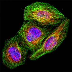
Eukaryotic cells are characterized by their internal membrane compartments (organelles), which allow different cellular processes to take place in a well-adapted environment. However, in order for the cell to function as a whole, the various membrane-bound organelles, as well as the plasma membrane, must communicate through vesicular and non-vesicular trafficking. Our research focuses on intracellular lipid trafficking pathways. Their disruption leads to various human pathologies such as cancer, diabetes and neurodegenerative diseases, and these pathways are also subverted for the propagation of numerous viruses, including Hepatitis C and SARS-CoV-2.

Keywords : Organelle, membrane trafficking, membrane contact site, polarity, lipid, endoplasmic reticulum, Golgi, plasma membrane

+33 (0)157278004 /  +33 (0)157278004     cathy.jackson(at)ijm.fr / jean-marc.verbavatz(at)ijm.fr

Eukaryotic cells are characterized by their internal membrane compartments (organelles), which allow different cellular processes to take place in a well-adapted environment. However, in order for the cell to function as a whole, the various membrane-bound organelles, including the endoplasmic reticulum (ER), Golgi apparatus, endosomes, lipid droplets, and mitochondria, as well as the plasma membrane, communicate through vesicular and non-vesicular trafficking (Jackson, Walch and Verbavatz, 2016; Kaczmarek, Verbavatz and Jackson, 2017; Jackson 2019). In vesicular trafficking, vesicles are transported from one compartment to their target, whereas non vesicular trafficking occurs at sites of close contact between organelles, named membrane contact sites (MCS), where the membranes of both organelles come together at a distance of 10-30 nm without fusing. Lipids can be transported by both vesicular and non-vesicular routes, but maintenance of the lipid composition of membranes requires lipid transfer at MCS (Jackson, Walch and Verbavatz, 2016). Disruption of lipid trafficking pathways leads to various human pathologies such as cancer, diabetes and neurodegenerative diseases, and these pathways are also subverted for the propagation of numerous viruses, including Hepatitis C and SARS-CoV-2.

Figure 1. Diagram illustrating the major organelles and membrane contact sites in a typical eukaryotic cell. ER, endoplasmic reticulum; PM, plasma membrane; LD, lipid droplet (Adapted from Jackson, Walch and Verbavatz, 2016).


We are using a multidisciplinary approach consisting of advanced imaging (light and electron microscopy, live cell imaging), biochemical approaches and proteomics analyses, to explore the following research questions:


  1. Arf family GTPase regulation of organelle dynamics.

Arf family proteins are crucial regulators of membrane dynamics in all eukaryotic cells, having essential functions at multiple organelles, in vesicular trafficking and at MCS. For a number of years, our group has studied the small G protein (GTPase) ADP ribosylation factor 1 (Arf1) and its activators (Donaldson and Jackson 2011), uncovering new functions for these regulators.

            Organelle dynamics in cells depends on microtubule motors that bind membrane compartments through adaptor proteins and pull them towards one or the other end of a microtubule. We discovered that the small G protein Arf1 and its activator GBF1 regulate the transport of mitochondria along microtubules and their spatial organization (Walch et al. 2018).

Lipid droplets (LDs) are the major energy storage depots of eukaryotic cells, which interface with membrane trafficking pathways. Our group uncovered a crucial role of Arf1 and its regulators in recruitment of LD-associated proteins to the LD surface (Donaldson and Jackson 2011, Bouvet et al. 2013, Jackson 2019). We have also described the mechanisms by which amphipathic helix-containing proteins such as perilipins are targeted to the LD surface (Donaldson and Jackson 2011, Copic et al. 2018).

            In an exciting recent development, Arf-related proteins have been identified in newly discovered archaea lineages (the Asgard archaea), suggesting that these proteins were present in the common ancestor of archaea and eukaryotes. Thus Asgard archaea appear to be the long-sought lineage from which eukaryotes emerged, i.e. the host that entered into endosymbiosis with the mitochondrial progenitor. We are collaborating with experts in bioinformatics, biophysics and structural biology, to determine if these predicted Asgard Arf-related proteins possess the hallmark features of eukaryotic Arf family GTPases, and to explore their potential roles in eukaryogenesis.


  1. Physiological functions of endoplasmic reticulum (ER) – organelle membrane contact sites (MCS)

Our studies have demonstrated functions of MCS in lipid transport from the ER to the plasma membrane, and their potential role in membrane growth (Moser von Filseck et al. 2015, Petkovic et al. 2014). The transport of lipids, including phosphoinositides at MCS, is an important determinant of the specific lipid composition of cell membranes (Jackson, Walch and Verbavatz, 2016), and it plays important roles in cellular rearrangements that occur during fundamental cell processes. Our group is currently studying the regulation and functions of MCS in processes such as cell division, cell migration, cell-cell contacts and apico-basal polarity and their implication in human disease.


2.1 MCS in cell division

Dramatic reorganizations of cellular organelles occur during cell division, essential for its successful completion. Improper cell division can result in numerous abnormalities, including aneuploidy and cancer. During division, membrane organelles undergo multiple steps of disassembly (nuclear membrane, Golgi apparatus…), fragmentation (mitochondria…) and spatial reorganization (mitochondria, ER…), followed by their reassembly at the end of mitosis. This is accompanied by coordinated changes in the distribution of phosphoinositides at the plasma membrane. We are studying the dynamic rearrangements of MCS in this process, the regulation of their function in lipid transport and their role in the choreography of membrane organelles during cell division.


2.2 MCS in epithelial cell polarity

The establishment and maintenance of cell polarity are crucial for the functions of epithelia, and are misregulated in human diseases such as cancer. In polarized epithelial cells, phosphoinositides are important determinants of the apical and basolateral plasma membrane identities. For example, the phosphoinositide PI(3,4,5)P3 is localized to the basolateral plasma membrane, but excluded from the apical membrane. Our goal is to decipher the role of MCS in determining the lipid composition of apical and basolateral membranes of epithelial cells and in the establishment and maintenance of cell polarity. Using cell biology approaches, we are investigating the membrane distribution of protein complexes at MCS, including lipid transfer proteins, and their function in the establishment of cell polarity.

Figure 2.  Junction between two epithelial MDCK cells showing the presence of multiple ER-PM contact sites (MCS, yellow arrows in the inset, right panel). ER, endoplasmic reticulum; PM, plasma membrane. Photos, L. Daunas and V. Proux-Gillardeaux.


2.3 MCS in cell motility

Cell motility highly relies on the distribution of phosphoinositides that act as lipidic messengers mediating cytoskeleton reshaping and adhesion dynamics. We investigate the role of VAPA, an ER-resident MCS tether, during cancer cell motility processes including collective and single cell migration, cell adhesion and mecano-sensing. Using high-resolution microscopy, quantitative imaging and optogenetic tools, we intend to define in space and time how lipid exchange taking place at VAPA-mediated MCS controls cell motility.

Figure 3. A. Colon adenocarcinoma Caco-2 cells migrating during a wound healing experiment. Here, a leader cell has been tracked for several hours (blue line). B. ER-PM contact sites identified by electron microscopy at the leading edge of Caco-2 migrating cells (yellow boxes). C. We use fluorescent probes to study the dynamics of MCS during cell motility. The picture has been taken at the tip of the leading edge of a migrating cell (green : ER marker ; magenta : ER-PM contact sites). Photos, M. Heuzé and H. Siegfried


Group Leaders:

Cathy JACKSON
Téléphone : +33 (0)157278004 / cathy.jackson (at) ijm.fr

Jean-Marc VERBAVATZ
Téléphone : +33 (0)157278004 / jean-marc.verbavatz (at) ijm.fr


Members :

HEUZÉ Mélina Researcher
PROUX-GILLARDEAUX Véronique Researcher
SIEGFRIED Hugo PhD Student
VERRAES Agathe Assistant engineer in biology
ALVES Marine Engineer
VERTUEUX Anais PhD Student
FARKOUH Georges Postdoctorant
NOVAK VANCLOVA Anna Postdoctorante
BEAUCOUSIN Julie Master 2

Lipid droplet biogenesis.

Jackson CL. Curr Opin Cell Biol. 2019 Aug;59:88-96. doi: 10.1016/j.ceb.2019.03.018. Epub 2019 May 7. PMID: 31075519 Review.

Correlative single-molecule localization microscopy and electron tomography reveals endosome nanoscale domains.

Franke C, Repnik U, Segeletz S, Brouilly N, Kalaidzidis Y, Verbavatz JM, Zerial M. Traffic. 2019 Aug;20(8):601-617. doi: 10.1111/tra.12671. PMID: 31206952

GBF1 and Arf1 interact with Miro and regulate mitochondrial positioning within cells.

Walch L, Pellier E, Leng W, Lakisic G, Gautreau A, Contremoulins V, Verbavatz JM#, Jackson CL#. Sci Rep. 2018 Nov 20;8(1):17121. doi: 10.1038/s41598-018-35190-0. PMID: 30459446 #co-corresponding authors

A giant amphipathic helix from a perilipin that is adapted for coating lipid droplets.

Čopič A, Antoine-Bally S, Giménez-Andrés M, La Torre Garay C, Antonny B, Manni MM, Pagnotta S, Guihot J, Jackson CL.Nat Commun. 2018 Apr 6;9(1):1332. doi: 10.1038/s41467-018-03717-8.PMID: 29626194

GBF1 and Arf1 function in vesicular trafficking, lipid homoeostasis and organelle dynamics.

Kaczmarek B, Verbavatz JM, Jackson CL. Biol Cell. 2017 Dec;109(12):391-399. doi: 10.1111/boc.201700042. Epub 2017 Nov 6. PMID: 28985001

Lipids and Their Trafficking: An Integral Part of Cellular Organization.

Jackson CL, Walch L, Verbavatz JM. Dev Cell. 2016 Oct 24;39(2):139-153. doi: 10.1016/j.devcel.2016.09.030. PMID: 27780039 Review.

The SNARE Sec22b has a non-fusogenic function in plasma membrane expansion.

Petkovic M, Jemaiel A, Daste F, Specht CG, Izeddin I, Vorkel D, Verbavatz JM, Darzacq X, Triller A, Pfenninger KH, Tareste D, Jackson CL#, Galli T#. Nat Cell Biol. 2014 May;16(5):434-44. doi: 10.1038/ncb2937. Epub 2014 Apr 6. PMID: 24705552  #co-corresponding authors

Targeting of the Arf-GEF GBF1 to lipid droplets and Golgi membranes.

Bouvet S, Golinelli-Cohen MP, Contremoulins V, Jackson CL.J Cell Sci. 2013 Oct 15;126(Pt 20):4794-805. doi: 10.1242/jcs.134254. Epub 2013 Aug 13.PMID: 23943872

Automated tracing of microtubules in electron tomograms of plastic embedded samples of Caenorhabditis elegans embryos.

Weber B, Greenan G, Prohaska S, Baum D, Hege HC, Müller-Reichert T, Hyman AA, Verbavatz JM.J Struct Biol. 2012 May;178(2):129-38. doi: 10.1016/j.jsb.2011.12.004. Epub 2011 Dec 13.PMID: 22182731

α-Synuclein and ALPS motifs are membrane curvature sensors whose contrasting chemistry mediates selective vesicle binding.

Pranke IM, Morello V, Bigay J, Gibson K, Verbavatz JM, Antonny B, Jackson CL. J Cell Biol. 2011 Jul 11;194(1):89-103. doi: 10.1083/jcb.201011118. PMID: 21746853

ARF family G proteins and their regulators: roles in membrane transport, development and disease.

Donaldson JG, Jackson CL.Nat Rev Mol Cell Biol. 2011 Jun;12(6):362-75. doi: 10.1038/nrm3117. Epub 2011 May 18.PMID: 21587297

Publications since 2017

2913254 L2N9KLHW items 1 0 date desc 8755 https://www.ijm.fr/wp-content/plugins/zotpress/
%7B%22status%22%3A%22success%22%2C%22updateneeded%22%3Afalse%2C%22instance%22%3A%22zotpress-4c84d17df7cbe09a9edd637319bb69e9%22%2C%22meta%22%3A%7B%22request_last%22%3A0%2C%22request_next%22%3A0%2C%22used_cache%22%3Atrue%7D%2C%22data%22%3A%5B%7B%22key%22%3A%22NHUW27MV%22%2C%22library%22%3A%7B%22id%22%3A2913254%7D%2C%22meta%22%3A%7B%22creatorSummary%22%3A%22Siegfried%20et%20al.%22%2C%22parsedDate%22%3A%222024-03-06%22%2C%22numChildren%22%3A1%7D%2C%22bib%22%3A%22%3Cdiv%20class%3D%5C%22csl-bib-body%5C%22%20style%3D%5C%22line-height%3A%202%3B%20padding-left%3A%201em%3B%20text-indent%3A-1em%3B%5C%22%3E%5Cn%20%20%3Cdiv%20class%3D%5C%22csl-entry%5C%22%3ESiegfried%2C%20H.%2C%20Farkouh%2C%20G.%2C%20Le%20Borgne%2C%20R.%2C%20Pioche-Durieu%2C%20C.%2C%20De%20Azevedo%20Laplace%2C%20T.%2C%20Verraes%2C%20A.%2C%20Daunas%2C%20L.%2C%20Verbavatz%2C%20J.-M.%2C%20%26amp%3B%20Heuz%26%23xE9%3B%2C%20M.%20L.%20%282024%29.%20The%20ER%20tether%20VAPA%20is%20required%20for%20proper%20cell%20motility%20and%20anchors%20ER-PM%20contact%20sites%20to%20focal%20adhesions.%20%3Ci%3EELife%3C%5C%2Fi%3E%2C%20%3Ci%3E13%3C%5C%2Fi%3E%2C%20e85962.%20%3Ca%20href%3D%27https%3A%5C%2F%5C%2Fdoi.org%5C%2F10.7554%5C%2FeLife.85962%27%3Ehttps%3A%5C%2F%5C%2Fdoi.org%5C%2F10.7554%5C%2FeLife.85962%3C%5C%2Fa%3E%3C%5C%2Fdiv%3E%5Cn%3C%5C%2Fdiv%3E%22%2C%22data%22%3A%7B%22itemType%22%3A%22journalArticle%22%2C%22title%22%3A%22The%20ER%20tether%20VAPA%20is%20required%20for%20proper%20cell%20motility%20and%20anchors%20ER-PM%20contact%20sites%20to%20focal%20adhesions%22%2C%22creators%22%3A%5B%7B%22creatorType%22%3A%22author%22%2C%22firstName%22%3A%22Hugo%22%2C%22lastName%22%3A%22Siegfried%22%7D%2C%7B%22creatorType%22%3A%22author%22%2C%22firstName%22%3A%22Georges%22%2C%22lastName%22%3A%22Farkouh%22%7D%2C%7B%22creatorType%22%3A%22author%22%2C%22firstName%22%3A%22R%5Cu00e9mi%22%2C%22lastName%22%3A%22Le%20Borgne%22%7D%2C%7B%22creatorType%22%3A%22author%22%2C%22firstName%22%3A%22Catherine%22%2C%22lastName%22%3A%22Pioche-Durieu%22%7D%2C%7B%22creatorType%22%3A%22author%22%2C%22firstName%22%3A%22Tha%5Cu00efs%22%2C%22lastName%22%3A%22De%20Azevedo%20Laplace%22%7D%2C%7B%22creatorType%22%3A%22author%22%2C%22firstName%22%3A%22Agathe%22%2C%22lastName%22%3A%22Verraes%22%7D%2C%7B%22creatorType%22%3A%22author%22%2C%22firstName%22%3A%22Lucien%22%2C%22lastName%22%3A%22Daunas%22%7D%2C%7B%22creatorType%22%3A%22author%22%2C%22firstName%22%3A%22Jean-Marc%22%2C%22lastName%22%3A%22Verbavatz%22%7D%2C%7B%22creatorType%22%3A%22author%22%2C%22firstName%22%3A%22M%5Cu00e9lina%20L.%22%2C%22lastName%22%3A%22Heuz%5Cu00e9%22%7D%5D%2C%22abstractNote%22%3A%22Cell%20motility%20processes%20highly%20depend%20on%20the%20membrane%20distribution%20of%20Phosphoinositides%2C%20giving%20rise%20to%20cytoskeleton%20reshaping%20and%20membrane%20trafficking%20events.%20Membrane%20contact%20sites%20serve%20as%20platforms%20for%20direct%20lipid%20exchange%20and%20calcium%20fluxes%20between%20two%20organelles.%20Here%2C%20we%20show%20that%20VAPA%2C%20an%20ER%20transmembrane%20contact%20site%20tether%2C%20plays%20a%20crucial%20role%20during%20cell%20motility.%20CaCo2%20adenocarcinoma%20epithelial%20cells%20depleted%20for%20VAPA%20exhibit%20several%20collective%20and%20individual%20motility%20defects%2C%20disorganized%20actin%20cytoskeleton%20and%20altered%20protrusive%20activity.%20During%20migration%2C%20VAPA%20is%20required%20for%20the%20maintenance%20of%20PI%284%29P%20and%20PI%284%2C5%29P2%20levels%20at%20the%20plasma%20membrane%2C%20but%20not%20for%20PI%284%29P%20homeostasis%20in%20the%20Golgi%20and%20endosomal%20compartments.%20Importantly%2C%20we%20show%20that%20VAPA%20regulates%20the%20dynamics%20of%20focal%20adhesions%20%28FA%29%20through%20its%20MSP%20domain%2C%20is%20essential%20to%20stabilize%20and%20anchor%20ventral%20ER-PM%20contact%20sites%20to%20FA%2C%20and%20mediates%20microtubule-dependent%20FA%20disassembly.%20To%20conclude%2C%20our%20results%20reveal%20unknown%20functions%20for%20VAPA-mediated%20membrane%20contact%20sites%20during%20cell%20motility%20and%20provide%20a%20dynamic%20picture%20of%20ER-PM%20contact%20sites%20connection%20with%20FA%20mediated%20by%20VAPA.%22%2C%22date%22%3A%222024-03-06%22%2C%22language%22%3A%22eng%22%2C%22DOI%22%3A%2210.7554%5C%2FeLife.85962%22%2C%22ISSN%22%3A%222050-084X%22%2C%22url%22%3A%22%22%2C%22collections%22%3A%5B%22L2N9KLHW%22%5D%2C%22dateModified%22%3A%222024-03-07T12%3A57%3A03Z%22%7D%7D%2C%7B%22key%22%3A%2284626TB7%22%2C%22library%22%3A%7B%22id%22%3A2913254%7D%2C%22meta%22%3A%7B%22creatorSummary%22%3A%22Jackson%20et%20al.%22%2C%22parsedDate%22%3A%222023-12-01%22%2C%22numChildren%22%3A2%7D%2C%22bib%22%3A%22%3Cdiv%20class%3D%5C%22csl-bib-body%5C%22%20style%3D%5C%22line-height%3A%202%3B%20padding-left%3A%201em%3B%20text-indent%3A-1em%3B%5C%22%3E%5Cn%20%20%3Cdiv%20class%3D%5C%22csl-entry%5C%22%3EJackson%2C%20C.%20L.%2C%20M%26%23xE9%3Bn%26%23xE9%3Btrey%2C%20J.%2C%20Sivia%2C%20M.%2C%20Dacks%2C%20J.%20B.%2C%20%26amp%3B%20Eli%26%23xE1%3B%26%23x161%3B%2C%20M.%20%282023%29.%20An%20evolutionary%20perspective%20on%20Arf%20family%20GTPases.%20%3Ci%3ECurrent%20Opinion%20in%20Cell%20Biology%3C%5C%2Fi%3E%2C%20%3Ci%3E85%3C%5C%2Fi%3E%2C%20102268.%20%3Ca%20href%3D%27https%3A%5C%2F%5C%2Fdoi.org%5C%2F10.1016%5C%2Fj.ceb.2023.102268%27%3Ehttps%3A%5C%2F%5C%2Fdoi.org%5C%2F10.1016%5C%2Fj.ceb.2023.102268%3C%5C%2Fa%3E%3C%5C%2Fdiv%3E%5Cn%3C%5C%2Fdiv%3E%22%2C%22data%22%3A%7B%22itemType%22%3A%22journalArticle%22%2C%22title%22%3A%22An%20evolutionary%20perspective%20on%20Arf%20family%20GTPases%22%2C%22creators%22%3A%5B%7B%22creatorType%22%3A%22author%22%2C%22firstName%22%3A%22Catherine%20L.%22%2C%22lastName%22%3A%22Jackson%22%7D%2C%7B%22creatorType%22%3A%22author%22%2C%22firstName%22%3A%22Julie%22%2C%22lastName%22%3A%22M%5Cu00e9n%5Cu00e9trey%22%7D%2C%7B%22creatorType%22%3A%22author%22%2C%22firstName%22%3A%22Mandeep%22%2C%22lastName%22%3A%22Sivia%22%7D%2C%7B%22creatorType%22%3A%22author%22%2C%22firstName%22%3A%22Joel%20B.%22%2C%22lastName%22%3A%22Dacks%22%7D%2C%7B%22creatorType%22%3A%22author%22%2C%22firstName%22%3A%22Marek%22%2C%22lastName%22%3A%22Eli%5Cu00e1%5Cu0161%22%7D%5D%2C%22abstractNote%22%3A%22The%20Arf%20family%20GTPases%20are%20regulators%20of%20eukaryotic%20cellular%20organization%2C%20functioning%20in%20the%20secretory%20and%20endocytic%20pathways%2C%20in%20cilia%20and%20flagella%2C%20in%20cytoskeleton%20dynamics%2C%20and%20in%20lipid%20metabolism.%20We%20describe%20the%20evolution%20of%20this%20protein%20family%20and%20its%20well-studied%20regulators.%20The%20last%20eukaryotic%20common%20ancestor%20had%20fifteen%20members%2C%20and%20the%20current%20complement%20of%20Arf%20GTPases%20has%20been%20sculpted%20by%20gene%20loss%20and%20gene%20duplications%20since%20that%20point.%20Some%20Arf%20family%20GTPases%20%28such%20as%20those%20that%20recruit%20vesicle%20coats%20in%20the%20secretory%20pathway%29%20are%20present%20in%20virtually%20all%20eukaryotes%2C%20whereas%20others%20%28such%20as%20those%20functioning%20in%20cilia%5C%2Fflagella%29%20have%20a%20more%20limited%20distribution.%20A%20challenge%20for%20the%20future%20is%20understanding%20the%20full%20spectrum%20of%20Arf%20family%20functions%20throughout%20eukaryotes.%22%2C%22date%22%3A%222023-12-01%22%2C%22language%22%3A%22%22%2C%22DOI%22%3A%2210.1016%5C%2Fj.ceb.2023.102268%22%2C%22ISSN%22%3A%220955-0674%22%2C%22url%22%3A%22https%3A%5C%2F%5C%2Fwww.sciencedirect.com%5C%2Fscience%5C%2Farticle%5C%2Fpii%5C%2FS0955067423001175%22%2C%22collections%22%3A%5B%22L2N9KLHW%22%5D%2C%22dateModified%22%3A%222023-12-14T10%3A32%3A13Z%22%7D%7D%2C%7B%22key%22%3A%22TKC7LLDL%22%2C%22library%22%3A%7B%22id%22%3A2913254%7D%2C%22meta%22%3A%7B%22creatorSummary%22%3A%22Ferreras%20et%20al.%22%2C%22parsedDate%22%3A%222023-02-02%22%2C%22numChildren%22%3A4%7D%2C%22bib%22%3A%22%3Cdiv%20class%3D%5C%22csl-bib-body%5C%22%20style%3D%5C%22line-height%3A%202%3B%20padding-left%3A%201em%3B%20text-indent%3A-1em%3B%5C%22%3E%5Cn%20%20%3Cdiv%20class%3D%5C%22csl-entry%5C%22%3EFerreras%2C%20S.%2C%20Singh%2C%20N.%20P.%2C%20Le%20Borgne%2C%20R.%2C%20Bun%2C%20P.%2C%20Binz%2C%20T.%2C%20Parton%2C%20R.%20G.%2C%20Verbavatz%2C%20J.-M.%2C%20Vannier%2C%20C.%2C%20%26amp%3B%20Galli%2C%20T.%20%282023%29.%20A%20synthetic%20organelle%20approach%20to%20probe%20SNARE-mediated%20membrane%20fusion%20in%20a%20bacterial%20host.%20%3Ci%3EThe%20Journal%20of%20Biological%20Chemistry%3C%5C%2Fi%3E%2C%20102974.%20%3Ca%20href%3D%27https%3A%5C%2F%5C%2Fdoi.org%5C%2F10.1016%5C%2Fj.jbc.2023.102974%27%3Ehttps%3A%5C%2F%5C%2Fdoi.org%5C%2F10.1016%5C%2Fj.jbc.2023.102974%3C%5C%2Fa%3E%3C%5C%2Fdiv%3E%5Cn%3C%5C%2Fdiv%3E%22%2C%22data%22%3A%7B%22itemType%22%3A%22journalArticle%22%2C%22title%22%3A%22A%20synthetic%20organelle%20approach%20to%20probe%20SNARE-mediated%20membrane%20fusion%20in%20a%20bacterial%20host%22%2C%22creators%22%3A%5B%7B%22creatorType%22%3A%22author%22%2C%22firstName%22%3A%22Soledad%22%2C%22lastName%22%3A%22Ferreras%22%7D%2C%7B%22creatorType%22%3A%22author%22%2C%22firstName%22%3A%22Neha%20Pratap%22%2C%22lastName%22%3A%22Singh%22%7D%2C%7B%22creatorType%22%3A%22author%22%2C%22firstName%22%3A%22R%5Cu00e9mi%22%2C%22lastName%22%3A%22Le%20Borgne%22%7D%2C%7B%22creatorType%22%3A%22author%22%2C%22firstName%22%3A%22Philippe%22%2C%22lastName%22%3A%22Bun%22%7D%2C%7B%22creatorType%22%3A%22author%22%2C%22firstName%22%3A%22Thomas%22%2C%22lastName%22%3A%22Binz%22%7D%2C%7B%22creatorType%22%3A%22author%22%2C%22firstName%22%3A%22Robert%20G.%22%2C%22lastName%22%3A%22Parton%22%7D%2C%7B%22creatorType%22%3A%22author%22%2C%22firstName%22%3A%22Jean-Marc%22%2C%22lastName%22%3A%22Verbavatz%22%7D%2C%7B%22creatorType%22%3A%22author%22%2C%22firstName%22%3A%22Christian%22%2C%22lastName%22%3A%22Vannier%22%7D%2C%7B%22creatorType%22%3A%22author%22%2C%22firstName%22%3A%22Thierry%22%2C%22lastName%22%3A%22Galli%22%7D%5D%2C%22abstractNote%22%3A%22In%20vivo%20and%20in%20vitro%20assays%2C%20particularly%20reconstitution%20using%20artificial%20membranes%20have%20established%20the%20role%20of%20synaptic%20soluble%20N-Ethylmaleimide%20sensitive%20attachment%20protein%20receptors%20%28SNAREs%29%20VAMP2%2C%20Syntaxin%201%2C%20and%20SNAP-25%20in%20membrane%20fusion.%20However%2C%20using%20artificial%20membranes%20requires%20challenging%20protein%20purifications%20that%20could%20be%20avoided%20in%20a%20cell-based%20assay.%20Here%20we%20developed%20a%20synthetic%20biological%20approach%20based%20on%20the%20generation%20of%20membrane%20cisternae%20by%20the%20integral%20membrane%20protein%20Caveolin%20in%20E.coli%20and%20co-expression%20of%20SNAREs.%20Syntaxin%201%5C%2FSNAP-25%5C%2FVAMP2%20complexes%20were%20formed%20and%20regulated%20by%20SNARE%20partner%20protein%20Munc-18a%20in%20the%20presence%20of%20Caveolin.%20Additionally%2C%20Syntaxin1%5C%2FSNAP-25%5C%2FVAMP2%20synthesis%20provoked%20increased%20length%20of%20E.coli%20only%20in%20the%20presence%20of%20Caveolin.%20We%20found%20that%20cell%20elongation%20required%20SNAP-25%20and%20was%20inhibited%20by%20tetanus%20neurotoxin.%20This%20elongation%20was%20not%20a%20result%20of%20cell%20division%20arrest.%20Furthermore%2C%20electron%20and%20super-resolution%20microscopies%20showed%20that%20synaptic%20SNAREs%20and%20Caveolin%20co-expression%20led%20to%20the%20partial%20loss%20of%20the%20cisternae%2C%20suggesting%20their%20fusion%20with%20the%20plasma%20membrane.%20In%20summary%2C%20we%20propose%20that%20this%20assay%20reconstitutes%20membrane%20fusion%20in%20a%20simple%20organism%20with%20an%20easy-to-observe%20phenotype%20and%20is%20amenable%20to%20structure-function%20studies%20of%20SNAREs.%22%2C%22date%22%3A%222023-02-02%22%2C%22language%22%3A%22eng%22%2C%22DOI%22%3A%2210.1016%5C%2Fj.jbc.2023.102974%22%2C%22ISSN%22%3A%221083-351X%22%2C%22url%22%3A%22%22%2C%22collections%22%3A%5B%22L2N9KLHW%22%2C%22I7CUV6U5%22%5D%2C%22dateModified%22%3A%222023-12-14T10%3A32%3A13Z%22%7D%7D%2C%7B%22key%22%3A%22BMQJ54DN%22%2C%22library%22%3A%7B%22id%22%3A2913254%7D%2C%22meta%22%3A%7B%22creatorSummary%22%3A%22Lachat%20et%20al.%22%2C%22parsedDate%22%3A%222022-06-30%22%2C%22numChildren%22%3A4%7D%2C%22bib%22%3A%22%3Cdiv%20class%3D%5C%22csl-bib-body%5C%22%20style%3D%5C%22line-height%3A%202%3B%20padding-left%3A%201em%3B%20text-indent%3A-1em%3B%5C%22%3E%5Cn%20%20%3Cdiv%20class%3D%5C%22csl-entry%5C%22%3ELachat%2C%20J.%2C%20Pascault%2C%20A.%2C%20Thibaut%2C%20D.%2C%20Le%20Borgne%2C%20R.%2C%20Verbavatz%2C%20J.-M.%2C%20%26amp%3B%20Weiner%2C%20A.%20%282022%29.%20Trans-cellular%20tunnels%20induced%20by%20the%20fungal%20pathogen%20Candida%20albicans%20facilitate%20invasion%20through%20successive%20epithelial%20cells%20without%20host%20damage.%20%3Ci%3ENature%20Communications%3C%5C%2Fi%3E%2C%20%3Ci%3E13%3C%5C%2Fi%3E%281%29%2C%203781.%20%3Ca%20href%3D%27https%3A%5C%2F%5C%2Fdoi.org%5C%2F10.1038%5C%2Fs41467-022-31237-z%27%3Ehttps%3A%5C%2F%5C%2Fdoi.org%5C%2F10.1038%5C%2Fs41467-022-31237-z%3C%5C%2Fa%3E%3C%5C%2Fdiv%3E%5Cn%3C%5C%2Fdiv%3E%22%2C%22data%22%3A%7B%22itemType%22%3A%22journalArticle%22%2C%22title%22%3A%22Trans-cellular%20tunnels%20induced%20by%20the%20fungal%20pathogen%20Candida%20albicans%20facilitate%20invasion%20through%20successive%20epithelial%20cells%20without%20host%20damage%22%2C%22creators%22%3A%5B%7B%22creatorType%22%3A%22author%22%2C%22firstName%22%3A%22Joy%22%2C%22lastName%22%3A%22Lachat%22%7D%2C%7B%22creatorType%22%3A%22author%22%2C%22firstName%22%3A%22Alice%22%2C%22lastName%22%3A%22Pascault%22%7D%2C%7B%22creatorType%22%3A%22author%22%2C%22firstName%22%3A%22Delphine%22%2C%22lastName%22%3A%22Thibaut%22%7D%2C%7B%22creatorType%22%3A%22author%22%2C%22firstName%22%3A%22R%5Cu00e9mi%22%2C%22lastName%22%3A%22Le%20Borgne%22%7D%2C%7B%22creatorType%22%3A%22author%22%2C%22firstName%22%3A%22Jean-Marc%22%2C%22lastName%22%3A%22Verbavatz%22%7D%2C%7B%22creatorType%22%3A%22author%22%2C%22firstName%22%3A%22Allon%22%2C%22lastName%22%3A%22Weiner%22%7D%5D%2C%22abstractNote%22%3A%22The%20opportunistic%20fungal%20pathogen%20Candida%20albicans%20is%20normally%20commensal%2C%20residing%20in%20the%20mucosa%20of%20most%20healthy%20individuals.%20In%20susceptible%20hosts%2C%20its%20filamentous%20hyphal%20form%20can%20invade%20epithelial%20layers%20leading%20to%20superficial%20or%20severe%20systemic%20infection.%20Although%20invasion%20is%20mainly%20intracellular%2C%20it%20causes%20no%20apparent%20damage%20to%20host%20cells%20at%20early%20stages%20of%20infection.%20Here%2C%20we%20investigate%20C.%20albicans%20invasion%20in%20vitro%20using%20live-cell%20imaging%20and%20the%20damage-sensitive%20reporter%20galectin-3.%20Quantitative%20single%20cell%20analysis%20shows%20that%20invasion%20can%20result%20in%20host%20membrane%20breaching%20at%20different%20stages%20and%20host%20cell%20death%2C%20or%20in%20traversal%20of%20host%20cells%20without%20membrane%20breaching.%20Membrane%20labelling%20and%20three-dimensional%20%27volume%27%20electron%20microscopy%20reveal%20that%20hyphae%20can%20traverse%20several%20host%20cells%20within%20trans-cellular%20tunnels%20that%20are%20progressively%20remodelled%20and%20may%20undergo%20%27inflations%27%20linked%20to%20host%20glycogen%20stores.%20Thus%2C%20C.%20albicans%20early%20invasion%20of%20epithelial%20tissues%20can%20lead%20to%20either%20host%20membrane%20breaching%20or%20trans-cellular%20tunnelling.%22%2C%22date%22%3A%222022-06-30%22%2C%22language%22%3A%22eng%22%2C%22DOI%22%3A%2210.1038%5C%2Fs41467-022-31237-z%22%2C%22ISSN%22%3A%222041-1723%22%2C%22url%22%3A%22%22%2C%22collections%22%3A%5B%22L2N9KLHW%22%2C%22I7CUV6U5%22%5D%2C%22dateModified%22%3A%222023-12-14T10%3A32%3A13Z%22%7D%7D%2C%7B%22key%22%3A%226QFCFER6%22%2C%22library%22%3A%7B%22id%22%3A2913254%7D%2C%22meta%22%3A%7B%22creatorSummary%22%3A%22Fernandes%20et%20al.%22%2C%22parsedDate%22%3A%222022-06%22%2C%22numChildren%22%3A3%7D%2C%22bib%22%3A%22%3Cdiv%20class%3D%5C%22csl-bib-body%5C%22%20style%3D%5C%22line-height%3A%202%3B%20padding-left%3A%201em%3B%20text-indent%3A-1em%3B%5C%22%3E%5Cn%20%20%3Cdiv%20class%3D%5C%22csl-entry%5C%22%3EFernandes%2C%20P.%2C%20Loubens%2C%20M.%2C%20Le%20Borgne%2C%20R.%2C%20Marinach%2C%20C.%2C%20Ardin%2C%20B.%2C%20Briquet%2C%20S.%2C%20Vincensini%2C%20L.%2C%20Hamada%2C%20S.%2C%20Hoareau-Coudert%2C%20B.%2C%20Verbavatz%2C%20J.-M.%2C%20Weiner%2C%20A.%2C%20%26amp%3B%20Silvie%2C%20O.%20%282022%29.%20The%20AMA1-RON%20complex%20drives%20Plasmodium%20sporozoite%20invasion%20in%20the%20mosquito%20and%20mammalian%20hosts.%20%3Ci%3EPLoS%20Pathogens%3C%5C%2Fi%3E%2C%20%3Ci%3E18%3C%5C%2Fi%3E%286%29%2C%20e1010643.%20%3Ca%20href%3D%27https%3A%5C%2F%5C%2Fdoi.org%5C%2F10.1371%5C%2Fjournal.ppat.1010643%27%3Ehttps%3A%5C%2F%5C%2Fdoi.org%5C%2F10.1371%5C%2Fjournal.ppat.1010643%3C%5C%2Fa%3E%3C%5C%2Fdiv%3E%5Cn%3C%5C%2Fdiv%3E%22%2C%22data%22%3A%7B%22itemType%22%3A%22journalArticle%22%2C%22title%22%3A%22The%20AMA1-RON%20complex%20drives%20Plasmodium%20sporozoite%20invasion%20in%20the%20mosquito%20and%20mammalian%20hosts%22%2C%22creators%22%3A%5B%7B%22creatorType%22%3A%22author%22%2C%22firstName%22%3A%22Priyanka%22%2C%22lastName%22%3A%22Fernandes%22%7D%2C%7B%22creatorType%22%3A%22author%22%2C%22firstName%22%3A%22Manon%22%2C%22lastName%22%3A%22Loubens%22%7D%2C%7B%22creatorType%22%3A%22author%22%2C%22firstName%22%3A%22R%5Cu00e9mi%22%2C%22lastName%22%3A%22Le%20Borgne%22%7D%2C%7B%22creatorType%22%3A%22author%22%2C%22firstName%22%3A%22Carine%22%2C%22lastName%22%3A%22Marinach%22%7D%2C%7B%22creatorType%22%3A%22author%22%2C%22firstName%22%3A%22B%5Cu00e9atrice%22%2C%22lastName%22%3A%22Ardin%22%7D%2C%7B%22creatorType%22%3A%22author%22%2C%22firstName%22%3A%22Sylvie%22%2C%22lastName%22%3A%22Briquet%22%7D%2C%7B%22creatorType%22%3A%22author%22%2C%22firstName%22%3A%22Laetitia%22%2C%22lastName%22%3A%22Vincensini%22%7D%2C%7B%22creatorType%22%3A%22author%22%2C%22firstName%22%3A%22Soumia%22%2C%22lastName%22%3A%22Hamada%22%7D%2C%7B%22creatorType%22%3A%22author%22%2C%22firstName%22%3A%22B%5Cu00e9n%5Cu00e9dicte%22%2C%22lastName%22%3A%22Hoareau-Coudert%22%7D%2C%7B%22creatorType%22%3A%22author%22%2C%22firstName%22%3A%22Jean-Marc%22%2C%22lastName%22%3A%22Verbavatz%22%7D%2C%7B%22creatorType%22%3A%22author%22%2C%22firstName%22%3A%22Allon%22%2C%22lastName%22%3A%22Weiner%22%7D%2C%7B%22creatorType%22%3A%22author%22%2C%22firstName%22%3A%22Olivier%22%2C%22lastName%22%3A%22Silvie%22%7D%5D%2C%22abstractNote%22%3A%22Plasmodium%20sporozoites%20that%20are%20transmitted%20by%20blood-feeding%20female%20Anopheles%20mosquitoes%20invade%20hepatocytes%20for%20an%20initial%20round%20of%20intracellular%20replication%2C%20leading%20to%20the%20release%20of%20merozoites%20that%20invade%20and%20multiply%20within%20red%20blood%20cells.%20Sporozoites%20and%20merozoites%20share%20a%20number%20of%20proteins%20that%20are%20expressed%20by%20both%20stages%2C%20including%20the%20Apical%20Membrane%20Antigen%201%20%28AMA1%29%20and%20the%20Rhoptry%20Neck%20Proteins%20%28RONs%29.%20Although%20AMA1%20and%20RONs%20are%20essential%20for%20merozoite%20invasion%20of%20erythrocytes%20during%20asexual%20blood%20stage%20replication%20of%20the%20parasite%2C%20their%20function%20in%20sporozoites%20was%20still%20unclear.%20Here%20we%20show%20that%20AMA1%20interacts%20with%20RONs%20in%20mature%20sporozoites.%20By%20using%20DiCre-mediated%20conditional%20gene%20deletion%20in%20P.%20berghei%2C%20we%20demonstrate%20that%20loss%20of%20AMA1%2C%20RON2%20or%20RON4%20in%20sporozoites%20impairs%20colonization%20of%20the%20mosquito%20salivary%20glands%20and%20invasion%20of%20mammalian%20hepatocytes%2C%20without%20affecting%20transcellular%20parasite%20migration.%20Three-dimensional%20electron%20microscopy%20data%20showed%20that%20sporozoites%20enter%20salivary%20gland%20cells%20through%20a%20ring-like%20structure%20and%20by%20forming%20a%20transient%20vacuole.%20The%20absence%20of%20a%20functional%20AMA1-RON%20complex%20led%20to%20an%20altered%20morphology%20of%20the%20entry%20junction%2C%20associated%20with%20epithelial%20cell%20damage.%20Our%20data%20establish%20that%20AMA1%20and%20RONs%20facilitate%20host%20cell%20invasion%20across%20Plasmodium%20invasive%20stages%2C%20and%20suggest%20that%20sporozoites%20use%20the%20AMA1-RON%20complex%20to%20efficiently%20and%20safely%20enter%20the%20mosquito%20salivary%20glands%20to%20ensure%20successful%20parasite%20transmission.%20These%20results%20open%20up%20the%20possibility%20of%20targeting%20the%20AMA1-RON%20complex%20for%20transmission-blocking%20antimalarial%20strategies.%22%2C%22date%22%3A%222022-06%22%2C%22language%22%3A%22eng%22%2C%22DOI%22%3A%2210.1371%5C%2Fjournal.ppat.1010643%22%2C%22ISSN%22%3A%221553-7374%22%2C%22url%22%3A%22%22%2C%22collections%22%3A%5B%22L2N9KLHW%22%2C%22I7CUV6U5%22%5D%2C%22dateModified%22%3A%222023-12-14T10%3A32%3A13Z%22%7D%7D%2C%7B%22key%22%3A%223DYY5CAP%22%2C%22library%22%3A%7B%22id%22%3A2913254%7D%2C%22meta%22%3A%7B%22creatorSummary%22%3A%22Xie%20et%20al.%22%2C%22parsedDate%22%3A%222022-02-22%22%2C%22numChildren%22%3A5%7D%2C%22bib%22%3A%22%3Cdiv%20class%3D%5C%22csl-bib-body%5C%22%20style%3D%5C%22line-height%3A%202%3B%20padding-left%3A%201em%3B%20text-indent%3A-1em%3B%5C%22%3E%5Cn%20%20%3Cdiv%20class%3D%5C%22csl-entry%5C%22%3EXie%2C%20J.%2C%20Najafi%2C%20J.%2C%20Le%20Borgne%2C%20R.%2C%20Verbavatz%2C%20J.-M.%2C%20Durieu%2C%20C.%2C%20Sall%26%23xE9%3B%2C%20J.%2C%20%26amp%3B%20Minc%2C%20N.%20%282022%29.%20Contribution%20of%20cytoplasm%20viscoelastic%20properties%20to%20mitotic%20spindle%20positioning.%20%3Ci%3EProceedings%20of%20the%20National%20Academy%20of%20Sciences%20of%20the%20United%20States%20of%20America%3C%5C%2Fi%3E%2C%20%3Ci%3E119%3C%5C%2Fi%3E%288%29%2C%20e2115593119.%20%3Ca%20href%3D%27https%3A%5C%2F%5C%2Fdoi.org%5C%2F10.1073%5C%2Fpnas.2115593119%27%3Ehttps%3A%5C%2F%5C%2Fdoi.org%5C%2F10.1073%5C%2Fpnas.2115593119%3C%5C%2Fa%3E%3C%5C%2Fdiv%3E%5Cn%3C%5C%2Fdiv%3E%22%2C%22data%22%3A%7B%22itemType%22%3A%22journalArticle%22%2C%22title%22%3A%22Contribution%20of%20cytoplasm%20viscoelastic%20properties%20to%20mitotic%20spindle%20positioning%22%2C%22creators%22%3A%5B%7B%22creatorType%22%3A%22author%22%2C%22firstName%22%3A%22Jing%22%2C%22lastName%22%3A%22Xie%22%7D%2C%7B%22creatorType%22%3A%22author%22%2C%22firstName%22%3A%22Javad%22%2C%22lastName%22%3A%22Najafi%22%7D%2C%7B%22creatorType%22%3A%22author%22%2C%22firstName%22%3A%22R%5Cu00e9mi%22%2C%22lastName%22%3A%22Le%20Borgne%22%7D%2C%7B%22creatorType%22%3A%22author%22%2C%22firstName%22%3A%22Jean-Marc%22%2C%22lastName%22%3A%22Verbavatz%22%7D%2C%7B%22creatorType%22%3A%22author%22%2C%22firstName%22%3A%22Catherine%22%2C%22lastName%22%3A%22Durieu%22%7D%2C%7B%22creatorType%22%3A%22author%22%2C%22firstName%22%3A%22Jeremy%22%2C%22lastName%22%3A%22Sall%5Cu00e9%22%7D%2C%7B%22creatorType%22%3A%22author%22%2C%22firstName%22%3A%22Nicolas%22%2C%22lastName%22%3A%22Minc%22%7D%5D%2C%22abstractNote%22%3A%22Cells%20are%20filled%20with%20macromolecules%20and%20polymer%20networks%20that%20set%20scale-dependent%20viscous%20and%20elastic%20properties%20to%20the%20cytoplasm.%20Although%20the%20role%20of%20these%20parameters%20in%20molecular%20diffusion%2C%20reaction%20kinetics%2C%20and%20cellular%20biochemistry%20is%20being%20increasingly%20recognized%2C%20their%20contributions%20to%20the%20motion%20and%20positioning%20of%20larger%20organelles%2C%20such%20as%20mitotic%20spindles%20for%20cell%20division%2C%20remain%20unknown.%20Here%2C%20using%20magnetic%20tweezers%20to%20displace%20and%20rotate%20mitotic%20spindles%20in%20living%20embryos%2C%20we%20uncovered%20that%20the%20cytoplasm%20can%20impart%20viscoelastic%20reactive%20forces%20that%20move%20spindles%2C%20or%20passive%20objects%20with%20similar%20size%2C%20back%20to%20their%20original%20positions.%20These%20forces%20are%20independent%20of%20cytoskeletal%20force%20generators%20yet%20reach%20hundreds%20of%20piconewtons%20and%20scale%20with%20cytoplasm%20crowding.%20Spindle%20motion%20shears%20and%20fluidizes%20the%20cytoplasm%2C%20dissipating%20elastic%20energy%20and%20limiting%20spindle%20recoils%20with%20functional%20implications%20for%20asymmetric%20and%20oriented%20divisions.%20These%20findings%20suggest%20that%20bulk%20cytoplasm%20material%20properties%20may%20constitute%20important%20control%20elements%20for%20the%20regulation%20of%20division%20positioning%20and%20cellular%20organization.%22%2C%22date%22%3A%222022-02-22%22%2C%22language%22%3A%22eng%22%2C%22DOI%22%3A%2210.1073%5C%2Fpnas.2115593119%22%2C%22ISSN%22%3A%221091-6490%22%2C%22url%22%3A%22%22%2C%22collections%22%3A%5B%22L2N9KLHW%22%2C%22I7CUV6U5%22%5D%2C%22dateModified%22%3A%222023-12-14T10%3A32%3A13Z%22%7D%7D%2C%7B%22key%22%3A%22QSXXNJ6L%22%2C%22library%22%3A%7B%22id%22%3A2913254%7D%2C%22meta%22%3A%7B%22creatorSummary%22%3A%22Dussouchaud%20et%20al.%22%2C%22parsedDate%22%3A%222022%22%2C%22numChildren%22%3A5%7D%2C%22bib%22%3A%22%3Cdiv%20class%3D%5C%22csl-bib-body%5C%22%20style%3D%5C%22line-height%3A%202%3B%20padding-left%3A%201em%3B%20text-indent%3A-1em%3B%5C%22%3E%5Cn%20%20%3Cdiv%20class%3D%5C%22csl-entry%5C%22%3EDussouchaud%2C%20A.%2C%20Jacob%2C%20J.%2C%20Secq%2C%20C.%2C%20Verbavatz%2C%20J.-M.%2C%20Moras%2C%20M.%2C%20Larghero%2C%20J.%2C%20Fader%2C%20C.%20M.%2C%20Ostuni%2C%20M.%20A.%2C%20%26amp%3B%20Lefevre%2C%20S.%20D.%20%282022%29.%20Transmission%20Electron%20Microscopy%20to%20Follow%20Ultrastructural%20Modifications%20of%20Erythroblasts%20Upon%20ex%20vivo%20Human%20Erythropoiesis.%20%3Ci%3EFrontiers%20in%20Physiology%3C%5C%2Fi%3E%2C%20%3Ci%3E12%3C%5C%2Fi%3E%2C%20791691.%20%3Ca%20href%3D%27https%3A%5C%2F%5C%2Fdoi.org%5C%2F10.3389%5C%2Ffphys.2021.791691%27%3Ehttps%3A%5C%2F%5C%2Fdoi.org%5C%2F10.3389%5C%2Ffphys.2021.791691%3C%5C%2Fa%3E%3C%5C%2Fdiv%3E%5Cn%3C%5C%2Fdiv%3E%22%2C%22data%22%3A%7B%22itemType%22%3A%22journalArticle%22%2C%22title%22%3A%22Transmission%20Electron%20Microscopy%20to%20Follow%20Ultrastructural%20Modifications%20of%20Erythroblasts%20Upon%20ex%20vivo%20Human%20Erythropoiesis%22%2C%22creators%22%3A%5B%7B%22creatorType%22%3A%22author%22%2C%22firstName%22%3A%22Alice%22%2C%22lastName%22%3A%22Dussouchaud%22%7D%2C%7B%22creatorType%22%3A%22author%22%2C%22firstName%22%3A%22Julieta%22%2C%22lastName%22%3A%22Jacob%22%7D%2C%7B%22creatorType%22%3A%22author%22%2C%22firstName%22%3A%22Charles%22%2C%22lastName%22%3A%22Secq%22%7D%2C%7B%22creatorType%22%3A%22author%22%2C%22firstName%22%3A%22Jean-Marc%22%2C%22lastName%22%3A%22Verbavatz%22%7D%2C%7B%22creatorType%22%3A%22author%22%2C%22firstName%22%3A%22Martina%22%2C%22lastName%22%3A%22Moras%22%7D%2C%7B%22creatorType%22%3A%22author%22%2C%22firstName%22%3A%22J%5Cu00e9r%5Cu00f4me%22%2C%22lastName%22%3A%22Larghero%22%7D%2C%7B%22creatorType%22%3A%22author%22%2C%22firstName%22%3A%22Claudio%20M.%22%2C%22lastName%22%3A%22Fader%22%7D%2C%7B%22creatorType%22%3A%22author%22%2C%22firstName%22%3A%22Mariano%20A.%22%2C%22lastName%22%3A%22Ostuni%22%7D%2C%7B%22creatorType%22%3A%22author%22%2C%22firstName%22%3A%22Sophie%20D.%22%2C%22lastName%22%3A%22Lefevre%22%7D%5D%2C%22abstractNote%22%3A%22Throughout%20mammal%20erythroid%20differentiation%2C%20erythroblasts%20undergo%20enucleation%20and%20organelle%20clearance%20becoming%20mature%20red%20blood%20cell.%20Organelles%20are%20cleared%20by%20autophagic%20pathways%20non-specifically%20targeting%20organelles%20and%20cytosolic%20content%20or%20by%20specific%20mitophagy%20targeting%20mitochondria.%20Mitochondrial%20functions%20are%20essential%20to%20coordinate%20metabolism%20reprogramming%2C%20cell%20death%2C%20and%20differentiation%20balance%2C%20and%20also%20synthesis%20of%20heme%2C%20the%20prosthetic%20group%20needed%20in%20hemoglobin%20assembly.%20In%20mammals%2C%20mitochondria%20subcellular%20localization%20and%20mitochondria%20interaction%20with%20other%20structures%20as%20endoplasmic%20reticulum%20and%20nucleus%20might%20be%20of%20importance%20for%20the%20removal%20of%20the%20nucleus%2C%20that%20is%2C%20the%20enucleation.%20Here%2C%20we%20aim%20to%20characterize%20by%20electron%20microscopy%20the%20changes%20in%20ultrastructure%20of%20cells%20over%20successive%20stages%20of%20human%20erythroblast%20differentiation.%20We%20focus%20on%20mitochondria%20to%20gain%20insights%20into%20intracellular%20localization%2C%20ultrastructure%2C%20and%20contact%20with%20other%20organelles.%20We%20found%20that%20mitochondria%20are%20progressively%20cleared%20with%20a%20significant%20switch%20between%20PolyE%20and%20OrthoE%20stages%2C%20acquiring%20a%20rounded%20shape%20and%20losing%20contact%20sites%20with%20both%20ER%20%28MAM%29%20and%20nucleus%20%28NAM%29.%20We%20studied%20intracellular%20vesicle%20trafficking%20and%20found%20that%20endosomes%20and%20MVBs%2C%20known%20to%20be%20involved%20in%20iron%20traffic%20and%20heme%20synthesis%2C%20are%20increased%20during%20BasoE%20to%20PolyE%20transition%3B%20autophagic%20structures%20such%20as%20autophagosomes%20increase%20from%20ProE%20to%20OrthoE%20stages.%20Finally%2C%20consistent%20with%20metabolic%20switch%2C%20glycogen%20accumulation%20was%20observed%20in%20OrthoE%20stage.%22%2C%22date%22%3A%222022%22%2C%22language%22%3A%22eng%22%2C%22DOI%22%3A%2210.3389%5C%2Ffphys.2021.791691%22%2C%22ISSN%22%3A%221664-042X%22%2C%22url%22%3A%22%22%2C%22collections%22%3A%5B%22L2N9KLHW%22%2C%22I7CUV6U5%22%5D%2C%22dateModified%22%3A%222023-12-14T10%3A32%3A13Z%22%7D%7D%2C%7B%22key%22%3A%222SJILF2M%22%2C%22library%22%3A%7B%22id%22%3A2913254%7D%2C%22meta%22%3A%7B%22creatorSummary%22%3A%22Nedara%20et%20al.%22%2C%22parsedDate%22%3A%222022%22%2C%22numChildren%22%3A3%7D%2C%22bib%22%3A%22%3Cdiv%20class%3D%5C%22csl-bib-body%5C%22%20style%3D%5C%22line-height%3A%202%3B%20padding-left%3A%201em%3B%20text-indent%3A-1em%3B%5C%22%3E%5Cn%20%20%3Cdiv%20class%3D%5C%22csl-entry%5C%22%3ENedara%2C%20K.%2C%20Reinhardt%2C%20C.%2C%20Lebraud%2C%20E.%2C%20Arena%2C%20G.%2C%20Gracia%2C%20C.%2C%20Buard%2C%20V.%2C%20Pioche-Durieu%2C%20C.%2C%20Castelli%2C%20F.%2C%20Colsch%2C%20B.%2C%20B%26%23xE9%3Bnit%2C%20P.%2C%20Rustin%2C%20P.%2C%20Albaud%2C%20B.%2C%20Gestraud%2C%20P.%2C%20Baulande%2C%20S.%2C%20Servant%2C%20N.%2C%20Deutsch%2C%20E.%2C%20Verbavatz%2C%20J.-M.%2C%20Brenner%2C%20C.%2C%20Milliat%2C%20F.%2C%20%26amp%3B%20Modjtahedi%2C%20N.%20%282022%29.%20Relevance%20of%20the%20TRIAP1%5C%2Fp53%20axis%20in%20colon%20cancer%20cell%20proliferation%20and%20adaptation%20to%20glutamine%20deprivation.%20%3Ci%3EFrontiers%20in%20Oncology%3C%5C%2Fi%3E%2C%20%3Ci%3E12%3C%5C%2Fi%3E%2C%20958155.%20%3Ca%20href%3D%27https%3A%5C%2F%5C%2Fdoi.org%5C%2F10.3389%5C%2Ffonc.2022.958155%27%3Ehttps%3A%5C%2F%5C%2Fdoi.org%5C%2F10.3389%5C%2Ffonc.2022.958155%3C%5C%2Fa%3E%3C%5C%2Fdiv%3E%5Cn%3C%5C%2Fdiv%3E%22%2C%22data%22%3A%7B%22itemType%22%3A%22journalArticle%22%2C%22title%22%3A%22Relevance%20of%20the%20TRIAP1%5C%2Fp53%20axis%20in%20colon%20cancer%20cell%20proliferation%20and%20adaptation%20to%20glutamine%20deprivation%22%2C%22creators%22%3A%5B%7B%22creatorType%22%3A%22author%22%2C%22firstName%22%3A%22Kenza%22%2C%22lastName%22%3A%22Nedara%22%7D%2C%7B%22creatorType%22%3A%22author%22%2C%22firstName%22%3A%22Camille%22%2C%22lastName%22%3A%22Reinhardt%22%7D%2C%7B%22creatorType%22%3A%22author%22%2C%22firstName%22%3A%22Emilie%22%2C%22lastName%22%3A%22Lebraud%22%7D%2C%7B%22creatorType%22%3A%22author%22%2C%22firstName%22%3A%22Giuseppe%22%2C%22lastName%22%3A%22Arena%22%7D%2C%7B%22creatorType%22%3A%22author%22%2C%22firstName%22%3A%22C%5Cu00e9line%22%2C%22lastName%22%3A%22Gracia%22%7D%2C%7B%22creatorType%22%3A%22author%22%2C%22firstName%22%3A%22Val%5Cu00e9rie%22%2C%22lastName%22%3A%22Buard%22%7D%2C%7B%22creatorType%22%3A%22author%22%2C%22firstName%22%3A%22Catherine%22%2C%22lastName%22%3A%22Pioche-Durieu%22%7D%2C%7B%22creatorType%22%3A%22author%22%2C%22firstName%22%3A%22Florence%22%2C%22lastName%22%3A%22Castelli%22%7D%2C%7B%22creatorType%22%3A%22author%22%2C%22firstName%22%3A%22Benoit%22%2C%22lastName%22%3A%22Colsch%22%7D%2C%7B%22creatorType%22%3A%22author%22%2C%22firstName%22%3A%22Paule%22%2C%22lastName%22%3A%22B%5Cu00e9nit%22%7D%2C%7B%22creatorType%22%3A%22author%22%2C%22firstName%22%3A%22Pierre%22%2C%22lastName%22%3A%22Rustin%22%7D%2C%7B%22creatorType%22%3A%22author%22%2C%22firstName%22%3A%22Benoit%22%2C%22lastName%22%3A%22Albaud%22%7D%2C%7B%22creatorType%22%3A%22author%22%2C%22firstName%22%3A%22Pierre%22%2C%22lastName%22%3A%22Gestraud%22%7D%2C%7B%22creatorType%22%3A%22author%22%2C%22firstName%22%3A%22Sylvain%22%2C%22lastName%22%3A%22Baulande%22%7D%2C%7B%22creatorType%22%3A%22author%22%2C%22firstName%22%3A%22Nicolas%22%2C%22lastName%22%3A%22Servant%22%7D%2C%7B%22creatorType%22%3A%22author%22%2C%22firstName%22%3A%22Eric%22%2C%22lastName%22%3A%22Deutsch%22%7D%2C%7B%22creatorType%22%3A%22author%22%2C%22firstName%22%3A%22Jean-Marc%22%2C%22lastName%22%3A%22Verbavatz%22%7D%2C%7B%22creatorType%22%3A%22author%22%2C%22firstName%22%3A%22Catherine%22%2C%22lastName%22%3A%22Brenner%22%7D%2C%7B%22creatorType%22%3A%22author%22%2C%22firstName%22%3A%22Fabien%22%2C%22lastName%22%3A%22Milliat%22%7D%2C%7B%22creatorType%22%3A%22author%22%2C%22firstName%22%3A%22Nazanine%22%2C%22lastName%22%3A%22Modjtahedi%22%7D%5D%2C%22abstractNote%22%3A%22Human%20TRIAP1%20%28TP53-regulated%20inhibitor%20of%20apoptosis%201%3B%20also%20known%20as%20p53CSV%20for%20p53-inducible%20cell%20survival%20factor%29%20is%20the%20homolog%20of%20yeast%20Mdm35%2C%20a%20well-known%20chaperone%20that%20interacts%20with%20the%20Ups%5C%2FPRELI%20family%20proteins%20and%20participates%20in%20the%20intramitochondrial%20transfer%20of%20lipids%20for%20the%20synthesis%20of%20cardiolipin%20%28CL%29%20and%20phosphatidylethanolamine.%20Although%20recent%20reports%20indicate%20that%20TRIAP1%20is%20a%20prosurvival%20factor%20abnormally%20overexpressed%20in%20various%20types%20of%20cancer%2C%20knowledge%20about%20its%20molecular%20and%20metabolic%20function%20in%20human%20cells%20is%20still%20elusive.%20It%20is%20therefore%20critical%20to%20understand%20the%20metabolic%20and%20proliferative%20advantages%20that%20TRIAP1%20expression%20provides%20to%20cancer%20cells.%20Here%2C%20in%20a%20colorectal%20cancer%20cell%20model%2C%20we%20report%20that%20the%20expression%20of%20TRIAP1%20supports%20cancer%20cell%20proliferation%20and%20tumorigenesis.%20Depletion%20of%20TRIAP1%20perturbed%20the%20mitochondrial%20ultrastructure%2C%20without%20a%20major%20impact%20on%20CL%20levels%20and%20mitochondrial%20activity.%20TRIAP1%20depletion%20caused%20extramitochondrial%20perturbations%20resulting%20in%20changes%20in%20the%20endoplasmic%20reticulum-dependent%20lipid%20homeostasis%20and%20induction%20of%20a%20p53-mediated%20stress%20response.%20Furthermore%2C%20we%20observed%20that%20TRIAP1%20depletion%20conferred%20a%20robust%20p53-mediated%20resistance%20to%20the%20metabolic%20stress%20caused%20by%20glutamine%20deprivation.%20These%20findings%20highlight%20the%20importance%20of%20TRIAP1%20in%20tumorigenesis%20and%20indicate%20that%20the%20loss%20of%20TRIAP1%20has%20extramitochondrial%20consequences%20that%20could%20impact%20on%20the%20metabolic%20plasticity%20of%20cancer%20cells%20and%20their%20response%20to%20conditions%20of%20nutrient%20deprivation.%22%2C%22date%22%3A%222022%22%2C%22language%22%3A%22eng%22%2C%22DOI%22%3A%2210.3389%5C%2Ffonc.2022.958155%22%2C%22ISSN%22%3A%222234-943X%22%2C%22url%22%3A%22%22%2C%22collections%22%3A%5B%22L2N9KLHW%22%2C%22I7CUV6U5%22%5D%2C%22dateModified%22%3A%222023-12-14T10%3A32%3A13Z%22%7D%7D%2C%7B%22key%22%3A%2248FA342T%22%2C%22library%22%3A%7B%22id%22%3A2913254%7D%2C%22meta%22%3A%7B%22creatorSummary%22%3A%22Velez-Aguilera%20et%20al.%22%2C%22parsedDate%22%3A%222020-10-08%22%2C%22numChildren%22%3A4%7D%2C%22bib%22%3A%22%3Cdiv%20class%3D%5C%22csl-bib-body%5C%22%20style%3D%5C%22line-height%3A%202%3B%20padding-left%3A%201em%3B%20text-indent%3A-1em%3B%5C%22%3E%5Cn%20%20%3Cdiv%20class%3D%5C%22csl-entry%5C%22%3EVelez-Aguilera%2C%20G.%2C%20Nkombo%20Nkoula%2C%20S.%2C%20Ossareh-Nazari%2C%20B.%2C%20Link%2C%20J.%2C%20Paouneskou%2C%20D.%2C%20Van%20Hove%2C%20L.%2C%20Joly%2C%20N.%2C%20Tavernier%2C%20N.%2C%20Verbavatz%2C%20J.-M.%2C%20Jantsch%2C%20V.%2C%20%26amp%3B%20Pintard%2C%20L.%20%282020%29.%20PLK-1%20promotes%20the%20merger%20of%20the%20parental%20genome%20into%20a%20single%20nucleus%20by%20triggering%20lamina%20disassembly.%20%3Ci%3EELife%3C%5C%2Fi%3E%2C%20%3Ci%3E9%3C%5C%2Fi%3E%2C%20e59510.%20%3Ca%20href%3D%27https%3A%5C%2F%5C%2Fdoi.org%5C%2F10.7554%5C%2FeLife.59510%27%3Ehttps%3A%5C%2F%5C%2Fdoi.org%5C%2F10.7554%5C%2FeLife.59510%3C%5C%2Fa%3E%3C%5C%2Fdiv%3E%5Cn%3C%5C%2Fdiv%3E%22%2C%22data%22%3A%7B%22itemType%22%3A%22journalArticle%22%2C%22title%22%3A%22PLK-1%20promotes%20the%20merger%20of%20the%20parental%20genome%20into%20a%20single%20nucleus%20by%20triggering%20lamina%20disassembly%22%2C%22creators%22%3A%5B%7B%22creatorType%22%3A%22author%22%2C%22firstName%22%3A%22Griselda%22%2C%22lastName%22%3A%22Velez-Aguilera%22%7D%2C%7B%22creatorType%22%3A%22author%22%2C%22firstName%22%3A%22Sylvia%22%2C%22lastName%22%3A%22Nkombo%20Nkoula%22%7D%2C%7B%22creatorType%22%3A%22author%22%2C%22firstName%22%3A%22Batool%22%2C%22lastName%22%3A%22Ossareh-Nazari%22%7D%2C%7B%22creatorType%22%3A%22author%22%2C%22firstName%22%3A%22Jana%22%2C%22lastName%22%3A%22Link%22%7D%2C%7B%22creatorType%22%3A%22author%22%2C%22firstName%22%3A%22Dimitra%22%2C%22lastName%22%3A%22Paouneskou%22%7D%2C%7B%22creatorType%22%3A%22author%22%2C%22firstName%22%3A%22Lucie%22%2C%22lastName%22%3A%22Van%20Hove%22%7D%2C%7B%22creatorType%22%3A%22author%22%2C%22firstName%22%3A%22Nicolas%22%2C%22lastName%22%3A%22Joly%22%7D%2C%7B%22creatorType%22%3A%22author%22%2C%22firstName%22%3A%22Nicolas%22%2C%22lastName%22%3A%22Tavernier%22%7D%2C%7B%22creatorType%22%3A%22author%22%2C%22firstName%22%3A%22Jean-Marc%22%2C%22lastName%22%3A%22Verbavatz%22%7D%2C%7B%22creatorType%22%3A%22author%22%2C%22firstName%22%3A%22Verena%22%2C%22lastName%22%3A%22Jantsch%22%7D%2C%7B%22creatorType%22%3A%22author%22%2C%22firstName%22%3A%22Lionel%22%2C%22lastName%22%3A%22Pintard%22%7D%5D%2C%22abstractNote%22%3A%22Life%20of%20sexually%20reproducing%20organisms%20starts%20with%20the%20fusion%20of%20the%20haploid%20egg%20and%20sperm%20gametes%20to%20form%20the%20genome%20of%20a%20new%20diploid%20organism.%20Using%20the%20newly%20fertilized%20Caenorhabditis%20elegans%20zygote%2C%20we%20show%20that%20the%20mitotic%20Polo-like%20kinase%20PLK-1%20phosphorylates%20the%20lamin%20LMN-1%20to%20promote%20timely%20lamina%20disassembly%20and%20subsequent%20merging%20of%20the%20parental%20genomes%20into%20a%20single%20nucleus%20after%20mitosis.%20Expression%20of%20non-phosphorylatable%20versions%20of%20LMN-1%2C%20which%20affect%20lamina%20depolymerization%20during%20mitosis%2C%20is%20sufficient%20to%20prevent%20the%20mixing%20of%20the%20parental%20chromosomes%20into%20a%20single%20nucleus%20in%20daughter%20cells.%20Finally%2C%20we%20recapitulate%20lamina%20depolymerization%20by%20PLK-1%20in%20vitro%20demonstrating%20that%20LMN-1%20is%20a%20direct%20PLK-1%20target.%20Our%20findings%20indicate%20that%20the%20timely%20removal%20of%20lamin%20is%20essential%20for%20the%20merging%20of%20parental%20chromosomes%20at%20the%20beginning%20of%20life%20in%20C.%20elegans%20and%20possibly%20also%20in%20humans%2C%20where%20a%20defect%20in%20this%20process%20might%20be%20fatal%20for%20embryo%20development.%22%2C%22date%22%3A%222020-10-08%22%2C%22language%22%3A%22eng%22%2C%22DOI%22%3A%2210.7554%5C%2FeLife.59510%22%2C%22ISSN%22%3A%222050-084X%22%2C%22url%22%3A%22%22%2C%22collections%22%3A%5B%22L2N9KLHW%22%2C%22I7CUV6U5%22%5D%2C%22dateModified%22%3A%222023-12-14T10%3A32%3A13Z%22%7D%7D%2C%7B%22key%22%3A%22L86IIJBZ%22%2C%22library%22%3A%7B%22id%22%3A2913254%7D%2C%22meta%22%3A%7B%22creatorSummary%22%3A%22Jackson%22%2C%22parsedDate%22%3A%222019-08-01%22%2C%22numChildren%22%3A2%7D%2C%22bib%22%3A%22%3Cdiv%20class%3D%5C%22csl-bib-body%5C%22%20style%3D%5C%22line-height%3A%202%3B%20padding-left%3A%201em%3B%20text-indent%3A-1em%3B%5C%22%3E%5Cn%20%20%3Cdiv%20class%3D%5C%22csl-entry%5C%22%3EJackson%2C%20C.%20L.%20%282019%29.%20Lipid%20droplet%20biogenesis.%20%3Ci%3ECurrent%20Opinion%20in%20Cell%20Biology%3C%5C%2Fi%3E%2C%20%3Ci%3E59%3C%5C%2Fi%3E%2C%2088%26%23x2013%3B96.%20%3Ca%20href%3D%27https%3A%5C%2F%5C%2Fdoi.org%5C%2F10.1016%5C%2Fj.ceb.2019.03.018%27%3Ehttps%3A%5C%2F%5C%2Fdoi.org%5C%2F10.1016%5C%2Fj.ceb.2019.03.018%3C%5C%2Fa%3E%3C%5C%2Fdiv%3E%5Cn%3C%5C%2Fdiv%3E%22%2C%22data%22%3A%7B%22itemType%22%3A%22journalArticle%22%2C%22title%22%3A%22Lipid%20droplet%20biogenesis%22%2C%22creators%22%3A%5B%7B%22creatorType%22%3A%22author%22%2C%22firstName%22%3A%22Catherine%20L%22%2C%22lastName%22%3A%22Jackson%22%7D%5D%2C%22abstractNote%22%3A%22Lipid%20droplets%20%28LDs%29%20store%20neutral%20lipids%20in%20their%20core%20as%20an%20energy%20source%20when%20nutrients%20are%20scarce.%20The%20center%20of%20an%20LD%20is%20hydrophobic%2C%20and%20hence%20it%20is%20surrounded%20by%20a%20phospholipid%20monolayer%2C%20unlike%20other%20organelles%20that%20have%20an%20aqueous%20interior%20and%20are%20bounded%20by%20a%20phospholipid%20bilayer.%20LDs%20arise%20from%20the%20ER%2C%20where%20neutral%20lipid%20synthesis%20enzymes%20are%20localized.%20A%20combination%20of%20biophysical%20analysis%20and%20modeling%2C%20in%20vitro%20reconstitution%20and%20cell%20biological%20analyses%20has%20provided%20a%20great%20deal%20of%20information%20over%20the%20past%20few%20years%20on%20the%20process%20of%20LD%20biogenesis%20from%20the%20ER.%20In%20addition%20to%20lipid%20composition%2C%20four%20protein%20families%20%28seipin%20proteins%2C%20perilipins%2C%20FIT%20proteins%20and%20ER%20shaping%20proteins%29%20are%20crucial%20for%20LD%20biogenesis.%20Recent%20studies%20have%20shown%20that%20LDs%20preferentially%20arise%2C%20along%20with%20peroxisomes%2C%20at%20special%20ER%20sites%20marked%20by%20the%20reticulon-like%20Pex30%5C%2FMCTP2%20protein.%20New%20functions%20for%20perilipins%20and%20FIT%20family%20proteins%20have%20been%20uncovered%2C%20and%20the%20cryo-electron%20microscopy%20structure%20of%20seipin%20coupled%20with%20high%20resolution%20imaging%20in%20cells%20has%20provided%20a%20more%20comprehensive%20picture%20of%20its%20function%20in%20LD%20biogenesis.%20Seipin%2C%20along%20with%20other%20proteins%20such%20as%20Rab18%20and%20its%20effector%20NRZ%2C%20have%20been%20shown%20to%20carry%20out%20their%20functions%20at%20least%20in%20part%20through%20regulation%20of%20ER%5Cu2013LD%20contact%20sites%2C%20whose%20establishment%20and%20maintenance%20have%20emerged%20as%20an%20essential%20component%20of%20LD%20biogenesis%20and%20maturation.%22%2C%22date%22%3A%222019-08-01%22%2C%22language%22%3A%22%22%2C%22DOI%22%3A%2210.1016%5C%2Fj.ceb.2019.03.018%22%2C%22ISSN%22%3A%220955-0674%22%2C%22url%22%3A%22https%3A%5C%2F%5C%2Fwww.sciencedirect.com%5C%2Fscience%5C%2Farticle%5C%2Fpii%5C%2FS0955067418301376%22%2C%22collections%22%3A%5B%22L2N9KLHW%22%5D%2C%22dateModified%22%3A%222023-12-14T10%3A32%3A13Z%22%7D%7D%2C%7B%22key%22%3A%22ZJ8CXBFU%22%2C%22library%22%3A%7B%22id%22%3A2913254%7D%2C%22meta%22%3A%7B%22creatorSummary%22%3A%22Franke%20et%20al.%22%2C%22parsedDate%22%3A%222019-08%22%2C%22numChildren%22%3A2%7D%2C%22bib%22%3A%22%3Cdiv%20class%3D%5C%22csl-bib-body%5C%22%20style%3D%5C%22line-height%3A%202%3B%20padding-left%3A%201em%3B%20text-indent%3A-1em%3B%5C%22%3E%5Cn%20%20%3Cdiv%20class%3D%5C%22csl-entry%5C%22%3EFranke%2C%20C.%2C%20Repnik%2C%20U.%2C%20Segeletz%2C%20S.%2C%20Brouilly%2C%20N.%2C%20Kalaidzidis%2C%20Y.%2C%20Verbavatz%2C%20J.-M.%2C%20%26amp%3B%20Zerial%2C%20M.%20%282019%29.%20Correlative%20single-molecule%20localization%20microscopy%20and%20electron%20tomography%20reveals%20endosome%20nanoscale%20domains.%20%3Ci%3ETraffic%20%28Copenhagen%2C%20Denmark%29%3C%5C%2Fi%3E%2C%20%3Ci%3E20%3C%5C%2Fi%3E%288%29%2C%20601%26%23x2013%3B617.%20%3Ca%20href%3D%27https%3A%5C%2F%5C%2Fdoi.org%5C%2F10.1111%5C%2Ftra.12671%27%3Ehttps%3A%5C%2F%5C%2Fdoi.org%5C%2F10.1111%5C%2Ftra.12671%3C%5C%2Fa%3E%3C%5C%2Fdiv%3E%5Cn%3C%5C%2Fdiv%3E%22%2C%22data%22%3A%7B%22itemType%22%3A%22journalArticle%22%2C%22title%22%3A%22Correlative%20single-molecule%20localization%20microscopy%20and%20electron%20tomography%20reveals%20endosome%20nanoscale%20domains%22%2C%22creators%22%3A%5B%7B%22creatorType%22%3A%22author%22%2C%22firstName%22%3A%22Christian%22%2C%22lastName%22%3A%22Franke%22%7D%2C%7B%22creatorType%22%3A%22author%22%2C%22firstName%22%3A%22Urska%22%2C%22lastName%22%3A%22Repnik%22%7D%2C%7B%22creatorType%22%3A%22author%22%2C%22firstName%22%3A%22Sandra%22%2C%22lastName%22%3A%22Segeletz%22%7D%2C%7B%22creatorType%22%3A%22author%22%2C%22firstName%22%3A%22Nicolas%22%2C%22lastName%22%3A%22Brouilly%22%7D%2C%7B%22creatorType%22%3A%22author%22%2C%22firstName%22%3A%22Yannis%22%2C%22lastName%22%3A%22Kalaidzidis%22%7D%2C%7B%22creatorType%22%3A%22author%22%2C%22firstName%22%3A%22Jean-Marc%22%2C%22lastName%22%3A%22Verbavatz%22%7D%2C%7B%22creatorType%22%3A%22author%22%2C%22firstName%22%3A%22Marino%22%2C%22lastName%22%3A%22Zerial%22%7D%5D%2C%22abstractNote%22%3A%22Many%20cellular%20organelles%2C%20including%20endosomes%2C%20show%20compartmentalization%20into%20distinct%20functional%20domains%2C%20which%2C%20however%2C%20cannot%20be%20resolved%20by%20diffraction-limited%20light%20microscopy.%20Single%20molecule%20localization%20microscopy%20%28SMLM%29%20offers%20nanoscale%20resolution%20but%20data%20interpretation%20is%20often%20inconclusive%20when%20the%20ultrastructural%20context%20is%20missing.%20Correlative%20light%20electron%20microscopy%20%28CLEM%29%20combining%20SMLM%20with%20electron%20microscopy%20%28EM%29%20enables%20correlation%20of%20functional%20subdomains%20of%20organelles%20in%20relation%20to%20their%20underlying%20ultrastructure%20at%20nanometer%20resolution.%20However%2C%20the%20specific%20demands%20for%20EM%20sample%20preparation%20and%20the%20requirements%20for%20fluorescent%20single-molecule%20photo-switching%20are%20opposed.%20Here%2C%20we%20developed%20a%20novel%20superCLEM%20workflow%20that%20combines%20triple-color%20SMLM%20%28dSTORM%20%26%20PALM%29%20and%20electron%20tomography%20using%20semi-thin%20Tokuyasu%20thawed%20cryosections.%20We%20applied%20the%20superCLEM%20approach%20to%20directly%20visualize%20nanoscale%20compartmentalization%20of%20endosomes%20in%20HeLa%20cells.%20Internalized%2C%20fluorescently%20labeled%20Transferrin%20and%20EGF%20were%20resolved%20into%20morphologically%20distinct%20domains%20within%20the%20same%20endosome.%20We%20found%20that%20the%20small%20GTPase%20Rab5%20is%20organized%20in%20nanodomains%20on%20the%20globular%20part%20of%20early%20endosomes.%20The%20simultaneous%20visualization%20of%20several%20proteins%20in%20functionally%20distinct%20endosomal%20sub-compartments%20demonstrates%20the%20potential%20of%20superCLEM%20to%20link%20the%20ultrastructure%20of%20organelles%20with%20their%20molecular%20organization%20at%20nanoscale%20resolution.%22%2C%22date%22%3A%222019-08%22%2C%22language%22%3A%22eng%22%2C%22DOI%22%3A%2210.1111%5C%2Ftra.12671%22%2C%22ISSN%22%3A%221600-0854%22%2C%22url%22%3A%22%22%2C%22collections%22%3A%5B%22L2N9KLHW%22%5D%2C%22dateModified%22%3A%222023-12-14T10%3A32%3A13Z%22%7D%7D%2C%7B%22key%22%3A%229SCTFWXM%22%2C%22library%22%3A%7B%22id%22%3A2913254%7D%2C%22meta%22%3A%7B%22creatorSummary%22%3A%22Lebsir%20et%20al.%22%2C%22parsedDate%22%3A%222019-03-15%22%2C%22numChildren%22%3A3%7D%2C%22bib%22%3A%22%3Cdiv%20class%3D%5C%22csl-bib-body%5C%22%20style%3D%5C%22line-height%3A%202%3B%20padding-left%3A%201em%3B%20text-indent%3A-1em%3B%5C%22%3E%5Cn%20%20%3Cdiv%20class%3D%5C%22csl-entry%5C%22%3ELebsir%2C%20N.%2C%20Goueslain%2C%20L.%2C%20Farhat%2C%20R.%2C%20Callens%2C%20N.%2C%20Dubuisson%2C%20J.%2C%20Jackson%2C%20C.%20L.%2C%20%26amp%3B%20Rouill%26%23xE9%3B%2C%20Y.%20%282019%29.%20Functional%20and%20Physical%20Interaction%20between%20the%20Arf%20Activator%20GBF1%20and%20Hepatitis%20C%20Virus%20NS3%20Protein.%20%3Ci%3EJournal%20of%20Virology%3C%5C%2Fi%3E%2C%20%3Ci%3E93%3C%5C%2Fi%3E%286%29%2C%20e01459-18.%20%3Ca%20href%3D%27https%3A%5C%2F%5C%2Fdoi.org%5C%2F10.1128%5C%2FJVI.01459-18%27%3Ehttps%3A%5C%2F%5C%2Fdoi.org%5C%2F10.1128%5C%2FJVI.01459-18%3C%5C%2Fa%3E%3C%5C%2Fdiv%3E%5Cn%3C%5C%2Fdiv%3E%22%2C%22data%22%3A%7B%22itemType%22%3A%22journalArticle%22%2C%22title%22%3A%22Functional%20and%20Physical%20Interaction%20between%20the%20Arf%20Activator%20GBF1%20and%20Hepatitis%20C%20Virus%20NS3%20Protein%22%2C%22creators%22%3A%5B%7B%22creatorType%22%3A%22author%22%2C%22firstName%22%3A%22Nadjet%22%2C%22lastName%22%3A%22Lebsir%22%7D%2C%7B%22creatorType%22%3A%22author%22%2C%22firstName%22%3A%22Lucie%22%2C%22lastName%22%3A%22Goueslain%22%7D%2C%7B%22creatorType%22%3A%22author%22%2C%22firstName%22%3A%22Rayan%22%2C%22lastName%22%3A%22Farhat%22%7D%2C%7B%22creatorType%22%3A%22author%22%2C%22firstName%22%3A%22Nathalie%22%2C%22lastName%22%3A%22Callens%22%7D%2C%7B%22creatorType%22%3A%22author%22%2C%22firstName%22%3A%22Jean%22%2C%22lastName%22%3A%22Dubuisson%22%7D%2C%7B%22creatorType%22%3A%22author%22%2C%22firstName%22%3A%22Catherine%20L.%22%2C%22lastName%22%3A%22Jackson%22%7D%2C%7B%22creatorType%22%3A%22author%22%2C%22firstName%22%3A%22Yves%22%2C%22lastName%22%3A%22Rouill%5Cu00e9%22%7D%5D%2C%22abstractNote%22%3A%22GBF1%20has%20emerged%20as%20a%20host%20factor%20required%20for%20the%20genome%20replication%20of%20RNA%20viruses%20of%20different%20families.%20During%20the%20hepatitis%20C%20virus%20%28HCV%29%20life%20cycle%2C%20GBF1%20performs%20a%20critical%20function%20at%20the%20onset%20of%20genome%20replication%20but%20is%20dispensable%20when%20the%20replication%20is%20established.%20To%20better%20understand%20how%20GBF1%20regulates%20HCV%20infection%2C%20we%20have%20looked%20for%20interactions%20between%20GBF1%20and%20HCV%20proteins.%20NS3%20was%20found%20to%20interact%20with%20GBF1%20in%20yeast%20two-hybrid%2C%20coimmunoprecipitation%2C%20and%20proximity%20ligation%20assays%20and%20to%20interfere%20with%20GBF1%20function%20and%20alter%20GBF1%20intracellular%20localization%20in%20cells%20expressing%20NS3.%20The%20interaction%20was%20mapped%20to%20the%20Sec7%20domain%20of%20GBF1%20and%20the%20protease%20domain%20of%20NS3.%20A%20reverse%20yeast%20two-hybrid%20screen%20to%20identify%20mutations%20altering%20NS3-GBF1%20interaction%20yielded%20an%20NS3%20mutant%20%28N77D%2C%20Con1%20strain%29%20that%20is%20nonreplicative%20despite%20conserved%20protease%20activity%20and%20does%20not%20interact%20with%20GBF1.%20The%20mutated%20residue%20is%20exposed%20at%20the%20surface%20of%20NS3%2C%20suggesting%20it%20is%20part%20of%20the%20domain%20of%20NS3%20that%20interacts%20with%20GBF1.%20The%20corresponding%20mutation%20in%20strain%20JFH-1%20%28S77D%29%20produces%20a%20similar%20phenotype.%20Our%20results%20provide%20evidence%20for%20an%20interaction%20between%20NS3%20and%20GBF1%20and%20suggest%20that%20an%20alteration%20of%20this%20interaction%20is%20detrimental%20to%20HCV%20genome%20replication.IMPORTANCE%20Single-stranded%2C%20positive-sense%20RNA%20viruses%20rely%20to%20a%20significant%20extent%20on%20host%20factors%20to%20achieve%20the%20replication%20of%20their%20genome.%20GBF1%20is%20such%20a%20cellular%20protein%20that%20is%20required%20for%20the%20replication%20of%20several%20RNA%20viruses%2C%20but%20its%20mechanism%20of%20action%20during%20viral%20infections%20is%20not%20yet%20defined.%20In%20this%20study%2C%20we%20investigated%20potential%20interactions%20that%20GBF1%20might%20engage%20in%20with%20proteins%20of%20HCV%2C%20a%20GBF1-dependent%20virus.%20We%20found%20that%20GBF1%20interacts%20with%20NS3%2C%20a%20nonstructural%20protein%20involved%20in%20HCV%20genome%20replication%2C%20and%20our%20results%20suggest%20that%20this%20interaction%20is%20important%20for%20GBF1%20function%20during%20HCV%20replication.%20Interestingly%2C%20GBF1%20interaction%20with%20HCV%20appears%20different%20from%20its%20interaction%20with%20enteroviruses%2C%20another%20group%20of%20GBF1-dependent%20RNA%20viruses%2C%20in%20keeping%20with%20the%20fact%20that%20HCV%20and%20enteroviruses%20use%20different%20functions%20of%20GBF1.%22%2C%22date%22%3A%222019-03-15%22%2C%22language%22%3A%22eng%22%2C%22DOI%22%3A%2210.1128%5C%2FJVI.01459-18%22%2C%22ISSN%22%3A%221098-5514%22%2C%22url%22%3A%22%22%2C%22collections%22%3A%5B%22L2N9KLHW%22%5D%2C%22dateModified%22%3A%222023-12-14T10%3A32%3A13Z%22%7D%7D%2C%7B%22key%22%3A%22J3ZNZ4XE%22%2C%22library%22%3A%7B%22id%22%3A2913254%7D%2C%22meta%22%3A%7B%22creatorSummary%22%3A%22Walch%20et%20al.%22%2C%22parsedDate%22%3A%222018-11-20%22%2C%22numChildren%22%3A3%7D%2C%22bib%22%3A%22%3Cdiv%20class%3D%5C%22csl-bib-body%5C%22%20style%3D%5C%22line-height%3A%202%3B%20padding-left%3A%201em%3B%20text-indent%3A-1em%3B%5C%22%3E%5Cn%20%20%3Cdiv%20class%3D%5C%22csl-entry%5C%22%3EWalch%2C%20L.%2C%20Pellier%2C%20E.%2C%20Leng%2C%20W.%2C%20Lakisic%2C%20G.%2C%20Gautreau%2C%20A.%2C%20Contremoulins%2C%20V.%2C%20Verbavatz%2C%20J.-M.%2C%20%26amp%3B%20Jackson%2C%20C.%20L.%20%282018%29.%20GBF1%20and%20Arf1%20interact%20with%20Miro%20and%20regulate%20mitochondrial%20positioning%20within%20cells.%20%3Ci%3EScientific%20Reports%3C%5C%2Fi%3E%2C%20%3Ci%3E8%3C%5C%2Fi%3E%281%29%2C%2017121.%20%3Ca%20href%3D%27https%3A%5C%2F%5C%2Fdoi.org%5C%2F10.1038%5C%2Fs41598-018-35190-0%27%3Ehttps%3A%5C%2F%5C%2Fdoi.org%5C%2F10.1038%5C%2Fs41598-018-35190-0%3C%5C%2Fa%3E%3C%5C%2Fdiv%3E%5Cn%3C%5C%2Fdiv%3E%22%2C%22data%22%3A%7B%22itemType%22%3A%22journalArticle%22%2C%22title%22%3A%22GBF1%20and%20Arf1%20interact%20with%20Miro%20and%20regulate%20mitochondrial%20positioning%20within%20cells%22%2C%22creators%22%3A%5B%7B%22creatorType%22%3A%22author%22%2C%22firstName%22%3A%22Laurence%22%2C%22lastName%22%3A%22Walch%22%7D%2C%7B%22creatorType%22%3A%22author%22%2C%22firstName%22%3A%22Emilie%22%2C%22lastName%22%3A%22Pellier%22%7D%2C%7B%22creatorType%22%3A%22author%22%2C%22firstName%22%3A%22Weihua%22%2C%22lastName%22%3A%22Leng%22%7D%2C%7B%22creatorType%22%3A%22author%22%2C%22firstName%22%3A%22Goran%22%2C%22lastName%22%3A%22Lakisic%22%7D%2C%7B%22creatorType%22%3A%22author%22%2C%22firstName%22%3A%22Alexis%22%2C%22lastName%22%3A%22Gautreau%22%7D%2C%7B%22creatorType%22%3A%22author%22%2C%22firstName%22%3A%22Vincent%22%2C%22lastName%22%3A%22Contremoulins%22%7D%2C%7B%22creatorType%22%3A%22author%22%2C%22firstName%22%3A%22Jean-Marc%22%2C%22lastName%22%3A%22Verbavatz%22%7D%2C%7B%22creatorType%22%3A%22author%22%2C%22firstName%22%3A%22Catherine%20L.%22%2C%22lastName%22%3A%22Jackson%22%7D%5D%2C%22abstractNote%22%3A%22The%20spatial%20organization%20of%20cells%20depends%20on%20coordination%20between%20cytoskeletal%20systems%20and%20intracellular%20organelles.%20The%20Arf1%20small%20G%20protein%20and%20its%20activator%20GBF1%20are%20important%20regulators%20of%20Golgi%20organization%2C%20maintaining%20its%20morphology%20and%20function.%20Here%20we%20show%20that%20GBF1%20and%20its%20substrate%20Arf1%20regulate%20the%20spatial%20organization%20of%20mitochondria%20in%20a%20microtubule-dependent%20manner.%20Miro%20is%20a%20mitochondrial%20membrane%20protein%20that%20interacts%20through%20adaptors%20with%20microtubule%20motor%20proteins%20such%20as%20cytoplasmic%20dynein%2C%20the%20major%20microtubule%20minus%20end%20directed%20motor.%20We%20demonstrate%20a%20physical%20interaction%20between%20GBF1%20and%20Miro%2C%20and%20also%20between%20the%20active%20GTP-bound%20form%20of%20Arf1%20and%20Miro.%20Inhibition%20of%20GBF1%2C%20inhibition%20of%20Arf1%20activation%2C%20or%20overexpression%20of%20Miro%2C%20caused%20a%20collapse%20of%20the%20mitochondrial%20network%20towards%20the%20centrosome.%20The%20change%20in%20mitochondrial%20morphology%20upon%20GBF1%20inhibition%20was%20due%20to%20a%20two-fold%20increase%20in%20the%20time%20engaged%20in%20retrograde%20movement%20compared%20to%20control%20conditions.%20Electron%20tomography%20revealed%20that%20GBF1%20inhibition%20also%20resulted%20in%20larger%20mitochondria%20with%20more%20complex%20morphology.%20Miro%20silencing%20or%20drug%20inhibition%20of%20cytoplasmic%20dynein%20activity%20blocked%20the%20GBF1-dependent%20repositioning%20of%20mitochondria.%20Our%20results%20show%20that%20blocking%20GBF1%20function%20promotes%20dynein-%20and%20Miro-dependent%20retrograde%20mitochondrial%20transport%20along%20microtubules%20towards%20the%20microtubule-organizing%20center%2C%20where%20they%20form%20an%20interconnected%20network.%22%2C%22date%22%3A%222018-11-20%22%2C%22language%22%3A%22eng%22%2C%22DOI%22%3A%2210.1038%5C%2Fs41598-018-35190-0%22%2C%22ISSN%22%3A%222045-2322%22%2C%22url%22%3A%22%22%2C%22collections%22%3A%5B%22L2N9KLHW%22%2C%22I7CUV6U5%22%5D%2C%22dateModified%22%3A%222023-12-14T10%3A32%3A13Z%22%7D%7D%2C%7B%22key%22%3A%22XCU2IIKY%22%2C%22library%22%3A%7B%22id%22%3A2913254%7D%2C%22meta%22%3A%7B%22creatorSummary%22%3A%22Jackson%22%2C%22parsedDate%22%3A%222018-06-18%22%2C%22numChildren%22%3A2%7D%2C%22bib%22%3A%22%3Cdiv%20class%3D%5C%22csl-bib-body%5C%22%20style%3D%5C%22line-height%3A%202%3B%20padding-left%3A%201em%3B%20text-indent%3A-1em%3B%5C%22%3E%5Cn%20%20%3Cdiv%20class%3D%5C%22csl-entry%5C%22%3EJackson%2C%20C.%20L.%20%282018%29.%20Membrane%20Trafficking%3A%20A%20Little%20Flexibility%20Helps%20Vesicles%20Get%20into%20Shape.%20%3Ci%3ECurrent%20Biology%3C%5C%2Fi%3E%2C%20%3Ci%3E28%3C%5C%2Fi%3E%2812%29%2C%20R706%26%23x2013%3BR709.%20%3Ca%20href%3D%27https%3A%5C%2F%5C%2Fdoi.org%5C%2F10.1016%5C%2Fj.cub.2018.04.068%27%3Ehttps%3A%5C%2F%5C%2Fdoi.org%5C%2F10.1016%5C%2Fj.cub.2018.04.068%3C%5C%2Fa%3E%3C%5C%2Fdiv%3E%5Cn%3C%5C%2Fdiv%3E%22%2C%22data%22%3A%7B%22itemType%22%3A%22journalArticle%22%2C%22title%22%3A%22Membrane%20Trafficking%3A%20A%20Little%20Flexibility%20Helps%20Vesicles%20Get%20into%20Shape%22%2C%22creators%22%3A%5B%7B%22creatorType%22%3A%22author%22%2C%22firstName%22%3A%22Catherine%20L.%22%2C%22lastName%22%3A%22Jackson%22%7D%5D%2C%22abstractNote%22%3A%22Formation%20of%20a%20transport%20vesicle%20in%20membrane%20trafficking%20pathways%20requires%20deformation%20of%20the%20membrane%20to%5Cu00a0form%20a%20highly%20curved%20structure.%20A%20recent%20study%20reveals%20a%20crucial%20function%20for%20the%20conical%20lipid%20lysophosphatidylinositol%20in%20reducing%20the%20bending%20rigidity%20of%20the%20membrane%20during%20COPII%20vesicle%20budding%20in%20the%20early%20secretory%20pathway.%22%2C%22date%22%3A%222018-06-18%22%2C%22language%22%3A%22%22%2C%22DOI%22%3A%2210.1016%5C%2Fj.cub.2018.04.068%22%2C%22ISSN%22%3A%220960-9822%22%2C%22url%22%3A%22https%3A%5C%2F%5C%2Fwww.sciencedirect.com%5C%2Fscience%5C%2Farticle%5C%2Fpii%5C%2FS0960982218305402%22%2C%22collections%22%3A%5B%22L2N9KLHW%22%5D%2C%22dateModified%22%3A%222023-12-14T10%3A32%3A13Z%22%7D%7D%2C%7B%22key%22%3A%22TYZXJBJW%22%2C%22library%22%3A%7B%22id%22%3A2913254%7D%2C%22meta%22%3A%7B%22creatorSummary%22%3A%22Magliozzi%20et%20al.%22%2C%22parsedDate%22%3A%222018-06-12%22%2C%22numChildren%22%3A0%7D%2C%22bib%22%3A%22%3Cdiv%20class%3D%5C%22csl-bib-body%5C%22%20style%3D%5C%22line-height%3A%202%3B%20padding-left%3A%201em%3B%20text-indent%3A-1em%3B%5C%22%3E%5Cn%20%20%3Cdiv%20class%3D%5C%22csl-entry%5C%22%3EMagliozzi%2C%20R.%2C%20Carrero%2C%20Z.%20I.%2C%20Low%2C%20T.%20Y.%2C%20Yuniati%2C%20L.%2C%20Valdes-Quezada%2C%20C.%2C%20Kruiswijk%2C%20F.%2C%20van%20Wijk%2C%20K.%2C%20Heck%2C%20A.%20J.%20R.%2C%20Jackson%2C%20C.%20L.%2C%20%26amp%3B%20Guardavaccaro%2C%20D.%20%282018%29.%20Inheritance%20of%20the%20Golgi%20Apparatus%20and%20Cytokinesis%20Are%20Controlled%20by%20Degradation%26%23xA0%3B%20of%20GBF1.%20%3Ci%3ECell%20Reports%3C%5C%2Fi%3E%2C%20%3Ci%3E23%3C%5C%2Fi%3E%2811%29%2C%203381-3391.e4.%20%3Ca%20href%3D%27https%3A%5C%2F%5C%2Fdoi.org%5C%2F10.1016%5C%2Fj.celrep.2018.05.031%27%3Ehttps%3A%5C%2F%5C%2Fdoi.org%5C%2F10.1016%5C%2Fj.celrep.2018.05.031%3C%5C%2Fa%3E%3C%5C%2Fdiv%3E%5Cn%3C%5C%2Fdiv%3E%22%2C%22data%22%3A%7B%22itemType%22%3A%22journalArticle%22%2C%22title%22%3A%22Inheritance%20of%20the%20Golgi%20Apparatus%20and%20Cytokinesis%20Are%20Controlled%20by%20Degradation%20%20of%20GBF1.%22%2C%22creators%22%3A%5B%7B%22creatorType%22%3A%22author%22%2C%22firstName%22%3A%22Roberto%22%2C%22lastName%22%3A%22Magliozzi%22%7D%2C%7B%22creatorType%22%3A%22author%22%2C%22firstName%22%3A%22Zunamys%20I.%22%2C%22lastName%22%3A%22Carrero%22%7D%2C%7B%22creatorType%22%3A%22author%22%2C%22firstName%22%3A%22Teck%20Yew%22%2C%22lastName%22%3A%22Low%22%7D%2C%7B%22creatorType%22%3A%22author%22%2C%22firstName%22%3A%22Laurensia%22%2C%22lastName%22%3A%22Yuniati%22%7D%2C%7B%22creatorType%22%3A%22author%22%2C%22firstName%22%3A%22Christian%22%2C%22lastName%22%3A%22Valdes-Quezada%22%7D%2C%7B%22creatorType%22%3A%22author%22%2C%22firstName%22%3A%22Flore%22%2C%22lastName%22%3A%22Kruiswijk%22%7D%2C%7B%22creatorType%22%3A%22author%22%2C%22firstName%22%3A%22Koen%22%2C%22lastName%22%3A%22van%20Wijk%22%7D%2C%7B%22creatorType%22%3A%22author%22%2C%22firstName%22%3A%22Albert%20J.%20R.%22%2C%22lastName%22%3A%22Heck%22%7D%2C%7B%22creatorType%22%3A%22author%22%2C%22firstName%22%3A%22Catherine%20L.%22%2C%22lastName%22%3A%22Jackson%22%7D%2C%7B%22creatorType%22%3A%22author%22%2C%22firstName%22%3A%22Daniele%22%2C%22lastName%22%3A%22Guardavaccaro%22%7D%5D%2C%22abstractNote%22%3A%22Although%20much%20is%20known%20about%20how%20chromosome%20segregation%20is%20coupled%20to%20cell%20division%2C%20how%20intracellular%20organelles%20partition%20during%20mitotic%20division%20is%20poorly%20understood.%20We%20report%20that%20the%20phosphorylation-dependent%20degradation%20of%20the%20ARFGEF%20GBF1%20regulates%20organelle%20trafficking%20during%20cell%20division.%20We%20show%20that%2C%20in%20mitosis%2C%20GBF1%20is%20phosphorylated%20on%20Ser292%20and%20Ser297%20by%20casein%20kinase-2%20%20allowing%20recognition%20by%20the%20F-box%20protein%20betaTrCP.%20GBF1%20interaction%20with%20betaTrCP%20recruits%20GBF1%20to%20the%20SCF%28betaTrCP%29%20ubiquitin%20ligase%20complex%2C%20triggering%20%20its%20degradation.%20Phosphorylation%20and%20degradation%20of%20GBF1%20occur%20along%20microtubules%20at%20the%20intercellular%20bridge%20of%20telophase%20cells%20and%20are%20required%20for%20Golgi%20membrane%20positioning%20and%20postmitotic%20Golgi%20reformation.%20Indeed%2C%20expression%20of%20a%20non-degradable%20GBF1%20mutant%20inhibits%20the%20transport%20of%20the%20Golgi%20cluster%20adjacent%20to%20the%20midbody%20toward%20the%20Golgi%20twin%20positioned%20next%20to%20the%20centrosome%20and%20results%20in%20defective%20Golgi%20reassembly%20and%20cytokinesis%20failure.%20These%20findings%20define%20a%20mechanism%20that%20controls%20postmitotic%20Golgi%20reassembly%20and%20inheritance.%22%2C%22date%22%3A%222018%20Jun%2012%22%2C%22language%22%3A%22eng%22%2C%22DOI%22%3A%2210.1016%5C%2Fj.celrep.2018.05.031%22%2C%22ISSN%22%3A%222211-1247%22%2C%22url%22%3A%22%22%2C%22collections%22%3A%5B%22L2N9KLHW%22%5D%2C%22dateModified%22%3A%222023-12-14T10%3A32%3A13Z%22%7D%7D%2C%7B%22key%22%3A%2299G9LALR%22%2C%22library%22%3A%7B%22id%22%3A2913254%7D%2C%22meta%22%3A%7B%22creatorSummary%22%3A%22%5Cu010copi%5Cu010d%20et%20al.%22%2C%22parsedDate%22%3A%222018-04-06%22%2C%22numChildren%22%3A2%7D%2C%22bib%22%3A%22%3Cdiv%20class%3D%5C%22csl-bib-body%5C%22%20style%3D%5C%22line-height%3A%202%3B%20padding-left%3A%201em%3B%20text-indent%3A-1em%3B%5C%22%3E%5Cn%20%20%3Cdiv%20class%3D%5C%22csl-entry%5C%22%3E%26%23x10C%3Bopi%26%23x10D%3B%2C%20A.%2C%20Antoine-Bally%2C%20S.%2C%20Gim%26%23xE9%3Bnez-Andr%26%23xE9%3Bs%2C%20M.%2C%20La%20Torre%20Garay%2C%20C.%2C%20Antonny%2C%20B.%2C%20Manni%2C%20M.%20M.%2C%20Pagnotta%2C%20S.%2C%20Guihot%2C%20J.%2C%20%26amp%3B%20Jackson%2C%20C.%20L.%20%282018%29.%20A%20giant%20amphipathic%20helix%20from%20a%20perilipin%20that%20is%20adapted%20for%20coating%20lipid%20droplets.%20%3Ci%3ENature%20Communications%3C%5C%2Fi%3E%2C%20%3Ci%3E9%3C%5C%2Fi%3E%281%29%2C%201332.%20%3Ca%20href%3D%27https%3A%5C%2F%5C%2Fdoi.org%5C%2F10.1038%5C%2Fs41467-018-03717-8%27%3Ehttps%3A%5C%2F%5C%2Fdoi.org%5C%2F10.1038%5C%2Fs41467-018-03717-8%3C%5C%2Fa%3E%3C%5C%2Fdiv%3E%5Cn%3C%5C%2Fdiv%3E%22%2C%22data%22%3A%7B%22itemType%22%3A%22journalArticle%22%2C%22title%22%3A%22A%20giant%20amphipathic%20helix%20from%20a%20perilipin%20that%20is%20adapted%20for%20coating%20lipid%20droplets%22%2C%22creators%22%3A%5B%7B%22creatorType%22%3A%22author%22%2C%22firstName%22%3A%22Alenka%22%2C%22lastName%22%3A%22%5Cu010copi%5Cu010d%22%7D%2C%7B%22creatorType%22%3A%22author%22%2C%22firstName%22%3A%22Sandra%22%2C%22lastName%22%3A%22Antoine-Bally%22%7D%2C%7B%22creatorType%22%3A%22author%22%2C%22firstName%22%3A%22Manuel%22%2C%22lastName%22%3A%22Gim%5Cu00e9nez-Andr%5Cu00e9s%22%7D%2C%7B%22creatorType%22%3A%22author%22%2C%22firstName%22%3A%22C%5Cu00e9sar%22%2C%22lastName%22%3A%22La%20Torre%20Garay%22%7D%2C%7B%22creatorType%22%3A%22author%22%2C%22firstName%22%3A%22Bruno%22%2C%22lastName%22%3A%22Antonny%22%7D%2C%7B%22creatorType%22%3A%22author%22%2C%22firstName%22%3A%22Marco%20M.%22%2C%22lastName%22%3A%22Manni%22%7D%2C%7B%22creatorType%22%3A%22author%22%2C%22firstName%22%3A%22Sophie%22%2C%22lastName%22%3A%22Pagnotta%22%7D%2C%7B%22creatorType%22%3A%22author%22%2C%22firstName%22%3A%22Jeanne%22%2C%22lastName%22%3A%22Guihot%22%7D%2C%7B%22creatorType%22%3A%22author%22%2C%22firstName%22%3A%22Catherine%20L.%22%2C%22lastName%22%3A%22Jackson%22%7D%5D%2C%22abstractNote%22%3A%22How%20proteins%20are%20targeted%20to%20lipid%20droplets%20%28LDs%29%20and%20distinguish%20the%20LD%20surface%20from%20the%20surfaces%20of%20other%20organelles%20is%20poorly%20understood%2C%20but%20many%20contain%20predicted%20amphipathic%20helices%20%28AHs%29%20that%20are%20involved%20in%20targeting.%20We%20have%20focused%20on%20human%20perilipin%204%20%28Plin4%29%2C%20which%20contains%20an%20AH%20that%20is%20exceptional%20in%20terms%20of%20length%20and%20repetitiveness.%20Using%20model%20cellular%20systems%2C%20we%20show%20that%20AH%20length%2C%20hydrophobicity%2C%20and%20charge%20are%20important%20for%20AH%20targeting%20to%20LDs%20and%20that%20these%20properties%20can%20compensate%20for%20one%20another%2C%20albeit%20at%20a%20loss%20of%20targeting%20specificity.%20Using%20synthetic%20lipids%2C%20we%20show%20that%20purified%20Plin4%20AH%20binds%20poorly%20to%20lipid%20bilayers%20but%20strongly%20interacts%20with%20pure%20triglycerides%2C%20acting%20as%20a%20coat%20and%20forming%20small%20oil%20droplets.%20Because%20Plin4%20overexpression%20alleviates%20LD%20instability%20under%20conditions%20where%20their%20coverage%20by%20phospholipids%20is%20limiting%2C%20we%20propose%20that%20the%20Plin4%20AH%20replaces%20the%20LD%20lipid%20monolayer%2C%20for%20example%20during%20LD%20growth.%22%2C%22date%22%3A%222018-04-06%22%2C%22language%22%3A%22eng%22%2C%22DOI%22%3A%2210.1038%5C%2Fs41467-018-03717-8%22%2C%22ISSN%22%3A%222041-1723%22%2C%22url%22%3A%22%22%2C%22collections%22%3A%5B%22L2N9KLHW%22%2C%2288QZGLGT%22%5D%2C%22dateModified%22%3A%222023-12-14T10%3A32%3A13Z%22%7D%7D%2C%7B%22key%22%3A%22CRXG9HZY%22%2C%22library%22%3A%7B%22id%22%3A2913254%7D%2C%22meta%22%3A%7B%22creatorSummary%22%3A%22Farhat%20et%20al.%22%2C%22parsedDate%22%3A%222018%22%2C%22numChildren%22%3A3%7D%2C%22bib%22%3A%22%3Cdiv%20class%3D%5C%22csl-bib-body%5C%22%20style%3D%5C%22line-height%3A%202%3B%20padding-left%3A%201em%3B%20text-indent%3A-1em%3B%5C%22%3E%5Cn%20%20%3Cdiv%20class%3D%5C%22csl-entry%5C%22%3EFarhat%2C%20R.%2C%20Ankavay%2C%20M.%2C%20Lebsir%2C%20N.%2C%20Gouttenoire%2C%20J.%2C%20Jackson%2C%20C.%20L.%2C%20Wychowski%2C%20C.%2C%20Moradpour%2C%20D.%2C%20Dubuisson%2C%20J.%2C%20Rouill%26%23xE9%3B%2C%20Y.%2C%20%26amp%3B%20Cocquerel%2C%20L.%20%282018%29.%20Identification%20of%20GBF1%20as%20a%20cellular%20factor%20required%20for%20hepatitis%20E%20virus%20RNA%20replication.%20%3Ci%3ECellular%20Microbiology%3C%5C%2Fi%3E%2C%20%3Ci%3E20%3C%5C%2Fi%3E%281%29%2C%20e12804.%20%3Ca%20href%3D%27https%3A%5C%2F%5C%2Fdoi.org%5C%2F10.1111%5C%2Fcmi.12804%27%3Ehttps%3A%5C%2F%5C%2Fdoi.org%5C%2F10.1111%5C%2Fcmi.12804%3C%5C%2Fa%3E%3C%5C%2Fdiv%3E%5Cn%3C%5C%2Fdiv%3E%22%2C%22data%22%3A%7B%22itemType%22%3A%22journalArticle%22%2C%22title%22%3A%22Identification%20of%20GBF1%20as%20a%20cellular%20factor%20required%20for%20hepatitis%20E%20virus%20RNA%20replication%22%2C%22creators%22%3A%5B%7B%22creatorType%22%3A%22author%22%2C%22firstName%22%3A%22Rayan%22%2C%22lastName%22%3A%22Farhat%22%7D%2C%7B%22creatorType%22%3A%22author%22%2C%22firstName%22%3A%22Maliki%22%2C%22lastName%22%3A%22Ankavay%22%7D%2C%7B%22creatorType%22%3A%22author%22%2C%22firstName%22%3A%22Nadjet%22%2C%22lastName%22%3A%22Lebsir%22%7D%2C%7B%22creatorType%22%3A%22author%22%2C%22firstName%22%3A%22J%5Cu00e9r%5Cu00f4me%22%2C%22lastName%22%3A%22Gouttenoire%22%7D%2C%7B%22creatorType%22%3A%22author%22%2C%22firstName%22%3A%22Catherine%20L.%22%2C%22lastName%22%3A%22Jackson%22%7D%2C%7B%22creatorType%22%3A%22author%22%2C%22firstName%22%3A%22Czeslaw%22%2C%22lastName%22%3A%22Wychowski%22%7D%2C%7B%22creatorType%22%3A%22author%22%2C%22firstName%22%3A%22Darius%22%2C%22lastName%22%3A%22Moradpour%22%7D%2C%7B%22creatorType%22%3A%22author%22%2C%22firstName%22%3A%22Jean%22%2C%22lastName%22%3A%22Dubuisson%22%7D%2C%7B%22creatorType%22%3A%22author%22%2C%22firstName%22%3A%22Yves%22%2C%22lastName%22%3A%22Rouill%5Cu00e9%22%7D%2C%7B%22creatorType%22%3A%22author%22%2C%22firstName%22%3A%22Laurence%22%2C%22lastName%22%3A%22Cocquerel%22%7D%5D%2C%22abstractNote%22%3A%22The%20hepatitis%20E%20virus%20%28HEV%29%20genome%20is%20a%20single-stranded%2C%20positive-sense%20RNA%20that%20encodes%20three%20proteins%20including%20the%20ORF1%20replicase.%20Mechanisms%20of%20HEV%20replication%20in%20host%20cells%20are%20unclear%2C%20and%20only%20a%20few%20cellular%20factors%20involved%20in%20this%20step%20have%20been%20identified%20so%20far.%20Here%2C%20we%20used%20brefeldin%20A%20%28BFA%29%20that%20blocks%20the%20activity%20of%20the%20cellular%20Arf%20guanine%20nucleotide%20exchange%20factors%20GBF1%2C%20BIG1%2C%20and%20BIG2%2C%20which%20play%20a%20major%20role%20in%20reshuffling%20of%20cellular%20membranes.%20We%20showed%20that%20BFA%20inhibits%20HEV%20replication%20in%20a%20dose-dependent%20manner.%20The%20use%20of%20siRNA%20and%20Golgicide%20A%20identified%20GBF1%20as%20a%20host%20factor%20critically%20involved%20in%20HEV%20replication.%20Experiments%20using%20cells%20expressing%20a%20mutation%20in%20the%20catalytic%20domain%20of%20GBF1%20and%20overexpression%20of%20wild%20type%20GBF1%20or%20a%20BFA-resistant%20GBF1%20mutant%20rescuing%20HEV%20replication%20in%20BFA-treated%20cells%2C%20confirmed%20that%20GBF1%20is%20the%20only%20BFA-sensitive%20factor%20required%20for%20HEV%20replication.%20We%20demonstrated%20that%20GBF1%20is%20likely%20required%20for%20the%20activity%20of%20HEV%20replication%20complexes.%20However%2C%20GBF1%20does%20not%20colocalise%20with%20the%20ORF1%20protein%2C%20and%20its%20subcellular%20distribution%20is%20unmodified%20upon%20infection%20or%20overexpression%20of%20viral%20proteins%2C%20indicating%20that%20GBF1%20is%20likely%20not%20recruited%20to%20replication%20sites.%20Together%2C%20our%20results%20suggest%20that%20HEV%20replication%20involves%20GBF1-regulated%20mechanisms.%22%2C%22date%22%3A%222018%22%2C%22language%22%3A%22en%22%2C%22DOI%22%3A%2210.1111%5C%2Fcmi.12804%22%2C%22ISSN%22%3A%221462-5822%22%2C%22url%22%3A%22https%3A%5C%2F%5C%2Fonlinelibrary.wiley.com%5C%2Fdoi%5C%2Fabs%5C%2F10.1111%5C%2Fcmi.12804%22%2C%22collections%22%3A%5B%22L2N9KLHW%22%5D%2C%22dateModified%22%3A%222023-12-14T10%3A32%3A13Z%22%7D%7D%2C%7B%22key%22%3A%22RPIGX5JA%22%2C%22library%22%3A%7B%22id%22%3A2913254%7D%2C%22meta%22%3A%7B%22creatorSummary%22%3A%22Jackson%22%2C%22parsedDate%22%3A%222018%22%2C%22numChildren%22%3A1%7D%2C%22bib%22%3A%22%3Cdiv%20class%3D%5C%22csl-bib-body%5C%22%20style%3D%5C%22line-height%3A%202%3B%20padding-left%3A%201em%3B%20text-indent%3A-1em%3B%5C%22%3E%5Cn%20%20%3Cdiv%20class%3D%5C%22csl-entry%5C%22%3EJackson%2C%20C.%20L.%20%282018%29.%20Activators%20and%20Effectors%20of%20the%20Small%20G%20Protein%20Arf1%20in%20Regulation%20of%20Golgi%20Dynamics%20During%20the%20Cell%20Division%20Cycle.%20%3Ci%3EFrontiers%20in%20Cell%20and%20Developmental%20Biology%3C%5C%2Fi%3E%2C%20%3Ci%3E6%3C%5C%2Fi%3E%2C%2029.%20%3Ca%20href%3D%27https%3A%5C%2F%5C%2Fdoi.org%5C%2F10.3389%5C%2Ffcell.2018.00029%27%3Ehttps%3A%5C%2F%5C%2Fdoi.org%5C%2F10.3389%5C%2Ffcell.2018.00029%3C%5C%2Fa%3E%3C%5C%2Fdiv%3E%5Cn%3C%5C%2Fdiv%3E%22%2C%22data%22%3A%7B%22itemType%22%3A%22journalArticle%22%2C%22title%22%3A%22Activators%20and%20Effectors%20of%20the%20Small%20G%20Protein%20Arf1%20in%20Regulation%20of%20Golgi%20Dynamics%20During%20the%20Cell%20Division%20Cycle%22%2C%22creators%22%3A%5B%7B%22creatorType%22%3A%22author%22%2C%22firstName%22%3A%22Catherine%20L.%22%2C%22lastName%22%3A%22Jackson%22%7D%5D%2C%22abstractNote%22%3A%22When%20eukaryotic%20cells%20divide%2C%20they%20must%20faithfully%20segregate%20not%20only%20the%20genetic%20material%20but%20also%20their%20membrane-bound%20organelles%20into%20each%20daughter%20cell.%20To%20assure%20correct%20partitioning%20of%20cellular%20contents%2C%20cells%20use%20regulatory%20mechanisms%20to%20verify%20that%20each%20stage%20of%20cell%20division%20has%20been%20correctly%20accomplished%20before%20proceeding%20to%20the%20next%20step.%20A%20great%20deal%20is%20known%20about%20mechanisms%20that%20regulate%20chromosome%20segregation%20during%20cell%20division%2C%20but%20we%20know%20much%20less%20about%20the%20mechanisms%20by%20which%20cellular%20organelles%20are%20partitioned%2C%20and%20how%20these%20processes%20are%20coordinated.%20The%20Golgi%20apparatus%2C%20the%20central%20sorting%20and%20modification%20station%20of%20the%20secretory%20pathway%2C%20disassembles%20during%20mitosis%2C%20a%20process%20that%20depends%20on%20Arf1%20and%20its%20regulators%20and%20effectors.%20Prior%20to%20total%20disassembly%2C%20the%20Golgi%20ribbon%20in%20mammalian%20cells%2C%20composed%20of%20alternating%20cisternal%20stacks%20and%20tubular%20networks%2C%20undergoes%20fission%20of%20the%20tubular%20networks%20to%20produce%20individual%20stacks.%20Failure%20to%20carry%20out%20this%20unlinking%20leads%20to%20cell%20division%20arrest%20at%20late%20G2%20prior%20to%20entering%20mitosis%2C%20an%20arrest%20that%20can%20be%20relieved%20by%20inhibition%20of%20Arf1%20activation.%20The%20level%20of%20active%20Arf1-GTP%20drops%20during%20mitosis%2C%20due%20to%20inactivation%20of%20the%20major%20Arf1%20guanine%20nucleotide%20exchange%20factor%20at%20the%20Golgi%2C%20GBF1.%20Expression%20of%20constitutively%20active%20Arf1%20prevents%20Golgi%20disassembly%2C%20and%20leads%20to%20defects%20in%20chromosome%20segregation%20and%20cytokinesis.%20In%20this%20review%2C%20we%20describe%20recent%20advances%20in%20understanding%20the%20functions%20of%20Arf1%20regulators%20and%20effectors%20in%20the%20crosstalk%20between%20Golgi%20structure%20and%20cell%20cycle%20regulation.%22%2C%22date%22%3A%222018%22%2C%22language%22%3A%22eng%22%2C%22DOI%22%3A%2210.3389%5C%2Ffcell.2018.00029%22%2C%22ISSN%22%3A%222296-634X%22%2C%22url%22%3A%22%22%2C%22collections%22%3A%5B%22L2N9KLHW%22%5D%2C%22dateModified%22%3A%222023-12-14T10%3A32%3A13Z%22%7D%7D%2C%7B%22key%22%3A%22ZI4MPWKG%22%2C%22library%22%3A%7B%22id%22%3A2913254%7D%2C%22meta%22%3A%7B%22creatorSummary%22%3A%22Kaczmarek%20et%20al.%22%2C%22parsedDate%22%3A%222017-12%22%2C%22numChildren%22%3A0%7D%2C%22bib%22%3A%22%3Cdiv%20class%3D%5C%22csl-bib-body%5C%22%20style%3D%5C%22line-height%3A%202%3B%20padding-left%3A%201em%3B%20text-indent%3A-1em%3B%5C%22%3E%5Cn%20%20%3Cdiv%20class%3D%5C%22csl-entry%5C%22%3EKaczmarek%2C%20B.%2C%20Verbavatz%2C%20J.-M.%2C%20%26amp%3B%20Jackson%2C%20C.%20L.%20%282017%29.%20GBF1%20and%20Arf1%20function%20in%20vesicular%20trafficking%2C%20lipid%20homoeostasis%20and%20organelle%20dynamics.%20%3Ci%3EBiology%20of%20the%20Cell%3C%5C%2Fi%3E%2C%20%3Ci%3E109%3C%5C%2Fi%3E%2812%29%2C%20391%26%23x2013%3B399.%20%3Ca%20href%3D%27https%3A%5C%2F%5C%2Fdoi.org%5C%2F10.1111%5C%2Fboc.201700042%27%3Ehttps%3A%5C%2F%5C%2Fdoi.org%5C%2F10.1111%5C%2Fboc.201700042%3C%5C%2Fa%3E%3C%5C%2Fdiv%3E%5Cn%3C%5C%2Fdiv%3E%22%2C%22data%22%3A%7B%22itemType%22%3A%22journalArticle%22%2C%22title%22%3A%22GBF1%20and%20Arf1%20function%20in%20vesicular%20trafficking%2C%20lipid%20homoeostasis%20and%20organelle%20dynamics.%22%2C%22creators%22%3A%5B%7B%22creatorType%22%3A%22author%22%2C%22firstName%22%3A%22Beata%22%2C%22lastName%22%3A%22Kaczmarek%22%7D%2C%7B%22creatorType%22%3A%22author%22%2C%22firstName%22%3A%22Jean-Marc%22%2C%22lastName%22%3A%22Verbavatz%22%7D%2C%7B%22creatorType%22%3A%22author%22%2C%22firstName%22%3A%22Catherine%20L.%22%2C%22lastName%22%3A%22Jackson%22%7D%5D%2C%22abstractNote%22%3A%22The%20ADP-ribosylation%20factor%20%28Arf%29%20small%20G%20proteins%20act%20as%20molecular%20switches%20to%20coordinate%20multiple%20downstream%20pathways%20that%20regulate%20membrane%20dynamics.%20Their%20activation%20is%20spatially%20and%20temporally%20controlled%20by%20the%20guanine%20nucleotide%20exchange%20factors%20%28GEFs%29.%20Members%20of%20the%20evolutionarily%20conserved%20GBF%5C%2FGea%20family%20of%20Arf%20GEFs%20are%20well%20known%20for%20their%20roles%20in%20formation%20of%20coat%20protein%20complex%20I%20%28COPI%29%20vesicles%2C%20essential%20for%20maintaining%20the%20structure%20and%20function%20of%20the%20Golgi%20apparatus.%20However%2C%20studies%20over%20the%20past%2010%20years%20have%20found%20new%20functions%20for%20these%20GEFs%2C%20along%20with%20their%20substrate%20Arf1%2C%20in%20lipid%20droplet%20metabolism%2C%20clathrin-independent%20endocytosis%2C%20signalling%20at%20the%20plasma%20membrane%2C%20mitochondrial%20dynamics%20and%20transport%20along%20microtubules.%20Here%2C%20we%20describe%20these%20%20different%20functions%2C%20focussing%20in%20particular%20on%20the%20emerging%20theme%20of%20GFB1%20and%20Arf1%20regulation%20of%20organelle%20movement%20on%20microtubules.%22%2C%22date%22%3A%222017%20Dec%22%2C%22language%22%3A%22eng%22%2C%22DOI%22%3A%2210.1111%5C%2Fboc.201700042%22%2C%22ISSN%22%3A%221768-322X%200248-4900%22%2C%22url%22%3A%22%22%2C%22collections%22%3A%5B%22L2N9KLHW%22%5D%2C%22dateModified%22%3A%222023-12-14T10%3A32%3A13Z%22%7D%7D%2C%7B%22key%22%3A%22FHKST3QM%22%2C%22library%22%3A%7B%22id%22%3A2913254%7D%2C%22meta%22%3A%7B%22creatorSummary%22%3A%22Laband%20et%20al.%22%2C%22parsedDate%22%3A%222017-11-14%22%2C%22numChildren%22%3A5%7D%2C%22bib%22%3A%22%3Cdiv%20class%3D%5C%22csl-bib-body%5C%22%20style%3D%5C%22line-height%3A%202%3B%20padding-left%3A%201em%3B%20text-indent%3A-1em%3B%5C%22%3E%5Cn%20%20%3Cdiv%20class%3D%5C%22csl-entry%5C%22%3ELaband%2C%20K.%2C%20Le%20Borgne%2C%20R.%2C%20Edwards%2C%20F.%2C%20Stefanutti%2C%20M.%2C%20Canman%2C%20J.%20C.%2C%20Verbavatz%2C%20J.-M.%2C%20%26amp%3B%20Dumont%2C%20J.%20%282017%29.%20Chromosome%20segregation%20occurs%20by%20microtubule%20pushing%20in%20oocytes.%20%3Ci%3ENature%20Communications%3C%5C%2Fi%3E%2C%20%3Ci%3E8%3C%5C%2Fi%3E%281%29%2C%201499.%20%3Ca%20href%3D%27https%3A%5C%2F%5C%2Fdoi.org%5C%2F10.1038%5C%2Fs41467-017-01539-8%27%3Ehttps%3A%5C%2F%5C%2Fdoi.org%5C%2F10.1038%5C%2Fs41467-017-01539-8%3C%5C%2Fa%3E%3C%5C%2Fdiv%3E%5Cn%3C%5C%2Fdiv%3E%22%2C%22data%22%3A%7B%22itemType%22%3A%22journalArticle%22%2C%22title%22%3A%22Chromosome%20segregation%20occurs%20by%20microtubule%20pushing%20in%20oocytes%22%2C%22creators%22%3A%5B%7B%22creatorType%22%3A%22author%22%2C%22firstName%22%3A%22Kimberley%22%2C%22lastName%22%3A%22Laband%22%7D%2C%7B%22creatorType%22%3A%22author%22%2C%22firstName%22%3A%22R%5Cu00e9mi%22%2C%22lastName%22%3A%22Le%20Borgne%22%7D%2C%7B%22creatorType%22%3A%22author%22%2C%22firstName%22%3A%22Frances%22%2C%22lastName%22%3A%22Edwards%22%7D%2C%7B%22creatorType%22%3A%22author%22%2C%22firstName%22%3A%22Marine%22%2C%22lastName%22%3A%22Stefanutti%22%7D%2C%7B%22creatorType%22%3A%22author%22%2C%22firstName%22%3A%22Julie%20C.%22%2C%22lastName%22%3A%22Canman%22%7D%2C%7B%22creatorType%22%3A%22author%22%2C%22firstName%22%3A%22Jean-Marc%22%2C%22lastName%22%3A%22Verbavatz%22%7D%2C%7B%22creatorType%22%3A%22author%22%2C%22firstName%22%3A%22Julien%22%2C%22lastName%22%3A%22Dumont%22%7D%5D%2C%22abstractNote%22%3A%22During%20cell%20division%2C%20spindle%20microtubules%20ensure%20an%20equal%20repartition%20of%20chromosomes%20between%20the%20two%20daughter%20cells.%20While%20the%20kinetochore-dependent%20mechanisms%20that%20drive%20mitotic%20chromosome%20segregation%20are%20well%20understood%2C%20in%20oocytes%20of%20most%20species%20atypical%20spindles%20assembled%20in%20absence%20of%20centrosomes%20entail%20poorly%20understood%20mechanisms%20of%20chromosome%20segregation.%20In%20particular%2C%20the%20structure%28s%29%20responsible%20for%20force%20generation%20during%20meiotic%20chromosome%20separation%20in%20oocytes%20is%20unclear.%20Using%20quantitative%20light%20microscopy%2C%20electron%20tomography%2C%20laser-mediated%20ablation%2C%20and%20genetic%20perturbations%20in%20the%20Caenorhabditis%20elegans%20oocyte%2C%20we%20studied%20the%20mechanism%20of%20chromosome%20segregation%20in%20meiosis.%20We%20find%20spindle%20poles%20are%20largely%20dispensable%2C%20and%20in%20fact%20act%20as%20brakes%20for%20chromosome%20segregation.%20Instead%2C%20our%20results%20suggest%20that%20CLS-2-dependent%20microtubules%20of%20the%20meiotic%20central%20spindle%2C%20located%20between%20the%20segregating%20chromosomes%20and%20aligned%20along%20the%20axis%20of%20segregation%2C%20are%20essential.%20Our%20results%20support%20a%20model%20in%20which%20inter-chromosomal%20microtubules%20of%20the%20central%20spindle%20push%20chromosomes%20apart%20during%20meiotic%20anaphase%20in%20oocytes.%22%2C%22date%22%3A%222017-11-14%22%2C%22language%22%3A%22eng%22%2C%22DOI%22%3A%2210.1038%5C%2Fs41467-017-01539-8%22%2C%22ISSN%22%3A%222041-1723%22%2C%22url%22%3A%22%22%2C%22collections%22%3A%5B%22L2N9KLHW%22%2C%22I7CUV6U5%22%5D%2C%22dateModified%22%3A%222023-12-14T10%3A32%3A13Z%22%7D%7D%2C%7B%22key%22%3A%226Q8SPJ9C%22%2C%22library%22%3A%7B%22id%22%3A2913254%7D%2C%22meta%22%3A%7B%22creatorSummary%22%3A%22M%5Cu00fcller%20et%20al.%22%2C%22parsedDate%22%3A%222017-02-02%22%2C%22numChildren%22%3A2%7D%2C%22bib%22%3A%22%3Cdiv%20class%3D%5C%22csl-bib-body%5C%22%20style%3D%5C%22line-height%3A%202%3B%20padding-left%3A%201em%3B%20text-indent%3A-1em%3B%5C%22%3E%5Cn%20%20%3Cdiv%20class%3D%5C%22csl-entry%5C%22%3EM%26%23xFC%3Bller%2C%20A.%2C%20Neukam%2C%20M.%2C%20Ivanova%2C%20A.%2C%20S%26%23xF6%3Bnmez%2C%20A.%2C%20M%26%23xFC%3Bnster%2C%20C.%2C%20Kretschmar%2C%20S.%2C%20Kalaidzidis%2C%20Y.%2C%20Kurth%2C%20T.%2C%20Verbavatz%2C%20J.-M.%2C%20%26amp%3B%20Solimena%2C%20M.%20%282017%29.%20A%20Global%20Approach%20for%20Quantitative%20Super%20Resolution%20and%20Electron%20Microscopy%20on%20Cryo%20and%20Epoxy%20Sections%20Using%20Self-labeling%20Protein%20Tags.%20%3Ci%3EScientific%20Reports%3C%5C%2Fi%3E%2C%20%3Ci%3E7%3C%5C%2Fi%3E%281%29%2C%2023.%20%3Ca%20href%3D%27https%3A%5C%2F%5C%2Fdoi.org%5C%2F10.1038%5C%2Fs41598-017-00033-x%27%3Ehttps%3A%5C%2F%5C%2Fdoi.org%5C%2F10.1038%5C%2Fs41598-017-00033-x%3C%5C%2Fa%3E%3C%5C%2Fdiv%3E%5Cn%3C%5C%2Fdiv%3E%22%2C%22data%22%3A%7B%22itemType%22%3A%22journalArticle%22%2C%22title%22%3A%22A%20Global%20Approach%20for%20Quantitative%20Super%20Resolution%20and%20Electron%20Microscopy%20on%20Cryo%20and%20Epoxy%20Sections%20Using%20Self-labeling%20Protein%20Tags%22%2C%22creators%22%3A%5B%7B%22creatorType%22%3A%22author%22%2C%22firstName%22%3A%22Andreas%22%2C%22lastName%22%3A%22M%5Cu00fcller%22%7D%2C%7B%22creatorType%22%3A%22author%22%2C%22firstName%22%3A%22Martin%22%2C%22lastName%22%3A%22Neukam%22%7D%2C%7B%22creatorType%22%3A%22author%22%2C%22firstName%22%3A%22Anna%22%2C%22lastName%22%3A%22Ivanova%22%7D%2C%7B%22creatorType%22%3A%22author%22%2C%22firstName%22%3A%22Anke%22%2C%22lastName%22%3A%22S%5Cu00f6nmez%22%7D%2C%7B%22creatorType%22%3A%22author%22%2C%22firstName%22%3A%22Carla%22%2C%22lastName%22%3A%22M%5Cu00fcnster%22%7D%2C%7B%22creatorType%22%3A%22author%22%2C%22firstName%22%3A%22Susanne%22%2C%22lastName%22%3A%22Kretschmar%22%7D%2C%7B%22creatorType%22%3A%22author%22%2C%22firstName%22%3A%22Yannis%22%2C%22lastName%22%3A%22Kalaidzidis%22%7D%2C%7B%22creatorType%22%3A%22author%22%2C%22firstName%22%3A%22Thomas%22%2C%22lastName%22%3A%22Kurth%22%7D%2C%7B%22creatorType%22%3A%22author%22%2C%22firstName%22%3A%22Jean-Marc%22%2C%22lastName%22%3A%22Verbavatz%22%7D%2C%7B%22creatorType%22%3A%22author%22%2C%22firstName%22%3A%22Michele%22%2C%22lastName%22%3A%22Solimena%22%7D%5D%2C%22abstractNote%22%3A%22Correlative%20light%20and%20electron%20microscopy%20%28CLEM%29%20is%20a%20powerful%20approach%20to%20investigate%20the%20molecular%20ultrastructure%20of%20labeled%20cell%20compartments.%20However%2C%20quantitative%20CLEM%20studies%20are%20rare%2C%20mainly%20due%20to%20small%20sample%20sizes%20and%20the%20sensitivity%20of%20fluorescent%20proteins%20to%20strong%20fixatives%20and%20contrasting%20reagents%20for%20EM.%20Here%2C%20we%20show%20that%20fusion%20of%20a%20self-labeling%20protein%20to%20insulin%20allows%20for%20the%20quantification%20of%20age-distinct%20insulin%20granule%20pools%20in%20pancreatic%20beta%20cells%20by%20a%20combination%20of%20super%20resolution%20and%20transmission%20electron%20microscopy%20on%20Tokuyasu%20cryosections.%20In%20contrast%20to%20fluorescent%20proteins%20like%20GFP%20organic%20dyes%20covalently%20bound%20to%20self-labeling%20proteins%20retain%20their%20fluorescence%20also%20in%20epoxy%20resin%20following%20high%20pressure%20freezing%20and%20freeze%20substitution%2C%20or%20remarkably%20even%20after%20strong%20chemical%20fixation.%20This%20enables%20for%20the%20assessment%20of%20age-defined%20granule%20morphology%20and%20degradation.%20Finally%2C%20we%20demonstrate%20that%20this%20CLEM%20protocol%20is%20highly%20versatile%2C%20being%20suitable%20for%20single%20and%20dual%20fluorescent%20labeling%20and%20detection%20of%20different%20proteins%20with%20optimal%20ultrastructure%20preservation%20and%20contrast.%22%2C%22date%22%3A%222017-02-02%22%2C%22language%22%3A%22eng%22%2C%22DOI%22%3A%2210.1038%5C%2Fs41598-017-00033-x%22%2C%22ISSN%22%3A%222045-2322%22%2C%22url%22%3A%22%22%2C%22collections%22%3A%5B%22L2N9KLHW%22%2C%22I7CUV6U5%22%5D%2C%22dateModified%22%3A%222023-12-14T10%3A32%3A13Z%22%7D%7D%2C%7B%22key%22%3A%22GBSUHPKT%22%2C%22library%22%3A%7B%22id%22%3A2913254%7D%2C%22meta%22%3A%7B%22creatorSummary%22%3A%22Jackson%20et%20al.%22%2C%22parsedDate%22%3A%222016-10-24%22%2C%22numChildren%22%3A0%7D%2C%22bib%22%3A%22%3Cdiv%20class%3D%5C%22csl-bib-body%5C%22%20style%3D%5C%22line-height%3A%202%3B%20padding-left%3A%201em%3B%20text-indent%3A-1em%3B%5C%22%3E%5Cn%20%20%3Cdiv%20class%3D%5C%22csl-entry%5C%22%3EJackson%2C%20C.%20L.%2C%20Walch%2C%20L.%2C%20%26amp%3B%20Verbavatz%2C%20J.-M.%20%282016%29.%20Lipids%20and%20Their%20Trafficking%3A%20An%20Integral%20Part%20of%20Cellular%20Organization.%20%3Ci%3EDevelopmental%20Cell%3C%5C%2Fi%3E%2C%20%3Ci%3E39%3C%5C%2Fi%3E%282%29%2C%20139%26%23x2013%3B153.%20%3Ca%20href%3D%27https%3A%5C%2F%5C%2Fdoi.org%5C%2F10.1016%5C%2Fj.devcel.2016.09.030%27%3Ehttps%3A%5C%2F%5C%2Fdoi.org%5C%2F10.1016%5C%2Fj.devcel.2016.09.030%3C%5C%2Fa%3E%3C%5C%2Fdiv%3E%5Cn%3C%5C%2Fdiv%3E%22%2C%22data%22%3A%7B%22itemType%22%3A%22journalArticle%22%2C%22title%22%3A%22Lipids%20and%20Their%20Trafficking%3A%20An%20Integral%20Part%20of%20Cellular%20Organization.%22%2C%22creators%22%3A%5B%7B%22creatorType%22%3A%22author%22%2C%22firstName%22%3A%22Catherine%20L.%22%2C%22lastName%22%3A%22Jackson%22%7D%2C%7B%22creatorType%22%3A%22author%22%2C%22firstName%22%3A%22Laurence%22%2C%22lastName%22%3A%22Walch%22%7D%2C%7B%22creatorType%22%3A%22author%22%2C%22firstName%22%3A%22Jean-Marc%22%2C%22lastName%22%3A%22Verbavatz%22%7D%5D%2C%22abstractNote%22%3A%22An%20evolutionarily%20conserved%20feature%20of%20cellular%20organelles%20is%20the%20distinct%20phospholipid%20composition%20of%20their%20bounding%20membranes%2C%20which%20is%20essential%20to%20their%20identity%20and%20function.%20Within%20eukaryotic%20cells%2C%20two%20major%20lipid%20territories%20can%20be%20discerned%2C%20one%20centered%20on%20the%20endoplasmic%20reticulum%20and%20characterized%20by%20membranes%20with%20lipid%20packing%20defects%2C%20the%20other%20comprising%20plasma-membrane-derived%20organelles%20and%20characterized%20by%20membrane%20charge.%20We%20discuss%20how%20this%20cellular%20lipid%20organization%20is%20maintained%2C%20how%20lipid%20flux%20is%20regulated%2C%20and%20how%20perturbations%20in%20cellular%20lipid%20homeostasis%20can%20lead%20to%20disease.%22%2C%22date%22%3A%222016%20Oct%2024%22%2C%22language%22%3A%22eng%22%2C%22DOI%22%3A%2210.1016%5C%2Fj.devcel.2016.09.030%22%2C%22ISSN%22%3A%221878-1551%201534-5807%22%2C%22url%22%3A%22%22%2C%22collections%22%3A%5B%22L2N9KLHW%22%5D%2C%22dateModified%22%3A%222023-12-14T10%3A32%3A13Z%22%7D%7D%2C%7B%22key%22%3A%229GP654GK%22%2C%22library%22%3A%7B%22id%22%3A2913254%7D%2C%22meta%22%3A%7B%22creatorSummary%22%3A%22Galmes%20et%20al.%22%2C%22parsedDate%22%3A%222016-06%22%2C%22numChildren%22%3A0%7D%2C%22bib%22%3A%22%3Cdiv%20class%3D%5C%22csl-bib-body%5C%22%20style%3D%5C%22line-height%3A%202%3B%20padding-left%3A%201em%3B%20text-indent%3A-1em%3B%5C%22%3E%5Cn%20%20%3Cdiv%20class%3D%5C%22csl-entry%5C%22%3EGalmes%2C%20R.%2C%20Houcine%2C%20A.%2C%20van%20Vliet%2C%20A.%20R.%2C%20Agostinis%2C%20P.%2C%20Jackson%2C%20C.%20L.%2C%20%26amp%3B%20Giordano%2C%20F.%20%282016%29.%20ORP5%5C%2FORP8%20localize%20to%20endoplasmic%20reticulum-mitochondria%20contacts%20and%20are%20involved%20in%20mitochondrial%20function.%20%3Ci%3EEMBO%20Reports%3C%5C%2Fi%3E%2C%20%3Ci%3E17%3C%5C%2Fi%3E%286%29%2C%20800%26%23x2013%3B810.%20%3Ca%20href%3D%27https%3A%5C%2F%5C%2Fdoi.org%5C%2F10.15252%5C%2Fembr.201541108%27%3Ehttps%3A%5C%2F%5C%2Fdoi.org%5C%2F10.15252%5C%2Fembr.201541108%3C%5C%2Fa%3E%3C%5C%2Fdiv%3E%5Cn%3C%5C%2Fdiv%3E%22%2C%22data%22%3A%7B%22itemType%22%3A%22journalArticle%22%2C%22title%22%3A%22ORP5%5C%2FORP8%20localize%20to%20endoplasmic%20reticulum-mitochondria%20contacts%20and%20are%20involved%20in%20mitochondrial%20function.%22%2C%22creators%22%3A%5B%7B%22creatorType%22%3A%22author%22%2C%22firstName%22%3A%22Romain%22%2C%22lastName%22%3A%22Galmes%22%7D%2C%7B%22creatorType%22%3A%22author%22%2C%22firstName%22%3A%22Audrey%22%2C%22lastName%22%3A%22Houcine%22%7D%2C%7B%22creatorType%22%3A%22author%22%2C%22firstName%22%3A%22Alexander%20R.%22%2C%22lastName%22%3A%22van%20Vliet%22%7D%2C%7B%22creatorType%22%3A%22author%22%2C%22firstName%22%3A%22Patrizia%22%2C%22lastName%22%3A%22Agostinis%22%7D%2C%7B%22creatorType%22%3A%22author%22%2C%22firstName%22%3A%22Catherine%20L.%22%2C%22lastName%22%3A%22Jackson%22%7D%2C%7B%22creatorType%22%3A%22author%22%2C%22firstName%22%3A%22Francesca%22%2C%22lastName%22%3A%22Giordano%22%7D%5D%2C%22abstractNote%22%3A%22The%20oxysterol-binding%20protein%20%28OSBP%29-related%20proteins%20ORP5%20and%20ORP8%20have%20been%20shown%20recently%20to%20transport%20phosphatidylserine%20%28PS%29%20from%20the%20endoplasmic%20reticulum%20%28ER%29%20to%20the%20plasma%20membrane%20%28PM%29%20at%20ER-PM%20contact%20sites.%20PS%20is%20also%20transferred%20from%20the%20ER%20to%20mitochondria%20where%20it%20acts%20as%20precursor%20for%20mitochondrial%20PE%20synthesis.%20Here%2C%20we%20show%20that%2C%20in%20addition%20to%20ER-PM%20contact%20sites%2C%20ORP5%20and%20ORP8%20are%20also%20localized%20to%20ER-mitochondria%20contacts%20and%20interact%20%20with%20the%20outer%20mitochondrial%20membrane%20protein%20PTPIP51.%20A%20functional%20lipid%20transfer%20%28ORD%29%20domain%20was%20required%20for%20this%20localization.%20Interestingly%2C%20ORP5%20and%20ORP8%20depletion%20leads%20to%20defects%20in%20mitochondria%20morphology%20and%20respiratory%20function.%22%2C%22date%22%3A%222016%20Jun%22%2C%22language%22%3A%22eng%22%2C%22DOI%22%3A%2210.15252%5C%2Fembr.201541108%22%2C%22ISSN%22%3A%221469-3178%201469-221X%22%2C%22url%22%3A%22%22%2C%22collections%22%3A%5B%22L2N9KLHW%22%5D%2C%22dateModified%22%3A%222023-12-14T10%3A32%3A13Z%22%7D%7D%2C%7B%22key%22%3A%22RSYQ5QR3%22%2C%22library%22%3A%7B%22id%22%3A2913254%7D%2C%22meta%22%3A%7B%22creatorSummary%22%3A%22Farhat%20et%20al.%22%2C%22parsedDate%22%3A%222016%22%2C%22numChildren%22%3A2%7D%2C%22bib%22%3A%22%3Cdiv%20class%3D%5C%22csl-bib-body%5C%22%20style%3D%5C%22line-height%3A%202%3B%20padding-left%3A%201em%3B%20text-indent%3A-1em%3B%5C%22%3E%5Cn%20%20%3Cdiv%20class%3D%5C%22csl-entry%5C%22%3EFarhat%2C%20R.%2C%20S%26%23xE9%3Bron%2C%20K.%2C%20Ferlin%2C%20J.%2C%20F%26%23xE9%3Bn%26%23xE9%3Bant%2C%20L.%2C%20Belouzard%2C%20S.%2C%20Goueslain%2C%20L.%2C%20Jackson%2C%20C.%20L.%2C%20Dubuisson%2C%20J.%2C%20%26amp%3B%20Rouill%26%23xE9%3B%2C%20Y.%20%282016%29.%20Identification%20of%20class%20II%20ADP-ribosylation%20factors%20as%20cellular%20factors%20required%20for%20hepatitis%20C%20virus%20replication.%20%3Ci%3ECellular%20Microbiology%3C%5C%2Fi%3E%2C%20%3Ci%3E18%3C%5C%2Fi%3E%288%29%2C%201121%26%23x2013%3B1133.%20%3Ca%20href%3D%27https%3A%5C%2F%5C%2Fdoi.org%5C%2F10.1111%5C%2Fcmi.12572%27%3Ehttps%3A%5C%2F%5C%2Fdoi.org%5C%2F10.1111%5C%2Fcmi.12572%3C%5C%2Fa%3E%3C%5C%2Fdiv%3E%5Cn%3C%5C%2Fdiv%3E%22%2C%22data%22%3A%7B%22itemType%22%3A%22journalArticle%22%2C%22title%22%3A%22Identification%20of%20class%20II%20ADP-ribosylation%20factors%20as%20cellular%20factors%20required%20for%20hepatitis%20C%20virus%20replication%22%2C%22creators%22%3A%5B%7B%22creatorType%22%3A%22author%22%2C%22firstName%22%3A%22Rayan%22%2C%22lastName%22%3A%22Farhat%22%7D%2C%7B%22creatorType%22%3A%22author%22%2C%22firstName%22%3A%22Karin%22%2C%22lastName%22%3A%22S%5Cu00e9ron%22%7D%2C%7B%22creatorType%22%3A%22author%22%2C%22firstName%22%3A%22Juliette%22%2C%22lastName%22%3A%22Ferlin%22%7D%2C%7B%22creatorType%22%3A%22author%22%2C%22firstName%22%3A%22Lucie%22%2C%22lastName%22%3A%22F%5Cu00e9n%5Cu00e9ant%22%7D%2C%7B%22creatorType%22%3A%22author%22%2C%22firstName%22%3A%22Sandrine%22%2C%22lastName%22%3A%22Belouzard%22%7D%2C%7B%22creatorType%22%3A%22author%22%2C%22firstName%22%3A%22Lucie%22%2C%22lastName%22%3A%22Goueslain%22%7D%2C%7B%22creatorType%22%3A%22author%22%2C%22firstName%22%3A%22Catherine%20L.%22%2C%22lastName%22%3A%22Jackson%22%7D%2C%7B%22creatorType%22%3A%22author%22%2C%22firstName%22%3A%22Jean%22%2C%22lastName%22%3A%22Dubuisson%22%7D%2C%7B%22creatorType%22%3A%22author%22%2C%22firstName%22%3A%22Yves%22%2C%22lastName%22%3A%22Rouill%5Cu00e9%22%7D%5D%2C%22abstractNote%22%3A%22GBF1%20is%20a%20host%20factor%20required%20for%20hepatitis%20C%20virus%20%28HCV%29%20replication.%20GBF1%20functions%20as%20a%20guanine%20nucleotide%20exchange%20factor%20for%20G-proteins%20of%20the%20Arf%20family%2C%20which%20regulate%20membrane%20dynamics%20in%20the%20early%20secretory%20pathway%20and%20the%20metabolism%20of%20cytoplasmic%20lipid%20droplets.%20Here%20we%20established%20that%20the%20Arf-guanine%20nucleotide%20exchange%20factor%20activity%20of%20GBF1%20is%20critical%20for%20its%20function%20in%20HCV%20replication%2C%20indicating%20that%20it%20promotes%20viral%20replication%20by%20activating%20one%20or%20more%20Arf%20family%20members.%20Arf%20involvement%20was%20confirmed%20with%20the%20use%20of%20two%20dominant%20negative%20Arf1%20mutants.%20However%2C%20siRNA-mediated%20depletion%20of%20Arf1%2C%20Arf3%20%28class%20I%20Arfs%29%2C%20Arf4%20or%20Arf5%20%28class%20II%20Arfs%29%2C%20which%20potentially%20interact%20with%20GBF1%2C%20did%20not%20significantly%20inhibit%20HCV%20infection.%20In%20contrast%2C%20the%20simultaneous%20depletion%20of%20both%20Arf4%20and%20Arf5%2C%20but%20not%20of%20any%20other%20Arf%20pair%2C%20imposed%20a%20significant%20inhibition%20of%20HCV%20infection.%20Interestingly%2C%20the%20simultaneous%20depletion%20of%20both%20Arf4%20and%20Arf5%20had%20no%20impact%20on%20the%20activity%20of%20the%20secretory%20pathway%20and%20induced%20a%20compaction%20of%20the%20Golgi%20and%20an%20accumulation%20of%20lipid%20droplets.%20A%20similar%20phenotype%20of%20lipid%20droplet%20accumulation%20was%20also%20observed%20when%20GBF1%20was%20inhibited%20by%20brefeldin%20A.%20In%20contrast%2C%20the%20simultaneous%20depletion%20of%20both%20Arf1%20and%20Arf4%20resulted%20in%20secretion%20inhibition%20and%20Golgi%20scattering%2C%20two%20actions%20reminiscent%20of%20GBF1%20inhibition.%20We%20conclude%20that%20GBF1%20could%20regulate%20different%20metabolic%20pathways%20through%20the%20activation%20of%20different%20pairs%20of%20Arf%20proteins.%22%2C%22date%22%3A%222016%22%2C%22language%22%3A%22en%22%2C%22DOI%22%3A%2210.1111%5C%2Fcmi.12572%22%2C%22ISSN%22%3A%221462-5822%22%2C%22url%22%3A%22https%3A%5C%2F%5C%2Fonlinelibrary.wiley.com%5C%2Fdoi%5C%2Fabs%5C%2F10.1111%5C%2Fcmi.12572%22%2C%22collections%22%3A%5B%22L2N9KLHW%22%5D%2C%22dateModified%22%3A%222023-12-14T10%3A32%3A13Z%22%7D%7D%2C%7B%22key%22%3A%22IXYIYWUP%22%2C%22library%22%3A%7B%22id%22%3A2913254%7D%2C%22meta%22%3A%7B%22creatorSummary%22%3A%22Moser%20von%20Filseck%20et%20al.%22%2C%22parsedDate%22%3A%222015-07-24%22%2C%22numChildren%22%3A1%7D%2C%22bib%22%3A%22%3Cdiv%20class%3D%5C%22csl-bib-body%5C%22%20style%3D%5C%22line-height%3A%202%3B%20padding-left%3A%201em%3B%20text-indent%3A-1em%3B%5C%22%3E%5Cn%20%20%3Cdiv%20class%3D%5C%22csl-entry%5C%22%3EMoser%20von%20Filseck%2C%20J.%2C%20%26%23x10C%3Bopi%26%23x10D%3B%2C%20A.%2C%20Delfosse%2C%20V.%2C%20Vanni%2C%20S.%2C%20Jackson%2C%20C.%20L.%2C%20Bourguet%2C%20W.%2C%20%26amp%3B%20Drin%2C%20G.%20%282015%29.%20Phosphatidylserine%20transport%20by%20ORP%5C%2FOsh%20proteins%20is%20driven%20by%20phosphatidylinositol%204-phosphate.%20%3Ci%3EScience%3C%5C%2Fi%3E%2C%20%3Ci%3E349%3C%5C%2Fi%3E%286246%29%2C%20432%26%23x2013%3B436.%20%3Ca%20href%3D%27https%3A%5C%2F%5C%2Fdoi.org%5C%2F10.1126%5C%2Fscience.aab1346%27%3Ehttps%3A%5C%2F%5C%2Fdoi.org%5C%2F10.1126%5C%2Fscience.aab1346%3C%5C%2Fa%3E%3C%5C%2Fdiv%3E%5Cn%3C%5C%2Fdiv%3E%22%2C%22data%22%3A%7B%22itemType%22%3A%22journalArticle%22%2C%22title%22%3A%22Phosphatidylserine%20transport%20by%20ORP%5C%2FOsh%20proteins%20is%20driven%20by%20phosphatidylinositol%204-phosphate%22%2C%22creators%22%3A%5B%7B%22creatorType%22%3A%22author%22%2C%22firstName%22%3A%22Joachim%22%2C%22lastName%22%3A%22Moser%20von%20Filseck%22%7D%2C%7B%22creatorType%22%3A%22author%22%2C%22firstName%22%3A%22Alenka%22%2C%22lastName%22%3A%22%5Cu010copi%5Cu010d%22%7D%2C%7B%22creatorType%22%3A%22author%22%2C%22firstName%22%3A%22Vanessa%22%2C%22lastName%22%3A%22Delfosse%22%7D%2C%7B%22creatorType%22%3A%22author%22%2C%22firstName%22%3A%22Stefano%22%2C%22lastName%22%3A%22Vanni%22%7D%2C%7B%22creatorType%22%3A%22author%22%2C%22firstName%22%3A%22Catherine%20L.%22%2C%22lastName%22%3A%22Jackson%22%7D%2C%7B%22creatorType%22%3A%22author%22%2C%22firstName%22%3A%22William%22%2C%22lastName%22%3A%22Bourguet%22%7D%2C%7B%22creatorType%22%3A%22author%22%2C%22firstName%22%3A%22Guillaume%22%2C%22lastName%22%3A%22Drin%22%7D%5D%2C%22abstractNote%22%3A%22In%20eukaryotic%20cells%2C%20phosphatidylserine%20%28PS%29%20is%20synthesized%20in%20the%20endoplasmic%20reticulum%20%28ER%29%20but%20is%20highly%20enriched%20in%20the%20plasma%20membrane%20%28PM%29%2C%20where%20it%20contributes%20negative%20charge%20and%20to%20specific%20recruitment%20of%20signaling%20proteins.%20This%20distribution%20relies%20on%20transport%20mechanisms%20whose%20nature%20remains%20elusive.%20Here%2C%20we%20found%20that%20the%20PS%20transporter%20Osh6p%20extracted%20phosphatidylinositol%204-phosphate%20%28PI4P%29%20and%20exchanged%20PS%20for%20PI4P%20between%20two%20membranes.%20We%20solved%20the%20crystal%20structure%20of%20Osh6p%3API4P%20complex%20and%20demonstrated%20that%20the%20transport%20of%20PS%20by%20Osh6p%20depends%20on%20PI4P%20recognition%20in%20vivo.%20Finally%2C%20we%20showed%20that%20the%20PI4P-phosphatase%20Sac1p%2C%20by%20maintaining%20a%20PI4P%20gradient%20at%20the%20ER%5C%2FPM%20interface%2C%20drove%20PS%20transport.%20Thus%2C%20PS%20transport%20by%20oxysterol-binding%20protein%5Cu2013related%20protein%20%28ORP%29%5C%2Foxysterol-binding%20homology%20%28Osh%29%20proteins%20is%20fueled%20by%20PI4P%20metabolism%20through%20PS%5C%2FPI4P%20exchange%20cycles.%22%2C%22date%22%3A%222015-07-24%22%2C%22language%22%3A%22%22%2C%22DOI%22%3A%2210.1126%5C%2Fscience.aab1346%22%2C%22ISSN%22%3A%22%22%2C%22url%22%3A%22https%3A%5C%2F%5C%2Fwww.science.org%5C%2Fdoi%5C%2F10.1126%5C%2Fscience.aab1346%22%2C%22collections%22%3A%5B%22L2N9KLHW%22%5D%2C%22dateModified%22%3A%222023-12-14T10%3A32%3A13Z%22%7D%7D%2C%7B%22key%22%3A%22Z3DSRFYW%22%2C%22library%22%3A%7B%22id%22%3A2913254%7D%2C%22meta%22%3A%7B%22creatorSummary%22%3A%22Moser%20von%20Filseck%20et%20al.%22%2C%22parsedDate%22%3A%222015-07-24%22%2C%22numChildren%22%3A2%7D%2C%22bib%22%3A%22%3Cdiv%20class%3D%5C%22csl-bib-body%5C%22%20style%3D%5C%22line-height%3A%202%3B%20padding-left%3A%201em%3B%20text-indent%3A-1em%3B%5C%22%3E%5Cn%20%20%3Cdiv%20class%3D%5C%22csl-entry%5C%22%3EMoser%20von%20Filseck%2C%20J.%2C%20Copic%2C%20A.%2C%20Delfosse%2C%20V.%2C%20Vanni%2C%20S.%2C%20Jackson%2C%20C.%20L.%2C%20Bourguet%2C%20W.%2C%20%26amp%3B%20Drin%2C%20G.%20%282015%29.%20INTRACELLULAR%20TRANSPORT.%20Phosphatidylserine%20transport%20by%20ORP%5C%2FOsh%20proteins%20is%20driven%20by%20phosphatidylinositol%204-phosphate.%20%3Ci%3EScience%20%28New%20York%2C%20N.Y.%29%3C%5C%2Fi%3E%2C%20%3Ci%3E349%3C%5C%2Fi%3E%286246%29%2C%20432%26%23x2013%3B436.%20%3Ca%20href%3D%27https%3A%5C%2F%5C%2Fdoi.org%5C%2F10.1126%5C%2Fscience.aab1346%27%3Ehttps%3A%5C%2F%5C%2Fdoi.org%5C%2F10.1126%5C%2Fscience.aab1346%3C%5C%2Fa%3E%3C%5C%2Fdiv%3E%5Cn%3C%5C%2Fdiv%3E%22%2C%22data%22%3A%7B%22itemType%22%3A%22journalArticle%22%2C%22title%22%3A%22INTRACELLULAR%20TRANSPORT.%20Phosphatidylserine%20transport%20by%20ORP%5C%2FOsh%20proteins%20is%20driven%20by%20phosphatidylinositol%204-phosphate.%22%2C%22creators%22%3A%5B%7B%22creatorType%22%3A%22author%22%2C%22firstName%22%3A%22Joachim%22%2C%22lastName%22%3A%22Moser%20von%20Filseck%22%7D%2C%7B%22creatorType%22%3A%22author%22%2C%22firstName%22%3A%22Alenka%22%2C%22lastName%22%3A%22Copic%22%7D%2C%7B%22creatorType%22%3A%22author%22%2C%22firstName%22%3A%22Vanessa%22%2C%22lastName%22%3A%22Delfosse%22%7D%2C%7B%22creatorType%22%3A%22author%22%2C%22firstName%22%3A%22Stefano%22%2C%22lastName%22%3A%22Vanni%22%7D%2C%7B%22creatorType%22%3A%22author%22%2C%22firstName%22%3A%22Catherine%20L.%22%2C%22lastName%22%3A%22Jackson%22%7D%2C%7B%22creatorType%22%3A%22author%22%2C%22firstName%22%3A%22William%22%2C%22lastName%22%3A%22Bourguet%22%7D%2C%7B%22creatorType%22%3A%22author%22%2C%22firstName%22%3A%22Guillaume%22%2C%22lastName%22%3A%22Drin%22%7D%5D%2C%22abstractNote%22%3A%22In%20eukaryotic%20cells%2C%20phosphatidylserine%20%28PS%29%20is%20synthesized%20in%20the%20endoplasmic%20reticulum%20%28ER%29%20but%20is%20highly%20enriched%20in%20the%20plasma%20membrane%20%28PM%29%2C%20where%20it%20contributes%20negative%20charge%20and%20to%20specific%20recruitment%20of%20signaling%20proteins.%20This%20distribution%20relies%20on%20transport%20mechanisms%20whose%20nature%20remains%20elusive.%20Here%2C%20we%20found%20that%20the%20PS%20transporter%20Osh6p%20extracted%20phosphatidylinositol%22%2C%22date%22%3A%222015%20Jul%2024%22%2C%22language%22%3A%22eng%22%2C%22DOI%22%3A%2210.1126%5C%2Fscience.aab1346%22%2C%22ISSN%22%3A%221095-9203%200036-8075%22%2C%22url%22%3A%22%22%2C%22collections%22%3A%5B%22L2N9KLHW%22%5D%2C%22dateModified%22%3A%222023-12-14T10%3A32%3A13Z%22%7D%7D%2C%7B%22key%22%3A%22K27ZS6FD%22%2C%22library%22%3A%7B%22id%22%3A2913254%7D%2C%22meta%22%3A%7B%22creatorSummary%22%3A%22Malinovska%20et%20al.%22%2C%22parsedDate%22%3A%222015-05-19%22%2C%22numChildren%22%3A2%7D%2C%22bib%22%3A%22%3Cdiv%20class%3D%5C%22csl-bib-body%5C%22%20style%3D%5C%22line-height%3A%202%3B%20padding-left%3A%201em%3B%20text-indent%3A-1em%3B%5C%22%3E%5Cn%20%20%3Cdiv%20class%3D%5C%22csl-entry%5C%22%3EMalinovska%2C%20L.%2C%20Palm%2C%20S.%2C%20Gibson%2C%20K.%2C%20Verbavatz%2C%20J.-M.%2C%20%26amp%3B%20Alberti%2C%20S.%20%282015%29.%20Dictyostelium%20discoideum%20has%20a%20highly%20Q%5C%2FN-rich%20proteome%20and%20shows%20an%20unusual%20resilience%20to%20protein%20aggregation.%20%3Ci%3EProceedings%20of%20the%20National%20Academy%20of%20Sciences%20of%20the%20United%20States%20of%20America%3C%5C%2Fi%3E%2C%20%3Ci%3E112%3C%5C%2Fi%3E%2820%29%2C%20E2620-2629.%20%3Ca%20href%3D%27https%3A%5C%2F%5C%2Fdoi.org%5C%2F10.1073%5C%2Fpnas.1504459112%27%3Ehttps%3A%5C%2F%5C%2Fdoi.org%5C%2F10.1073%5C%2Fpnas.1504459112%3C%5C%2Fa%3E%3C%5C%2Fdiv%3E%5Cn%3C%5C%2Fdiv%3E%22%2C%22data%22%3A%7B%22itemType%22%3A%22journalArticle%22%2C%22title%22%3A%22Dictyostelium%20discoideum%20has%20a%20highly%20Q%5C%2FN-rich%20proteome%20and%20shows%20an%20unusual%20resilience%20to%20protein%20aggregation.%22%2C%22creators%22%3A%5B%7B%22creatorType%22%3A%22author%22%2C%22firstName%22%3A%22Liliana%22%2C%22lastName%22%3A%22Malinovska%22%7D%2C%7B%22creatorType%22%3A%22author%22%2C%22firstName%22%3A%22Sandra%22%2C%22lastName%22%3A%22Palm%22%7D%2C%7B%22creatorType%22%3A%22author%22%2C%22firstName%22%3A%22Kimberley%22%2C%22lastName%22%3A%22Gibson%22%7D%2C%7B%22creatorType%22%3A%22author%22%2C%22firstName%22%3A%22Jean-Marc%22%2C%22lastName%22%3A%22Verbavatz%22%7D%2C%7B%22creatorType%22%3A%22author%22%2C%22firstName%22%3A%22Simon%22%2C%22lastName%22%3A%22Alberti%22%7D%5D%2C%22abstractNote%22%3A%22Many%20protein-misfolding%20diseases%20are%20caused%20by%20proteins%20carrying%20prion-like%20domains.%20These%20proteins%20show%20sequence%20similarity%20to%20yeast%20prion%20proteins%2C%20which%20can%20interconvert%20between%20an%20intrinsically%20disordered%20and%20an%20aggregated%20prion%20state.%20The%20natural%20presence%20of%20prions%20in%20yeast%20has%20provided%20important%20insight%20into%20disease%20mechanisms%20and%20cellular%20proteostasis.%20However%2C%20little%20is%20known%20about%20prions%20in%20other%20organisms%2C%20and%20it%20is%20not%20yet%20clear%20whether%20the%20findings%20in%20yeast%20%20can%20be%20generalized.%20Using%20bioinformatics%20tools%2C%20we%20show%20that%20Dictyostelium%20discoideum%20has%20the%20highest%20content%20of%20prion-like%20proteins%20of%20all%20organisms%20investigated%20to%20date%2C%20suggesting%20that%20its%20proteome%20has%20a%20high%20overall%20aggregation%20propensity.%20To%20study%20mechanisms%20regulating%20these%20proteins%2C%20we%20analyze%20the%20behavior%20of%20several%20well-characterized%20prion-like%20proteins%2C%20such%20as%20an%20expanded%20version%20of%20human%20huntingtin%20exon%201%20%28Q103%29%20and%20the%20prion%20domain%20of%20the%20yeast%20prion%20protein%20Sup35%20%28NM%29%2C%20in%20D.%20discoideum.%20We%20find%20that%20these%20proteins%20remain%20soluble%20%20and%20are%20innocuous%20to%20D.%20discoideum%2C%20in%20contrast%20to%20other%20organisms%2C%20where%20they%20form%20cytotoxic%20cytosolic%20aggregates.%20However%2C%20when%20exposed%20to%20conditions%20that%20compromise%20molecular%20chaperones%2C%20these%20proteins%20aggregate%20and%20become%20cytotoxic.%20We%20show%20that%20the%20disaggregase%20Hsp101%2C%20a%20molecular%20chaperone%20of%20the%20Hsp100%20family%2C%20dissolves%20heat-induced%20aggregates%20and%20promotes%20thermotolerance.%20Furthermore%2C%20prion-like%20proteins%20accumulate%20in%20the%20nucleus%2C%20where%20they%20are%20targeted%20by%20the%20ubiquitin-proteasome%20system.%20Our%20data%20suggest%20that%20D.%20discoideum%20has%20undergone%20specific%20adaptations%20that%20increase%20the%20proteostatic%20capacity%20of%20this%20organism%20and%20allow%20for%20an%20efficient%20regulation%20of%20its%20prion-like%20proteome.%22%2C%22date%22%3A%222015%20May%2019%22%2C%22language%22%3A%22eng%22%2C%22DOI%22%3A%2210.1073%5C%2Fpnas.1504459112%22%2C%22ISSN%22%3A%221091-6490%200027-8424%22%2C%22url%22%3A%22%22%2C%22collections%22%3A%5B%22L2N9KLHW%22%2C%22I7CUV6U5%22%5D%2C%22dateModified%22%3A%222023-12-14T10%3A32%3A13Z%22%7D%7D%2C%7B%22key%22%3A%22A9K63D8D%22%2C%22library%22%3A%7B%22id%22%3A2913254%7D%2C%22meta%22%3A%7B%22creatorSummary%22%3A%22Walch%20et%20al.%22%2C%22parsedDate%22%3A%222015-03-23%22%2C%22numChildren%22%3A2%7D%2C%22bib%22%3A%22%3Cdiv%20class%3D%5C%22csl-bib-body%5C%22%20style%3D%5C%22line-height%3A%202%3B%20padding-left%3A%201em%3B%20text-indent%3A-1em%3B%5C%22%3E%5Cn%20%20%3Cdiv%20class%3D%5C%22csl-entry%5C%22%3EWalch%2C%20L.%2C%20%26%23x10C%3Bopi%26%23x10D%3B%2C%20A.%2C%20%26amp%3B%20Jackson%2C%20C.%20L.%20%282015%29.%20Fatty%20acid%20metabolism%20meets%20organelle%20dynamics.%20%3Ci%3EDevelopmental%20Cell%3C%5C%2Fi%3E%2C%20%3Ci%3E32%3C%5C%2Fi%3E%286%29%2C%20657%26%23x2013%3B658.%20%3Ca%20href%3D%27https%3A%5C%2F%5C%2Fdoi.org%5C%2F10.1016%5C%2Fj.devcel.2015.03.008%27%3Ehttps%3A%5C%2F%5C%2Fdoi.org%5C%2F10.1016%5C%2Fj.devcel.2015.03.008%3C%5C%2Fa%3E%3C%5C%2Fdiv%3E%5Cn%3C%5C%2Fdiv%3E%22%2C%22data%22%3A%7B%22itemType%22%3A%22journalArticle%22%2C%22title%22%3A%22Fatty%20acid%20metabolism%20meets%20organelle%20dynamics%22%2C%22creators%22%3A%5B%7B%22creatorType%22%3A%22author%22%2C%22firstName%22%3A%22Laurence%22%2C%22lastName%22%3A%22Walch%22%7D%2C%7B%22creatorType%22%3A%22author%22%2C%22firstName%22%3A%22Alenka%22%2C%22lastName%22%3A%22%5Cu010copi%5Cu010d%22%7D%2C%7B%22creatorType%22%3A%22author%22%2C%22firstName%22%3A%22Catherine%20L.%22%2C%22lastName%22%3A%22Jackson%22%7D%5D%2C%22abstractNote%22%3A%22Upon%20nutrient%20deprivation%2C%20cells%20metabolize%20fatty%20acids%20%28FAs%29%20in%20mitochondria%20to%20supply%20energy%2C%20but%20how%20FAs%2C%20stored%20as%20triacylglycerols%20in%20lipid%20droplets%2C%20reach%20mitochondria%20has%20been%20mysterious.%20Rambold%20et%20al.%20%282015%29%20now%20show%20that%20FA%20mobilization%20depends%20on%20triacylglycerol%20lipolysis%2C%20whereas%20autophagy%20feeds%20the%20lipid%20droplet%20pool%20for%20continued%20fueling%20of%20mitochondria.%22%2C%22date%22%3A%222015-03-23%22%2C%22language%22%3A%22eng%22%2C%22DOI%22%3A%2210.1016%5C%2Fj.devcel.2015.03.008%22%2C%22ISSN%22%3A%221878-1551%22%2C%22url%22%3A%22%22%2C%22collections%22%3A%5B%22L2N9KLHW%22%5D%2C%22dateModified%22%3A%222023-12-14T10%3A32%3A13Z%22%7D%7D%2C%7B%22key%22%3A%225XDTA3MG%22%2C%22library%22%3A%7B%22id%22%3A2913254%7D%2C%22meta%22%3A%7B%22creatorSummary%22%3A%22Jackson%20and%20Bouvet%22%2C%22parsedDate%22%3A%222014-10-01%22%2C%22numChildren%22%3A2%7D%2C%22bib%22%3A%22%3Cdiv%20class%3D%5C%22csl-bib-body%5C%22%20style%3D%5C%22line-height%3A%202%3B%20padding-left%3A%201em%3B%20text-indent%3A-1em%3B%5C%22%3E%5Cn%20%20%3Cdiv%20class%3D%5C%22csl-entry%5C%22%3EJackson%2C%20C.%20L.%2C%20%26amp%3B%20Bouvet%2C%20S.%20%282014%29.%20Arfs%20at%20a%20glance.%20%3Ci%3EJournal%20of%20Cell%20Science%3C%5C%2Fi%3E%2C%20%3Ci%3E127%3C%5C%2Fi%3E%28Pt%2019%29%2C%204103%26%23x2013%3B4109.%20%3Ca%20href%3D%27https%3A%5C%2F%5C%2Fdoi.org%5C%2F10.1242%5C%2Fjcs.144899%27%3Ehttps%3A%5C%2F%5C%2Fdoi.org%5C%2F10.1242%5C%2Fjcs.144899%3C%5C%2Fa%3E%3C%5C%2Fdiv%3E%5Cn%3C%5C%2Fdiv%3E%22%2C%22data%22%3A%7B%22itemType%22%3A%22journalArticle%22%2C%22title%22%3A%22Arfs%20at%20a%20glance.%22%2C%22creators%22%3A%5B%7B%22creatorType%22%3A%22author%22%2C%22firstName%22%3A%22Catherine%20L.%22%2C%22lastName%22%3A%22Jackson%22%7D%2C%7B%22creatorType%22%3A%22author%22%2C%22firstName%22%3A%22Samuel%22%2C%22lastName%22%3A%22Bouvet%22%7D%5D%2C%22abstractNote%22%3A%22The%20Arf%20small%20G%20proteins%20regulate%20protein%20and%20lipid%20trafficking%20in%20eukaryotic%20cells%20through%20a%20regulated%20cycle%20of%20GTP%20binding%20and%20hydrolysis.%20In%20their%20GTP-bound%20form%2C%20Arf%20proteins%20recruit%20a%20specific%20set%20of%20protein%20effectors%20to%20the%20membrane%20surface.%20These%20effectors%20function%20in%20vesicle%20formation%20and%20tethering%2C%20non-vesicular%20lipid%20transport%20and%20cytoskeletal%20regulation.%20Beyond%20fundamental%20membrane%20trafficking%20roles%2C%20Arf%20proteins%20also%20regulate%20mitosis%2C%20plasma%20membrane%20signaling%2C%20cilary%20trafficking%20and%20lipid%20droplet%20function.%20Tight%20spatial%20and%20temporal%20regulation%20of%20the%20relatively%20small%20number%20of%20Arf%20proteins%20is%20achieved%20by%20their%20guanine%20nucleotide-exchange%20factors%20%28GEFs%29%20and%20GTPase-activating%20proteins%20%28GAPs%29%2C%20which%20catalyze%20GTP%20binding%20and%20hydrolysis%2C%20respectively.%20A%20unifying%20function%20of%20Arf%20proteins%2C%20performed%20in%20conjunction%20with%20their%20regulators%20and%20effectors%2C%20is%20sensing%2C%20modulating%20and%20transporting%20the%20lipids%20that%20make%20up%20cellular%20membranes.%20In%20this%20Cell%20Science%20at%20a%20Glance%20article%20and%20the%20accompanying%20poster%2C%20we%20discuss%20the%20unique%20features%20of%20Arf%20small%20G%20proteins%2C%20their%20functions%20in%20vesicular%20and%20lipid%20trafficking%20in%20cells%2C%20and%20how%20these%20functions%20are%20modulated%20by%20their%20regulators%2C%20the%20GEFs%20and%20GAPs.%20We%20also%20discuss%20how%20these%20Arf%20functions%20are%20subverted%20by%20human%20pathogens%20and%20disease%20states.%22%2C%22date%22%3A%222014%20Oct%201%22%2C%22language%22%3A%22eng%22%2C%22DOI%22%3A%2210.1242%5C%2Fjcs.144899%22%2C%22ISSN%22%3A%221477-9137%200021-9533%22%2C%22url%22%3A%22%22%2C%22collections%22%3A%5B%22L2N9KLHW%22%5D%2C%22dateModified%22%3A%222023-12-14T10%3A32%3A13Z%22%7D%7D%2C%7B%22key%22%3A%22VR5RYCH3%22%2C%22library%22%3A%7B%22id%22%3A2913254%7D%2C%22meta%22%3A%7B%22creatorSummary%22%3A%22Petkovic%20et%20al.%22%2C%22parsedDate%22%3A%222014-05%22%2C%22numChildren%22%3A2%7D%2C%22bib%22%3A%22%3Cdiv%20class%3D%5C%22csl-bib-body%5C%22%20style%3D%5C%22line-height%3A%202%3B%20padding-left%3A%201em%3B%20text-indent%3A-1em%3B%5C%22%3E%5Cn%20%20%3Cdiv%20class%3D%5C%22csl-entry%5C%22%3EPetkovic%2C%20M.%2C%20Jemaiel%2C%20A.%2C%20Daste%2C%20F.%2C%20Specht%2C%20C.%20G.%2C%20Izeddin%2C%20I.%2C%20Vorkel%2C%20D.%2C%20Verbavatz%2C%20J.-M.%2C%20Darzacq%2C%20X.%2C%20Triller%2C%20A.%2C%20Pfenninger%2C%20K.%20H.%2C%20Tareste%2C%20D.%2C%20Jackson%2C%20C.%20L.%2C%20%26amp%3B%20Galli%2C%20T.%20%282014%29.%20The%20SNARE%20Sec22b%20has%20a%20non-fusogenic%20function%20in%20plasma%20membrane%20expansion.%20%3Ci%3ENature%20Cell%20Biology%3C%5C%2Fi%3E%2C%20%3Ci%3E16%3C%5C%2Fi%3E%285%29%2C%20434%26%23x2013%3B444.%20%3Ca%20href%3D%27https%3A%5C%2F%5C%2Fdoi.org%5C%2F10.1038%5C%2Fncb2937%27%3Ehttps%3A%5C%2F%5C%2Fdoi.org%5C%2F10.1038%5C%2Fncb2937%3C%5C%2Fa%3E%3C%5C%2Fdiv%3E%5Cn%3C%5C%2Fdiv%3E%22%2C%22data%22%3A%7B%22itemType%22%3A%22journalArticle%22%2C%22title%22%3A%22The%20SNARE%20Sec22b%20has%20a%20non-fusogenic%20function%20in%20plasma%20membrane%20expansion.%22%2C%22creators%22%3A%5B%7B%22creatorType%22%3A%22author%22%2C%22firstName%22%3A%22Maja%22%2C%22lastName%22%3A%22Petkovic%22%7D%2C%7B%22creatorType%22%3A%22author%22%2C%22firstName%22%3A%22Aymen%22%2C%22lastName%22%3A%22Jemaiel%22%7D%2C%7B%22creatorType%22%3A%22author%22%2C%22firstName%22%3A%22Frederic%22%2C%22lastName%22%3A%22Daste%22%7D%2C%7B%22creatorType%22%3A%22author%22%2C%22firstName%22%3A%22Christian%20G.%22%2C%22lastName%22%3A%22Specht%22%7D%2C%7B%22creatorType%22%3A%22author%22%2C%22firstName%22%3A%22Ignacio%22%2C%22lastName%22%3A%22Izeddin%22%7D%2C%7B%22creatorType%22%3A%22author%22%2C%22firstName%22%3A%22Daniela%22%2C%22lastName%22%3A%22Vorkel%22%7D%2C%7B%22creatorType%22%3A%22author%22%2C%22firstName%22%3A%22Jean-Marc%22%2C%22lastName%22%3A%22Verbavatz%22%7D%2C%7B%22creatorType%22%3A%22author%22%2C%22firstName%22%3A%22Xavier%22%2C%22lastName%22%3A%22Darzacq%22%7D%2C%7B%22creatorType%22%3A%22author%22%2C%22firstName%22%3A%22Antoine%22%2C%22lastName%22%3A%22Triller%22%7D%2C%7B%22creatorType%22%3A%22author%22%2C%22firstName%22%3A%22Karl%20H.%22%2C%22lastName%22%3A%22Pfenninger%22%7D%2C%7B%22creatorType%22%3A%22author%22%2C%22firstName%22%3A%22David%22%2C%22lastName%22%3A%22Tareste%22%7D%2C%7B%22creatorType%22%3A%22author%22%2C%22firstName%22%3A%22Catherine%20L.%22%2C%22lastName%22%3A%22Jackson%22%7D%2C%7B%22creatorType%22%3A%22author%22%2C%22firstName%22%3A%22Thierry%22%2C%22lastName%22%3A%22Galli%22%7D%5D%2C%22abstractNote%22%3A%22Development%20of%20the%20nervous%20system%20requires%20extensive%20axonal%20and%20dendritic%20growth%20%20during%20which%20neurons%20massively%20increase%20their%20surface%20area.%20Here%20we%20report%20that%20the%20endoplasmic%20reticulum%20%28ER%29-resident%20SNARE%20Sec22b%20has%20a%20conserved%20non-fusogenic%20function%20in%20plasma%20membrane%20expansion.%20Sec22b%20is%20closely%20apposed%20to%20the%20plasma%20membrane%20SNARE%20syntaxin1.%20Sec22b%20forms%20a%20trans-SNARE%20complex%20with%20syntaxin1%20that%20does%20not%20include%20SNAP23%5C%2F25%5C%2F29%2C%20and%20does%20not%20mediate%20fusion.%20Insertion%20of%20a%20long%20rigid%20linker%20between%20the%20SNARE%20and%20transmembrane%20domains%20of%20Sec22b%20extends%20the%20distance%20between%20the%20ER%20and%20plasma%20membrane%2C%20and%20impairs%20neurite%20growth%20but%20not%20the%20secretion%20of%20VSV-G.%20In%20yeast%2C%20Sec22%20interacts%20with%20lipid%20transfer%20proteins%2C%20and%20inhibition%20of%20Sec22%20leads%20to%20defects%20in%20lipid%20metabolism%20at%20contact%20sites%20between%20the%20ER%20and%20plasma%20membrane.%20These%20results%20suggest%20that%20close%20apposition%20of%20the%20ER%20and%20plasma%20membrane%20mediated%20by%20Sec22%20and%20plasma%20membrane%20syntaxins%20generates%20a%20non-fusogenic%20SNARE%20bridge%20contributing%20to%20%20plasma%20membrane%20expansion%2C%20probably%20through%20non-vesicular%20lipid%20transfer.%22%2C%22date%22%3A%222014%20May%22%2C%22language%22%3A%22eng%22%2C%22DOI%22%3A%2210.1038%5C%2Fncb2937%22%2C%22ISSN%22%3A%221476-4679%201465-7392%22%2C%22url%22%3A%22%22%2C%22collections%22%3A%5B%22L2N9KLHW%22%2C%22I7CUV6U5%22%5D%2C%22dateModified%22%3A%222023-12-14T10%3A32%3A13Z%22%7D%7D%2C%7B%22key%22%3A%227YZICTAX%22%2C%22library%22%3A%7B%22id%22%3A2913254%7D%2C%22meta%22%3A%7B%22creatorSummary%22%3A%22Petrovska%20et%20al.%22%2C%22parsedDate%22%3A%222014-04-25%22%2C%22numChildren%22%3A2%7D%2C%22bib%22%3A%22%3Cdiv%20class%3D%5C%22csl-bib-body%5C%22%20style%3D%5C%22line-height%3A%202%3B%20padding-left%3A%201em%3B%20text-indent%3A-1em%3B%5C%22%3E%5Cn%20%20%3Cdiv%20class%3D%5C%22csl-entry%5C%22%3EPetrovska%2C%20I.%2C%20Nuske%2C%20E.%2C%20Munder%2C%20M.%20C.%2C%20Kulasegaran%2C%20G.%2C%20Malinovska%2C%20L.%2C%20Kroschwald%2C%20S.%2C%20Richter%2C%20D.%2C%20Fahmy%2C%20K.%2C%20Gibson%2C%20K.%2C%20Verbavatz%2C%20J.-M.%2C%20%26amp%3B%20Alberti%2C%20S.%20%282014%29.%20Filament%20formation%20by%20metabolic%20enzymes%20is%20a%20specific%20adaptation%20to%20an%20advanced%20state%20of%20cellular%20starvation.%20%3Ci%3EELife%3C%5C%2Fi%3E.%20%3Ca%20href%3D%27https%3A%5C%2F%5C%2Fdoi.org%5C%2F10.7554%5C%2FeLife.02409%27%3Ehttps%3A%5C%2F%5C%2Fdoi.org%5C%2F10.7554%5C%2FeLife.02409%3C%5C%2Fa%3E%3C%5C%2Fdiv%3E%5Cn%3C%5C%2Fdiv%3E%22%2C%22data%22%3A%7B%22itemType%22%3A%22journalArticle%22%2C%22title%22%3A%22Filament%20formation%20by%20metabolic%20enzymes%20is%20a%20specific%20adaptation%20to%20an%20advanced%20state%20of%20cellular%20starvation.%22%2C%22creators%22%3A%5B%7B%22creatorType%22%3A%22author%22%2C%22firstName%22%3A%22Ivana%22%2C%22lastName%22%3A%22Petrovska%22%7D%2C%7B%22creatorType%22%3A%22author%22%2C%22firstName%22%3A%22Elisabeth%22%2C%22lastName%22%3A%22Nuske%22%7D%2C%7B%22creatorType%22%3A%22author%22%2C%22firstName%22%3A%22Matthias%20C.%22%2C%22lastName%22%3A%22Munder%22%7D%2C%7B%22creatorType%22%3A%22author%22%2C%22firstName%22%3A%22Gayathrie%22%2C%22lastName%22%3A%22Kulasegaran%22%7D%2C%7B%22creatorType%22%3A%22author%22%2C%22firstName%22%3A%22Liliana%22%2C%22lastName%22%3A%22Malinovska%22%7D%2C%7B%22creatorType%22%3A%22author%22%2C%22firstName%22%3A%22Sonja%22%2C%22lastName%22%3A%22Kroschwald%22%7D%2C%7B%22creatorType%22%3A%22author%22%2C%22firstName%22%3A%22Doris%22%2C%22lastName%22%3A%22Richter%22%7D%2C%7B%22creatorType%22%3A%22author%22%2C%22firstName%22%3A%22Karim%22%2C%22lastName%22%3A%22Fahmy%22%7D%2C%7B%22creatorType%22%3A%22author%22%2C%22firstName%22%3A%22Kimberley%22%2C%22lastName%22%3A%22Gibson%22%7D%2C%7B%22creatorType%22%3A%22author%22%2C%22firstName%22%3A%22Jean-Marc%22%2C%22lastName%22%3A%22Verbavatz%22%7D%2C%7B%22creatorType%22%3A%22author%22%2C%22firstName%22%3A%22Simon%22%2C%22lastName%22%3A%22Alberti%22%7D%5D%2C%22abstractNote%22%3A%22One%20of%20the%20key%20questions%20in%20biology%20is%20how%20the%20metabolism%20of%20a%20cell%20responds%20to%20changes%20in%20the%20environment.%20In%20budding%20yeast%2C%20starvation%20causes%20a%20drop%20in%20intracellular%20pH%2C%20but%20the%20functional%20role%20of%20this%20pH%20change%20is%20not%20well%20understood.%20Here%2C%20we%20show%20that%20the%20enzyme%20glutamine%20synthetase%20%28Gln1%29%20forms%20filaments%20at%20low%20pH%20and%20that%20filament%20formation%20leads%20to%20enzymatic%20inactivation.%20%20Filament%20formation%20by%20Gln1%20is%20a%20highly%20cooperative%20process%2C%20strongly%20dependent%20on%20macromolecular%20crowding%2C%20and%20involves%20back-to-back%20stacking%20of%20cylindrical%20homo-decamers%20into%20filaments%20that%20associate%20laterally%20to%20form%20higher%20order%20fibrils.%20Other%20metabolic%20enzymes%20also%20assemble%20into%20filaments%20at%20low%20pH.%20Hence%2C%20we%20propose%20that%20filament%20formation%20is%20a%20general%20mechanism%20to%20inactivate%20and%20store%20key%20metabolic%20enzymes%20during%20a%20state%20of%20advanced%20cellular%20starvation.%20These%20findings%20have%20broad%20implications%20for%20understanding%20the%20interplay%20between%20nutritional%20stress%2C%20the%20metabolism%20and%20the%20physical%20organization%20of%20a%20cell.%22%2C%22date%22%3A%222014%20Apr%2025%22%2C%22language%22%3A%22eng%22%2C%22DOI%22%3A%2210.7554%5C%2FeLife.02409%22%2C%22ISSN%22%3A%222050-084X%202050-084X%22%2C%22url%22%3A%22%22%2C%22collections%22%3A%5B%22L2N9KLHW%22%2C%22I7CUV6U5%22%5D%2C%22dateModified%22%3A%222023-12-14T10%3A32%3A13Z%22%7D%7D%2C%7B%22key%22%3A%22C3QS4TMA%22%2C%22library%22%3A%7B%22id%22%3A2913254%7D%2C%22meta%22%3A%7B%22creatorSummary%22%3A%22Weber%20et%20al.%22%2C%22parsedDate%22%3A%222014%22%2C%22numChildren%22%3A2%7D%2C%22bib%22%3A%22%3Cdiv%20class%3D%5C%22csl-bib-body%5C%22%20style%3D%5C%22line-height%3A%202%3B%20padding-left%3A%201em%3B%20text-indent%3A-1em%3B%5C%22%3E%5Cn%20%20%3Cdiv%20class%3D%5C%22csl-entry%5C%22%3EWeber%2C%20B.%2C%20Tranfield%2C%20E.%20M.%2C%20Hoog%2C%20J.%20L.%2C%20Baum%2C%20D.%2C%20Antony%2C%20C.%2C%20Hyman%2C%20T.%2C%20Verbavatz%2C%20J.-M.%2C%20%26amp%3B%20Prohaska%2C%20S.%20%282014%29.%20Automated%20stitching%20of%20microtubule%20centerlines%20across%20serial%20electron%20tomograms.%20%3Ci%3EPloS%20One%3C%5C%2Fi%3E%2C%20%3Ci%3E9%3C%5C%2Fi%3E%2812%29%2C%20e113222.%20%3Ca%20href%3D%27https%3A%5C%2F%5C%2Fdoi.org%5C%2F10.1371%5C%2Fjournal.pone.0113222%27%3Ehttps%3A%5C%2F%5C%2Fdoi.org%5C%2F10.1371%5C%2Fjournal.pone.0113222%3C%5C%2Fa%3E%3C%5C%2Fdiv%3E%5Cn%3C%5C%2Fdiv%3E%22%2C%22data%22%3A%7B%22itemType%22%3A%22journalArticle%22%2C%22title%22%3A%22Automated%20stitching%20of%20microtubule%20centerlines%20across%20serial%20electron%20tomograms.%22%2C%22creators%22%3A%5B%7B%22creatorType%22%3A%22author%22%2C%22firstName%22%3A%22Britta%22%2C%22lastName%22%3A%22Weber%22%7D%2C%7B%22creatorType%22%3A%22author%22%2C%22firstName%22%3A%22Erin%20M.%22%2C%22lastName%22%3A%22Tranfield%22%7D%2C%7B%22creatorType%22%3A%22author%22%2C%22firstName%22%3A%22Johanna%20L.%22%2C%22lastName%22%3A%22Hoog%22%7D%2C%7B%22creatorType%22%3A%22author%22%2C%22firstName%22%3A%22Daniel%22%2C%22lastName%22%3A%22Baum%22%7D%2C%7B%22creatorType%22%3A%22author%22%2C%22firstName%22%3A%22Claude%22%2C%22lastName%22%3A%22Antony%22%7D%2C%7B%22creatorType%22%3A%22author%22%2C%22firstName%22%3A%22Tony%22%2C%22lastName%22%3A%22Hyman%22%7D%2C%7B%22creatorType%22%3A%22author%22%2C%22firstName%22%3A%22Jean-Marc%22%2C%22lastName%22%3A%22Verbavatz%22%7D%2C%7B%22creatorType%22%3A%22author%22%2C%22firstName%22%3A%22Steffen%22%2C%22lastName%22%3A%22Prohaska%22%7D%5D%2C%22abstractNote%22%3A%22Tracing%20microtubule%20centerlines%20in%20serial%20section%20electron%20tomography%20requires%20microtubules%20to%20be%20stitched%20across%20sections%2C%20that%20is%20lines%20from%20different%20sections%20need%20to%20be%20aligned%2C%20endpoints%20need%20to%20be%20matched%20at%20section%20boundaries%20to%20establish%20a%20correspondence%20between%20neighboring%20sections%2C%20and%20corresponding%20lines%20need%20to%20be%20connected%20across%20multiple%20sections.%20We%20present%20computational%20methods%20for%20these%20tasks%3A%201%29%20An%20initial%20alignment%20is%20computed%20using%20a%20distance%20compatibility%20graph.%202%29%20A%20fine%20alignment%20is%20then%20computed%20with%20a%20probabilistic%20variant%20of%20the%20iterative%20closest%20points%20algorithm%2C%20which%20we%20extended%20to%20handle%20the%20orientation%20of%20lines%20by%20introducing%20a%20periodic%20random%20variable%20to%20the%20probabilistic%20formulation.%203%29%20Endpoint%20correspondence%20is%20established%20by%20formulating%20a%20matching%20problem%20in%20terms%20of%20a%20Markov%20random%20field%20and%20computing%20the%20best%20matching%20with%20belief%20propagation.%20Belief%20propagation%20is%20not%20generally%20guaranteed%20to%20converge%20to%20a%20minimum.%20We%20show%20how%20convergence%20can%20be%20achieved%2C%20nonetheless%2C%20with%20minimal%20manual%20input.%20In%20addition%20to%20stitching%20microtubule%20centerlines%2C%20the%20correspondence%20is%20also%20applied%20to%20transform%20and%20merge%20the%20electron%20tomograms.%20We%20applied%20the%20proposed%20methods%20to%20samples%20from%20the%20mitotic%20spindle%20in%20C.%20elegans%2C%20the%20meiotic%20spindle%20in%20X.%20laevis%2C%20and%20sub-pellicular%20microtubule%20arrays%20in%20T.%20brucei.%20The%20methods%20were%20able%20to%20stitch%20microtubules%20across%20section%20boundaries%20in%20good%20agreement%20with%20experts%27%20opinions%20for%20the%20spindle%20samples.%20Results%2C%20however%2C%20were%20not%20satisfactory%20for%20the%20microtubule%20arrays.%20For%20certain%20experiments%2C%20such%20as%20an%20analysis%20of%20the%20spindle%2C%20the%20proposed%20methods%20can%20replace%20manual%20expert%20tracing%20and%20thus%20enable%20the%20analysis%20of%20microtubules%20over%20long%20distances%20with%20reasonable%20manual%20effort.%22%2C%22date%22%3A%222014%22%2C%22language%22%3A%22eng%22%2C%22DOI%22%3A%2210.1371%5C%2Fjournal.pone.0113222%22%2C%22ISSN%22%3A%221932-6203%201932-6203%22%2C%22url%22%3A%22%22%2C%22collections%22%3A%5B%22L2N9KLHW%22%2C%22I7CUV6U5%22%5D%2C%22dateModified%22%3A%222023-12-14T10%3A32%3A13Z%22%7D%7D%2C%7B%22key%22%3A%22DI75D9BP%22%2C%22library%22%3A%7B%22id%22%3A2913254%7D%2C%22meta%22%3A%7B%22creatorSummary%22%3A%22Gibson%20et%20al.%22%2C%22parsedDate%22%3A%222014%22%2C%22numChildren%22%3A2%7D%2C%22bib%22%3A%22%3Cdiv%20class%3D%5C%22csl-bib-body%5C%22%20style%3D%5C%22line-height%3A%202%3B%20padding-left%3A%201em%3B%20text-indent%3A-1em%3B%5C%22%3E%5Cn%20%20%3Cdiv%20class%3D%5C%22csl-entry%5C%22%3EGibson%2C%20K.%20H.%2C%20Vorkel%2C%20D.%2C%20Meissner%2C%20J.%2C%20%26amp%3B%20Verbavatz%2C%20J.-M.%20%282014%29.%20Fluorescing%20the%20electron%3A%20strategies%20in%20correlative%20experimental%20design.%20%3Ci%3EMethods%20in%20Cell%20Biology%3C%5C%2Fi%3E%2C%20%3Ci%3E124%3C%5C%2Fi%3E%2C%2023%26%23x2013%3B54.%20%3Ca%20href%3D%27https%3A%5C%2F%5C%2Fdoi.org%5C%2F10.1016%5C%2FB978-0-12-801075-4.00002-1%27%3Ehttps%3A%5C%2F%5C%2Fdoi.org%5C%2F10.1016%5C%2FB978-0-12-801075-4.00002-1%3C%5C%2Fa%3E%3C%5C%2Fdiv%3E%5Cn%3C%5C%2Fdiv%3E%22%2C%22data%22%3A%7B%22itemType%22%3A%22journalArticle%22%2C%22title%22%3A%22Fluorescing%20the%20electron%3A%20strategies%20in%20correlative%20experimental%20design%22%2C%22creators%22%3A%5B%7B%22creatorType%22%3A%22author%22%2C%22firstName%22%3A%22Kimberley%20H.%22%2C%22lastName%22%3A%22Gibson%22%7D%2C%7B%22creatorType%22%3A%22author%22%2C%22firstName%22%3A%22Daniela%22%2C%22lastName%22%3A%22Vorkel%22%7D%2C%7B%22creatorType%22%3A%22author%22%2C%22firstName%22%3A%22Jana%22%2C%22lastName%22%3A%22Meissner%22%7D%2C%7B%22creatorType%22%3A%22author%22%2C%22firstName%22%3A%22Jean-Marc%22%2C%22lastName%22%3A%22Verbavatz%22%7D%5D%2C%22abstractNote%22%3A%22Correlative%20light%20and%20electron%20microscopy%20%28CLEM%29%20encompasses%20a%20growing%20number%20of%20imaging%20techniques%20aiming%20to%20combine%20the%20benefits%20of%20light%20microscopy%2C%20which%20allows%20routine%20labeling%20of%20molecules%20and%20live-cell%20imaging%20of%20fluorescently%20tagged%20proteins%20with%20the%20resolution%20and%20ultrastructural%20detail%20provided%20by%20electron%20microscopy%20%28EM%29.%20Here%20we%20review%20three%20different%20strategies%20that%20are%20commonly%20used%20in%20CLEM%20and%20we%20illustrate%20each%20approach%20with%20one%20detailed%20example%20of%20their%20application.%20The%20focus%20is%20on%20different%20options%20for%20sample%20preparation%20with%20their%20respective%20benefits%20as%20well%20as%20on%20the%20imaging%20workflows%20that%20can%20be%20used.%20The%20three%20strategies%20cover%3A%20%281%29%20the%20combination%20of%20live-cell%20imaging%20with%20the%20high%20resolution%20of%20EM%20%28time-resolved%20CLEM%29%2C%20%282%29%20the%20need%20to%20identify%20a%20fluorescent%20cell%20of%20interest%20for%20further%20exploration%20by%20EM%20%28cell%20sorting%29%2C%20and%20%283%29%20the%20subcellular%20correlation%20of%20a%20fluorescent%20feature%20in%20a%20cell%20with%20its%20associated%20ultrastructural%20features%20%28spatial%20CLEM%29.%20Finally%2C%20we%20discuss%20future%20directions%20for%20CLEM%20exploring%20the%20possibilities%20for%20combining%20super-resolution%20microscopy%20with%20EM.%22%2C%22date%22%3A%222014%22%2C%22language%22%3A%22eng%22%2C%22DOI%22%3A%2210.1016%5C%2FB978-0-12-801075-4.00002-1%22%2C%22ISSN%22%3A%220091-679X%22%2C%22url%22%3A%22%22%2C%22collections%22%3A%5B%22L2N9KLHW%22%5D%2C%22dateModified%22%3A%222023-12-14T10%3A32%3A13Z%22%7D%7D%2C%7B%22key%22%3A%22P2BHFRG8%22%2C%22library%22%3A%7B%22id%22%3A2913254%7D%2C%22meta%22%3A%7B%22creatorSummary%22%3A%22Jackson%22%2C%22parsedDate%22%3A%222014%22%2C%22numChildren%22%3A1%7D%2C%22bib%22%3A%22%3Cdiv%20class%3D%5C%22csl-bib-body%5C%22%20style%3D%5C%22line-height%3A%202%3B%20padding-left%3A%201em%3B%20text-indent%3A-1em%3B%5C%22%3E%5Cn%20%20%3Cdiv%20class%3D%5C%22csl-entry%5C%22%3EJackson%2C%20C.%20L.%20%282014%29.%20Arf%20Proteins%20and%20Their%20Regulators%3A%20At%20the%20Interface%20Between%20Membrane%20Lipids%20and%20the%20Protein%20Trafficking%20Machinery.%20In%20A.%20Wittinghofer%20%28Ed.%29%2C%20%3Ci%3ERas%20Superfamily%20Small%20G%20Proteins%3A%20Biology%20and%20Mechanisms%202%3A%20Transport%3C%5C%2Fi%3E%20%28pp.%20151%26%23x2013%3B180%29.%20Springer%20International%20Publishing.%20%3Ca%20href%3D%27https%3A%5C%2F%5C%2Fdoi.org%5C%2F10.1007%5C%2F978-3-319-07761-1_8%27%3Ehttps%3A%5C%2F%5C%2Fdoi.org%5C%2F10.1007%5C%2F978-3-319-07761-1_8%3C%5C%2Fa%3E%3C%5C%2Fdiv%3E%5Cn%3C%5C%2Fdiv%3E%22%2C%22data%22%3A%7B%22itemType%22%3A%22bookSection%22%2C%22title%22%3A%22Arf%20Proteins%20and%20Their%20Regulators%3A%20At%20the%20Interface%20Between%20Membrane%20Lipids%20and%20the%20Protein%20Trafficking%20Machinery%22%2C%22creators%22%3A%5B%7B%22creatorType%22%3A%22author%22%2C%22firstName%22%3A%22Catherine%20L.%22%2C%22lastName%22%3A%22Jackson%22%7D%2C%7B%22creatorType%22%3A%22editor%22%2C%22firstName%22%3A%22Alfred%22%2C%22lastName%22%3A%22Wittinghofer%22%7D%5D%2C%22abstractNote%22%3A%22The%20Arf%20small%20GTP-binding%20%28G%29%20proteins%20regulate%20membrane%20traffic%20and%20organelle%20structure%20in%20eukaryotic%20cells%20through%20a%20regulated%20cycle%20of%20GTP%20binding%20and%20hydrolysis.%20The%20first%20function%20identified%20for%20Arf%20proteins%20was%20recruitment%20of%20cytosolic%20coat%20complexes%20to%20membranes%20to%20mediate%20vesicle%20formation.%20However%2C%20subsequent%20studies%20have%20uncovered%20additional%20functions%2C%20including%20roles%20in%20plasma%20membrane%20signalling%20pathways%2C%20cytoskeleton%20regulation%2C%20lipid%20droplet%20function%2C%20and%20non-vesicular%20lipid%20transport.%20In%20contrast%20to%20other%20families%20of%20G%20proteins%2C%20there%20are%20only%20a%20few%20Arf%20proteins%20in%20each%20organism%2C%20yet%20they%20function%20specifically%20at%20many%20different%20cellular%20locations.%20Part%20of%20this%20specificity%20is%20achieved%20by%20formation%20of%20complexes%20with%20their%20guanine%20nucleotide-exchange%20factors%20%28GEFs%29%20and%20GTPase%20activating%20proteins%20%28GAPs%29%20that%20catalyse%20GTP%20binding%20and%20hydrolysis%2C%20respectively.%20Because%20these%20regulators%20outnumber%20their%20Arf%20substrates%20by%20at%20least%203-to-1%2C%20an%20important%20aspect%20of%20understanding%20Arf%20function%20is%20elucidating%20the%20mechanisms%20by%20which%20a%20single%20Arf%20protein%20is%20incorporated%20into%20different%20GEF%2C%20GAP%2C%20and%20effector%20complexes.%20New%20insights%20into%20these%20mechanisms%20have%20come%20from%20recent%20studies%20showing%20GEF%5Cu2013effector%20interactions%2C%20Arf%20activation%20cascades%2C%20and%20positive%20feedback%20loops.%20A%20unifying%20theme%20in%20the%20function%20of%20Arf%20proteins%2C%20carried%20out%20in%20conjunction%20with%20their%20regulators%20and%20effectors%2C%20is%20sensing%20and%20modulating%20the%20properties%20of%20the%20lipids%20that%20make%20up%20cellular%20membranes.%22%2C%22bookTitle%22%3A%22Ras%20Superfamily%20Small%20G%20Proteins%3A%20Biology%20and%20Mechanisms%202%3A%20Transport%22%2C%22date%22%3A%222014%22%2C%22language%22%3A%22en%22%2C%22ISBN%22%3A%22978-3-319-07761-1%22%2C%22url%22%3A%22https%3A%5C%2F%5C%2Fdoi.org%5C%2F10.1007%5C%2F978-3-319-07761-1_8%22%2C%22collections%22%3A%5B%22L2N9KLHW%22%5D%2C%22dateModified%22%3A%222023-12-14T10%3A32%3A13Z%22%7D%7D%2C%7B%22key%22%3A%225CFIN863%22%2C%22library%22%3A%7B%22id%22%3A2913254%7D%2C%22meta%22%3A%7B%22creatorSummary%22%3A%22Bouvet%20et%20al.%22%2C%22parsedDate%22%3A%222013-10-15%22%2C%22numChildren%22%3A4%7D%2C%22bib%22%3A%22%3Cdiv%20class%3D%5C%22csl-bib-body%5C%22%20style%3D%5C%22line-height%3A%202%3B%20padding-left%3A%201em%3B%20text-indent%3A-1em%3B%5C%22%3E%5Cn%20%20%3Cdiv%20class%3D%5C%22csl-entry%5C%22%3EBouvet%2C%20S.%2C%20Golinelli-Cohen%2C%20M.-P.%2C%20Contremoulins%2C%20V.%2C%20%26amp%3B%20Jackson%2C%20C.%20L.%20%282013%29.%20Targeting%20of%20the%20Arf-GEF%20GBF1%20to%20lipid%20droplets%20and%20Golgi%20membranes.%20%3Ci%3EJournal%20of%20Cell%20Science%3C%5C%2Fi%3E%2C%20%3Ci%3E126%3C%5C%2Fi%3E%28Pt%2020%29%2C%204794%26%23x2013%3B4805.%20%3Ca%20href%3D%27https%3A%5C%2F%5C%2Fdoi.org%5C%2F10.1242%5C%2Fjcs.134254%27%3Ehttps%3A%5C%2F%5C%2Fdoi.org%5C%2F10.1242%5C%2Fjcs.134254%3C%5C%2Fa%3E%3C%5C%2Fdiv%3E%5Cn%3C%5C%2Fdiv%3E%22%2C%22data%22%3A%7B%22itemType%22%3A%22journalArticle%22%2C%22title%22%3A%22Targeting%20of%20the%20Arf-GEF%20GBF1%20to%20lipid%20droplets%20and%20Golgi%20membranes%22%2C%22creators%22%3A%5B%7B%22creatorType%22%3A%22author%22%2C%22firstName%22%3A%22Samuel%22%2C%22lastName%22%3A%22Bouvet%22%7D%2C%7B%22creatorType%22%3A%22author%22%2C%22firstName%22%3A%22Marie-Pierre%22%2C%22lastName%22%3A%22Golinelli-Cohen%22%7D%2C%7B%22creatorType%22%3A%22author%22%2C%22firstName%22%3A%22Vincent%22%2C%22lastName%22%3A%22Contremoulins%22%7D%2C%7B%22creatorType%22%3A%22author%22%2C%22firstName%22%3A%22Catherine%20L.%22%2C%22lastName%22%3A%22Jackson%22%7D%5D%2C%22abstractNote%22%3A%22Lipid%20droplet%20metabolism%20and%20secretory%20pathway%20trafficking%20both%20require%20activation%20of%20the%20Arf1%20small%20G%20protein.%20The%20spatiotemporal%20regulation%20of%20Arf1%20activation%20is%20mediated%20by%20guanine%20nucleotide%20exchange%20factors%20%28GEFs%29%20of%20the%20GBF%20and%20BIG%20families%2C%20but%20the%20mechanisms%20of%20their%20localization%20to%20multiple%20sites%20within%20cells%20are%20poorly%20understood.%20Here%20we%20show%20that%20GBF1%20has%20a%20lipid-binding%20domain%20%28HDS1%29%20immediately%20downstream%20of%20the%20catalytic%20Sec7%20domain%2C%20which%20mediates%20association%20with%20both%20lipid%20droplets%20and%20Golgi%20membranes%20in%20cells%2C%20and%20with%20bilayer%20liposomes%20and%20artificial%20droplets%20in%20vitro.%20An%20amphipathic%20helix%20within%20HDS1%20is%20necessary%20and%20sufficient%20for%20lipid%20binding%2C%20both%20in%20vitro%20and%20in%20cells.%20The%20HDS1%20domain%20of%20GBF1%20is%20stably%20associated%20with%20lipid%20droplets%20in%20cells%2C%20and%20the%20catalytic%20Sec7%20domain%20inhibits%20this%20potent%20lipid-droplet-binding%20capacity.%20Additional%20sequences%20upstream%20of%20the%20Sec7%20domain-HDS1%20tandem%20are%20required%20for%20localization%20to%20Golgi%20membranes.%20This%20mechanism%20provides%20insight%20into%20crosstalk%20between%20lipid%20droplet%20function%20and%20secretory%20trafficking.%22%2C%22date%22%3A%222013-10-15%22%2C%22language%22%3A%22eng%22%2C%22DOI%22%3A%2210.1242%5C%2Fjcs.134254%22%2C%22ISSN%22%3A%221477-9137%22%2C%22url%22%3A%22%22%2C%22collections%22%3A%5B%22L2N9KLHW%22%2C%22I7CUV6U5%22%5D%2C%22dateModified%22%3A%222023-12-14T10%3A32%3A13Z%22%7D%7D%2C%7B%22key%22%3A%22V8HM2FTF%22%2C%22library%22%3A%7B%22id%22%3A2913254%7D%2C%22meta%22%3A%7B%22creatorSummary%22%3A%22Pranke%20et%20al.%22%2C%22parsedDate%22%3A%222011-07-11%22%2C%22numChildren%22%3A2%7D%2C%22bib%22%3A%22%3Cdiv%20class%3D%5C%22csl-bib-body%5C%22%20style%3D%5C%22line-height%3A%202%3B%20padding-left%3A%201em%3B%20text-indent%3A-1em%3B%5C%22%3E%5Cn%20%20%3Cdiv%20class%3D%5C%22csl-entry%5C%22%3EPranke%2C%20I.%20M.%2C%20Morello%2C%20V.%2C%20Bigay%2C%20J.%2C%20Gibson%2C%20K.%2C%20Verbavatz%2C%20J.-M.%2C%20Antonny%2C%20B.%2C%20%26amp%3B%20Jackson%2C%20C.%20L.%20%282011%29.%20%26%23x3B1%3B-Synuclein%20and%20ALPS%20motifs%20are%20membrane%20curvature%20sensors%20whose%20contrasting%20chemistry%20mediates%20selective%20vesicle%20binding.%20%3Ci%3EThe%20Journal%20of%20Cell%20Biology%3C%5C%2Fi%3E%2C%20%3Ci%3E194%3C%5C%2Fi%3E%281%29%2C%2089%26%23x2013%3B103.%20%3Ca%20href%3D%27https%3A%5C%2F%5C%2Fdoi.org%5C%2F10.1083%5C%2Fjcb.201011118%27%3Ehttps%3A%5C%2F%5C%2Fdoi.org%5C%2F10.1083%5C%2Fjcb.201011118%3C%5C%2Fa%3E%3C%5C%2Fdiv%3E%5Cn%3C%5C%2Fdiv%3E%22%2C%22data%22%3A%7B%22itemType%22%3A%22journalArticle%22%2C%22title%22%3A%22%5Cu03b1-Synuclein%20and%20ALPS%20motifs%20are%20membrane%20curvature%20sensors%20whose%20contrasting%20chemistry%20mediates%20selective%20vesicle%20binding%22%2C%22creators%22%3A%5B%7B%22creatorType%22%3A%22author%22%2C%22firstName%22%3A%22Iwona%20M.%22%2C%22lastName%22%3A%22Pranke%22%7D%2C%7B%22creatorType%22%3A%22author%22%2C%22firstName%22%3A%22Vincent%22%2C%22lastName%22%3A%22Morello%22%7D%2C%7B%22creatorType%22%3A%22author%22%2C%22firstName%22%3A%22Jo%5Cu00eblle%22%2C%22lastName%22%3A%22Bigay%22%7D%2C%7B%22creatorType%22%3A%22author%22%2C%22firstName%22%3A%22Kimberley%22%2C%22lastName%22%3A%22Gibson%22%7D%2C%7B%22creatorType%22%3A%22author%22%2C%22firstName%22%3A%22Jean-Marc%22%2C%22lastName%22%3A%22Verbavatz%22%7D%2C%7B%22creatorType%22%3A%22author%22%2C%22firstName%22%3A%22Bruno%22%2C%22lastName%22%3A%22Antonny%22%7D%2C%7B%22creatorType%22%3A%22author%22%2C%22firstName%22%3A%22Catherine%20L.%22%2C%22lastName%22%3A%22Jackson%22%7D%5D%2C%22abstractNote%22%3A%22Membrane%20curvature%20sensors%20have%20diverse%20structures%20and%20chemistries%2C%20suggesting%20that%20they%20might%20have%20the%20intrinsic%20capacity%20to%20discriminate%20between%20different%20types%20of%20vesicles%20in%20cells.%20In%20this%20paper%2C%20we%20compare%20the%20in%20vitro%20and%20in%20vivo%20membrane-binding%20properties%20of%20two%20curvature%20sensors%20that%20form%20very%20different%20amphipathic%20helices%3A%20the%20amphipathic%20lipid-packing%20sensor%20%28ALPS%29%20motif%20of%20a%20Golgi%20vesicle%20tether%20and%20the%20synaptic%20vesicle%20protein%20%5Cu03b1-synuclein%2C%20a%20causative%20agent%20of%20Parkinson%27s%20disease.%20We%20demonstrate%20the%20mechanism%20by%20which%20%5Cu03b1-synuclein%20senses%20membrane%20curvature.%20Unlike%20ALPS%20motifs%2C%20%5Cu03b1-synuclein%20has%20a%20poorly%20developed%20hydrophobic%20face%2C%20and%20this%20feature%20explains%20its%20dual%20sensitivity%20to%20negatively%20charged%20lipids%20and%20to%20membrane%20curvature.%20When%20expressed%20in%20yeast%20cells%2C%20these%20two%20curvature%20sensors%20were%20targeted%20to%20different%20classes%20of%20vesicles%2C%20those%20of%20the%20early%20secretory%20pathway%20for%20ALPS%20motifs%20and%20to%20negatively%20charged%20endocytic%5C%2Fpost-Golgi%20vesicles%20in%20the%20case%20of%20%5Cu03b1-synuclein.%20Through%20structures%20with%20complementary%20chemistries%2C%20%5Cu03b1-synuclein%20and%20ALPS%20motifs%20target%20distinct%20vesicles%20in%20cells%20by%20direct%20interaction%20with%20different%20lipid%20environments.%22%2C%22date%22%3A%222011-07-11%22%2C%22language%22%3A%22eng%22%2C%22DOI%22%3A%2210.1083%5C%2Fjcb.201011118%22%2C%22ISSN%22%3A%221540-8140%22%2C%22url%22%3A%22%22%2C%22collections%22%3A%5B%22L2N9KLHW%22%5D%2C%22dateModified%22%3A%222023-12-14T10%3A32%3A13Z%22%7D%7D%2C%7B%22key%22%3A%22733QTAW6%22%2C%22library%22%3A%7B%22id%22%3A2913254%7D%2C%22meta%22%3A%7B%22creatorSummary%22%3A%22Renaud%20et%20al.%22%2C%22numChildren%22%3A2%7D%2C%22bib%22%3A%22%3Cdiv%20class%3D%5C%22csl-bib-body%5C%22%20style%3D%5C%22line-height%3A%202%3B%20padding-left%3A%201em%3B%20text-indent%3A-1em%3B%5C%22%3E%5Cn%20%20%3Cdiv%20class%3D%5C%22csl-entry%5C%22%3ERenaud%2C%20O.%2C%20Aulner%2C%20N.%2C%20Salles%2C%20A.%2C%20Halidi%2C%20N.%2C%20Brunstein%2C%20M.%2C%20Mallet%2C%20A.%2C%20Aumayr%2C%20K.%2C%20Terjung%2C%20S.%2C%20Levy%2C%20D.%2C%20Lippens%2C%20S.%2C%20Verbavatz%2C%20J.-M.%2C%20Heuser%2C%20T.%2C%20Santarella-Mellwig%2C%20R.%2C%20Tinevez%2C%20J.-Y.%2C%20Woller%2C%20T.%2C%20Botzki%2C%20A.%2C%20Cawthorne%2C%20C.%2C%20Consortium%2C%20T.%20C.%2C%20%26amp%3B%20Munck%2C%20S.%20%28n.d.%29.%20Staying%20on%20track%20%26%23x2013%3B%20Keeping%20things%20running%20in%20a%20high-end%20scientific%20imaging%20core%20facility.%20%3Ci%3EJournal%20of%20Microscopy%3C%5C%2Fi%3E%2C%20%3Ci%3En%5C%2Fa%3C%5C%2Fi%3E%28n%5C%2Fa%29.%20%3Ca%20href%3D%27https%3A%5C%2F%5C%2Fdoi.org%5C%2F10.1111%5C%2Fjmi.13304%27%3Ehttps%3A%5C%2F%5C%2Fdoi.org%5C%2F10.1111%5C%2Fjmi.13304%3C%5C%2Fa%3E%3C%5C%2Fdiv%3E%5Cn%3C%5C%2Fdiv%3E%22%2C%22data%22%3A%7B%22itemType%22%3A%22journalArticle%22%2C%22title%22%3A%22Staying%20on%20track%20%5Cu2013%20Keeping%20things%20running%20in%20a%20high-end%20scientific%20imaging%20core%20facility%22%2C%22creators%22%3A%5B%7B%22creatorType%22%3A%22author%22%2C%22firstName%22%3A%22Oliver%22%2C%22lastName%22%3A%22Renaud%22%7D%2C%7B%22creatorType%22%3A%22author%22%2C%22firstName%22%3A%22Nathalie%22%2C%22lastName%22%3A%22Aulner%22%7D%2C%7B%22creatorType%22%3A%22author%22%2C%22firstName%22%3A%22Audrey%22%2C%22lastName%22%3A%22Salles%22%7D%2C%7B%22creatorType%22%3A%22author%22%2C%22firstName%22%3A%22Nadia%22%2C%22lastName%22%3A%22Halidi%22%7D%2C%7B%22creatorType%22%3A%22author%22%2C%22firstName%22%3A%22Maia%22%2C%22lastName%22%3A%22Brunstein%22%7D%2C%7B%22creatorType%22%3A%22author%22%2C%22firstName%22%3A%22Adeline%22%2C%22lastName%22%3A%22Mallet%22%7D%2C%7B%22creatorType%22%3A%22author%22%2C%22firstName%22%3A%22Karin%22%2C%22lastName%22%3A%22Aumayr%22%7D%2C%7B%22creatorType%22%3A%22author%22%2C%22firstName%22%3A%22Stefan%22%2C%22lastName%22%3A%22Terjung%22%7D%2C%7B%22creatorType%22%3A%22author%22%2C%22firstName%22%3A%22Daniel%22%2C%22lastName%22%3A%22Levy%22%7D%2C%7B%22creatorType%22%3A%22author%22%2C%22firstName%22%3A%22Saskia%22%2C%22lastName%22%3A%22Lippens%22%7D%2C%7B%22creatorType%22%3A%22author%22%2C%22firstName%22%3A%22Jean-Marc%22%2C%22lastName%22%3A%22Verbavatz%22%7D%2C%7B%22creatorType%22%3A%22author%22%2C%22firstName%22%3A%22Thomas%22%2C%22lastName%22%3A%22Heuser%22%7D%2C%7B%22creatorType%22%3A%22author%22%2C%22firstName%22%3A%22Rachel%22%2C%22lastName%22%3A%22Santarella-Mellwig%22%7D%2C%7B%22creatorType%22%3A%22author%22%2C%22firstName%22%3A%22Jean-Yves%22%2C%22lastName%22%3A%22Tinevez%22%7D%2C%7B%22creatorType%22%3A%22author%22%2C%22firstName%22%3A%22Tatiana%22%2C%22lastName%22%3A%22Woller%22%7D%2C%7B%22creatorType%22%3A%22author%22%2C%22firstName%22%3A%22Alexander%22%2C%22lastName%22%3A%22Botzki%22%7D%2C%7B%22creatorType%22%3A%22author%22%2C%22firstName%22%3A%22Christopher%22%2C%22lastName%22%3A%22Cawthorne%22%7D%2C%7B%22creatorType%22%3A%22author%22%2C%22firstName%22%3A%22The%20Core4Life%22%2C%22lastName%22%3A%22Consortium%22%7D%2C%7B%22creatorType%22%3A%22author%22%2C%22firstName%22%3A%22Sebastian%22%2C%22lastName%22%3A%22Munck%22%7D%5D%2C%22abstractNote%22%3A%22Modern%20life%20science%20research%20is%20a%20collaborative%20effort.%20Few%20research%20groups%20can%20single-handedly%20support%20the%20necessary%20equipment%2C%20expertise%20and%20personnel%20needed%20for%20the%20ever-expanding%20portfolio%20of%20technologies%20that%20are%20required%20across%20multiple%20disciplines%20in%20today%27s%20life%20science%20endeavours.%20Thus%2C%20research%20institutes%20are%20increasingly%20setting%20up%20scientific%20core%20facilities%20to%20provide%20access%20and%20specialised%20support%20for%20cutting-edge%20technologies.%20Maintaining%20the%20momentum%20needed%20to%20carry%20out%20leading%20research%20while%20ensuring%20high-quality%20daily%20operations%20is%20an%20ongoing%20challenge%2C%20regardless%20of%20the%20resources%20allocated%20to%20establish%20such%20facilities.%20Here%2C%20we%20outline%20and%20discuss%20the%20range%20of%20activities%20required%20to%20keep%20things%20running%20once%20a%20scientific%20imaging%20core%20facility%20has%20been%20established.%20These%20include%20managing%20a%20wide%20range%20of%20equipment%20and%20users%2C%20handling%20repairs%20and%20service%20contracts%2C%20planning%20for%20equipment%20upgrades%2C%20renewals%2C%20or%20decommissioning%2C%20and%20continuously%20upskilling%20while%20balancing%20innovation%20and%20consolidation.%22%2C%22date%22%3A%22%22%2C%22language%22%3A%22en%22%2C%22DOI%22%3A%2210.1111%5C%2Fjmi.13304%22%2C%22ISSN%22%3A%221365-2818%22%2C%22url%22%3A%22https%3A%5C%2F%5C%2Fonlinelibrary.wiley.com%5C%2Fdoi%5C%2Fabs%5C%2F10.1111%5C%2Fjmi.13304%22%2C%22collections%22%3A%5B%22L2N9KLHW%22%5D%2C%22dateModified%22%3A%222024-04-26T11%3A48%3A53Z%22%7D%7D%5D%7D
Siegfried, H., Farkouh, G., Le Borgne, R., Pioche-Durieu, C., De Azevedo Laplace, T., Verraes, A., Daunas, L., Verbavatz, J.-M., & Heuzé, M. L. (2024). The ER tether VAPA is required for proper cell motility and anchors ER-PM contact sites to focal adhesions. ELife, 13, e85962. https://doi.org/10.7554/eLife.85962
Jackson, C. L., Ménétrey, J., Sivia, M., Dacks, J. B., & Eliáš, M. (2023). An evolutionary perspective on Arf family GTPases. Current Opinion in Cell Biology, 85, 102268. https://doi.org/10.1016/j.ceb.2023.102268
Ferreras, S., Singh, N. P., Le Borgne, R., Bun, P., Binz, T., Parton, R. G., Verbavatz, J.-M., Vannier, C., & Galli, T. (2023). A synthetic organelle approach to probe SNARE-mediated membrane fusion in a bacterial host. The Journal of Biological Chemistry, 102974. https://doi.org/10.1016/j.jbc.2023.102974
Lachat, J., Pascault, A., Thibaut, D., Le Borgne, R., Verbavatz, J.-M., & Weiner, A. (2022). Trans-cellular tunnels induced by the fungal pathogen Candida albicans facilitate invasion through successive epithelial cells without host damage. Nature Communications, 13(1), 3781. https://doi.org/10.1038/s41467-022-31237-z
Fernandes, P., Loubens, M., Le Borgne, R., Marinach, C., Ardin, B., Briquet, S., Vincensini, L., Hamada, S., Hoareau-Coudert, B., Verbavatz, J.-M., Weiner, A., & Silvie, O. (2022). The AMA1-RON complex drives Plasmodium sporozoite invasion in the mosquito and mammalian hosts. PLoS Pathogens, 18(6), e1010643. https://doi.org/10.1371/journal.ppat.1010643
Xie, J., Najafi, J., Le Borgne, R., Verbavatz, J.-M., Durieu, C., Sallé, J., & Minc, N. (2022). Contribution of cytoplasm viscoelastic properties to mitotic spindle positioning. Proceedings of the National Academy of Sciences of the United States of America, 119(8), e2115593119. https://doi.org/10.1073/pnas.2115593119
Dussouchaud, A., Jacob, J., Secq, C., Verbavatz, J.-M., Moras, M., Larghero, J., Fader, C. M., Ostuni, M. A., & Lefevre, S. D. (2022). Transmission Electron Microscopy to Follow Ultrastructural Modifications of Erythroblasts Upon ex vivo Human Erythropoiesis. Frontiers in Physiology, 12, 791691. https://doi.org/10.3389/fphys.2021.791691
Nedara, K., Reinhardt, C., Lebraud, E., Arena, G., Gracia, C., Buard, V., Pioche-Durieu, C., Castelli, F., Colsch, B., Bénit, P., Rustin, P., Albaud, B., Gestraud, P., Baulande, S., Servant, N., Deutsch, E., Verbavatz, J.-M., Brenner, C., Milliat, F., & Modjtahedi, N. (2022). Relevance of the TRIAP1/p53 axis in colon cancer cell proliferation and adaptation to glutamine deprivation. Frontiers in Oncology, 12, 958155. https://doi.org/10.3389/fonc.2022.958155
Velez-Aguilera, G., Nkombo Nkoula, S., Ossareh-Nazari, B., Link, J., Paouneskou, D., Van Hove, L., Joly, N., Tavernier, N., Verbavatz, J.-M., Jantsch, V., & Pintard, L. (2020). PLK-1 promotes the merger of the parental genome into a single nucleus by triggering lamina disassembly. ELife, 9, e59510. https://doi.org/10.7554/eLife.59510
Jackson, C. L. (2019). Lipid droplet biogenesis. Current Opinion in Cell Biology, 59, 88–96. https://doi.org/10.1016/j.ceb.2019.03.018
Franke, C., Repnik, U., Segeletz, S., Brouilly, N., Kalaidzidis, Y., Verbavatz, J.-M., & Zerial, M. (2019). Correlative single-molecule localization microscopy and electron tomography reveals endosome nanoscale domains. Traffic (Copenhagen, Denmark), 20(8), 601–617. https://doi.org/10.1111/tra.12671
Lebsir, N., Goueslain, L., Farhat, R., Callens, N., Dubuisson, J., Jackson, C. L., & Rouillé, Y. (2019). Functional and Physical Interaction between the Arf Activator GBF1 and Hepatitis C Virus NS3 Protein. Journal of Virology, 93(6), e01459-18. https://doi.org/10.1128/JVI.01459-18
Walch, L., Pellier, E., Leng, W., Lakisic, G., Gautreau, A., Contremoulins, V., Verbavatz, J.-M., & Jackson, C. L. (2018). GBF1 and Arf1 interact with Miro and regulate mitochondrial positioning within cells. Scientific Reports, 8(1), 17121. https://doi.org/10.1038/s41598-018-35190-0
Jackson, C. L. (2018). Membrane Trafficking: A Little Flexibility Helps Vesicles Get into Shape. Current Biology, 28(12), R706–R709. https://doi.org/10.1016/j.cub.2018.04.068
Magliozzi, R., Carrero, Z. I., Low, T. Y., Yuniati, L., Valdes-Quezada, C., Kruiswijk, F., van Wijk, K., Heck, A. J. R., Jackson, C. L., & Guardavaccaro, D. (2018). Inheritance of the Golgi Apparatus and Cytokinesis Are Controlled by Degradation  of GBF1. Cell Reports, 23(11), 3381-3391.e4. https://doi.org/10.1016/j.celrep.2018.05.031
Čopič, A., Antoine-Bally, S., Giménez-Andrés, M., La Torre Garay, C., Antonny, B., Manni, M. M., Pagnotta, S., Guihot, J., & Jackson, C. L. (2018). A giant amphipathic helix from a perilipin that is adapted for coating lipid droplets. Nature Communications, 9(1), 1332. https://doi.org/10.1038/s41467-018-03717-8
Farhat, R., Ankavay, M., Lebsir, N., Gouttenoire, J., Jackson, C. L., Wychowski, C., Moradpour, D., Dubuisson, J., Rouillé, Y., & Cocquerel, L. (2018). Identification of GBF1 as a cellular factor required for hepatitis E virus RNA replication. Cellular Microbiology, 20(1), e12804. https://doi.org/10.1111/cmi.12804
Jackson, C. L. (2018). Activators and Effectors of the Small G Protein Arf1 in Regulation of Golgi Dynamics During the Cell Division Cycle. Frontiers in Cell and Developmental Biology, 6, 29. https://doi.org/10.3389/fcell.2018.00029
Kaczmarek, B., Verbavatz, J.-M., & Jackson, C. L. (2017). GBF1 and Arf1 function in vesicular trafficking, lipid homoeostasis and organelle dynamics. Biology of the Cell, 109(12), 391–399. https://doi.org/10.1111/boc.201700042
Laband, K., Le Borgne, R., Edwards, F., Stefanutti, M., Canman, J. C., Verbavatz, J.-M., & Dumont, J. (2017). Chromosome segregation occurs by microtubule pushing in oocytes. Nature Communications, 8(1), 1499. https://doi.org/10.1038/s41467-017-01539-8
Müller, A., Neukam, M., Ivanova, A., Sönmez, A., Münster, C., Kretschmar, S., Kalaidzidis, Y., Kurth, T., Verbavatz, J.-M., & Solimena, M. (2017). A Global Approach for Quantitative Super Resolution and Electron Microscopy on Cryo and Epoxy Sections Using Self-labeling Protein Tags. Scientific Reports, 7(1), 23. https://doi.org/10.1038/s41598-017-00033-x
Jackson, C. L., Walch, L., & Verbavatz, J.-M. (2016). Lipids and Their Trafficking: An Integral Part of Cellular Organization. Developmental Cell, 39(2), 139–153. https://doi.org/10.1016/j.devcel.2016.09.030
Galmes, R., Houcine, A., van Vliet, A. R., Agostinis, P., Jackson, C. L., & Giordano, F. (2016). ORP5/ORP8 localize to endoplasmic reticulum-mitochondria contacts and are involved in mitochondrial function. EMBO Reports, 17(6), 800–810. https://doi.org/10.15252/embr.201541108
Farhat, R., Séron, K., Ferlin, J., Fénéant, L., Belouzard, S., Goueslain, L., Jackson, C. L., Dubuisson, J., & Rouillé, Y. (2016). Identification of class II ADP-ribosylation factors as cellular factors required for hepatitis C virus replication. Cellular Microbiology, 18(8), 1121–1133. https://doi.org/10.1111/cmi.12572
Moser von Filseck, J., Čopič, A., Delfosse, V., Vanni, S., Jackson, C. L., Bourguet, W., & Drin, G. (2015). Phosphatidylserine transport by ORP/Osh proteins is driven by phosphatidylinositol 4-phosphate. Science, 349(6246), 432–436. https://doi.org/10.1126/science.aab1346
Moser von Filseck, J., Copic, A., Delfosse, V., Vanni, S., Jackson, C. L., Bourguet, W., & Drin, G. (2015). INTRACELLULAR TRANSPORT. Phosphatidylserine transport by ORP/Osh proteins is driven by phosphatidylinositol 4-phosphate. Science (New York, N.Y.), 349(6246), 432–436. https://doi.org/10.1126/science.aab1346
Malinovska, L., Palm, S., Gibson, K., Verbavatz, J.-M., & Alberti, S. (2015). Dictyostelium discoideum has a highly Q/N-rich proteome and shows an unusual resilience to protein aggregation. Proceedings of the National Academy of Sciences of the United States of America, 112(20), E2620-2629. https://doi.org/10.1073/pnas.1504459112
Walch, L., Čopič, A., & Jackson, C. L. (2015). Fatty acid metabolism meets organelle dynamics. Developmental Cell, 32(6), 657–658. https://doi.org/10.1016/j.devcel.2015.03.008
Jackson, C. L., & Bouvet, S. (2014). Arfs at a glance. Journal of Cell Science, 127(Pt 19), 4103–4109. https://doi.org/10.1242/jcs.144899
Petkovic, M., Jemaiel, A., Daste, F., Specht, C. G., Izeddin, I., Vorkel, D., Verbavatz, J.-M., Darzacq, X., Triller, A., Pfenninger, K. H., Tareste, D., Jackson, C. L., & Galli, T. (2014). The SNARE Sec22b has a non-fusogenic function in plasma membrane expansion. Nature Cell Biology, 16(5), 434–444. https://doi.org/10.1038/ncb2937
Petrovska, I., Nuske, E., Munder, M. C., Kulasegaran, G., Malinovska, L., Kroschwald, S., Richter, D., Fahmy, K., Gibson, K., Verbavatz, J.-M., & Alberti, S. (2014). Filament formation by metabolic enzymes is a specific adaptation to an advanced state of cellular starvation. ELife. https://doi.org/10.7554/eLife.02409
Weber, B., Tranfield, E. M., Hoog, J. L., Baum, D., Antony, C., Hyman, T., Verbavatz, J.-M., & Prohaska, S. (2014). Automated stitching of microtubule centerlines across serial electron tomograms. PloS One, 9(12), e113222. https://doi.org/10.1371/journal.pone.0113222
Gibson, K. H., Vorkel, D., Meissner, J., & Verbavatz, J.-M. (2014). Fluorescing the electron: strategies in correlative experimental design. Methods in Cell Biology, 124, 23–54. https://doi.org/10.1016/B978-0-12-801075-4.00002-1
Jackson, C. L. (2014). Arf Proteins and Their Regulators: At the Interface Between Membrane Lipids and the Protein Trafficking Machinery. In A. Wittinghofer (Ed.), Ras Superfamily Small G Proteins: Biology and Mechanisms 2: Transport (pp. 151–180). Springer International Publishing. https://doi.org/10.1007/978-3-319-07761-1_8
Bouvet, S., Golinelli-Cohen, M.-P., Contremoulins, V., & Jackson, C. L. (2013). Targeting of the Arf-GEF GBF1 to lipid droplets and Golgi membranes. Journal of Cell Science, 126(Pt 20), 4794–4805. https://doi.org/10.1242/jcs.134254
Pranke, I. M., Morello, V., Bigay, J., Gibson, K., Verbavatz, J.-M., Antonny, B., & Jackson, C. L. (2011). α-Synuclein and ALPS motifs are membrane curvature sensors whose contrasting chemistry mediates selective vesicle binding. The Journal of Cell Biology, 194(1), 89–103. https://doi.org/10.1083/jcb.201011118
Renaud, O., Aulner, N., Salles, A., Halidi, N., Brunstein, M., Mallet, A., Aumayr, K., Terjung, S., Levy, D., Lippens, S., Verbavatz, J.-M., Heuser, T., Santarella-Mellwig, R., Tinevez, J.-Y., Woller, T., Botzki, A., Cawthorne, C., Consortium, T. C., & Munck, S. (n.d.). Staying on track – Keeping things running in a high-end scientific imaging core facility. Journal of Microscopy, n/a(n/a). https://doi.org/10.1111/jmi.13304


Book chapter since 2017

2913254 G9YGEXAV items 1 0 date desc 8755 https://www.ijm.fr/wp-content/plugins/zotpress/
%7B%22status%22%3A%22success%22%2C%22updateneeded%22%3Afalse%2C%22instance%22%3A%22zotpress-22264456fdfc7477d350b035d9da1c34%22%2C%22meta%22%3A%7B%22request_last%22%3A0%2C%22request_next%22%3A0%2C%22used_cache%22%3Atrue%7D%2C%22data%22%3A%5B%7B%22key%22%3A%22VQELHGWH%22%2C%22library%22%3A%7B%22id%22%3A2913254%7D%2C%22meta%22%3A%7B%22creatorSummary%22%3A%22Jackson%20and%20McNiven%22%2C%22parsedDate%22%3A%222020%22%2C%22numChildren%22%3A1%7D%2C%22bib%22%3A%22%3Cdiv%20class%3D%5C%22csl-bib-body%5C%22%20style%3D%5C%22line-height%3A%202%3B%20padding-left%3A%201em%3B%20text-indent%3A-1em%3B%5C%22%3E%5Cn%20%20%3Cdiv%20class%3D%5C%22csl-entry%5C%22%3EJackson%2C%20C.%20L.%2C%20%26amp%3B%20McNiven%2C%20M.%20A.%20%282020%29.%20The%20Hepatocellular%20Secretory%20Pathway.%20In%20%3Ci%3EThe%20Liver%3C%5C%2Fi%3E%20%28pp.%2075%26%23x2013%3B85%29.%20John%20Wiley%20%26amp%3B%20Sons%2C%20Ltd.%20https%3A%5C%2F%5C%2Fdoi.org%5C%2F10.1002%5C%2F9781119436812.ch7%3C%5C%2Fdiv%3E%5Cn%3C%5C%2Fdiv%3E%22%2C%22data%22%3A%7B%22itemType%22%3A%22bookSection%22%2C%22title%22%3A%22The%20Hepatocellular%20Secretory%20Pathway%22%2C%22creators%22%3A%5B%7B%22creatorType%22%3A%22author%22%2C%22firstName%22%3A%22Catherine%20L.%22%2C%22lastName%22%3A%22Jackson%22%7D%2C%7B%22creatorType%22%3A%22author%22%2C%22firstName%22%3A%22Mark%20A.%22%2C%22lastName%22%3A%22McNiven%22%7D%5D%2C%22abstractNote%22%3A%22This%20chapter%20focuses%20on%20the%20molecular%20mechanisms%20by%20which%20nascent%20proteins%20and%20larger%20cargos%20are%20sequestered%2C%20packaged%20into%20vesicle%20carriers%2C%20and%20targeted%20to%20specific%20hepatocellular%20destinations%20during%20the%20secretory%20process.%20The%20secretory%20pathway%20begins%20with%20synthesis%20of%20proteins%20at%20the%20endoplasmic%20reticulum.%20The%20chapter%20describes%20some%20of%20the%20molecular%20components%20required%20to%20direct%20nascent%20proteins%20and%20more%20complex%20lipoprotein%20particles%20from%20the%20endoplasmic%20reticulum%20%28ER%29%20to%20the%20Golgi%20apparatus%20and%20then%20to%20their%20final%20destination.%20Coat%20protomer%20I%20coated%20vesicles%20are%20required%20for%20intra-Golgi%20trafficking%20and%20for%20recycling%20from%20the%20Golgi%20apparatus%20back%20to%20the%20ER.%20The%20first%20cis-Golgi%20element%20and%20the%20trans-Golgi%20network%20are%20tubular%20membrane%20meshworks%20at%20the%20entry%20and%20exit%20sides%20of%20the%20Golgi%20apparatus%2C%20respectively.%20Transport%20in%20the%20anterograde%20direction%20from%20the%20ER%20to%20the%20Golgi%20apparatus%20is%20mediated%20by%20coat%20protomer%20II%20coated%20vesicles.%22%2C%22bookTitle%22%3A%22The%20Liver%22%2C%22date%22%3A%222020%22%2C%22language%22%3A%22en%22%2C%22ISBN%22%3A%22978-1-119-43681-2%22%2C%22url%22%3A%22https%3A%5C%2F%5C%2Fonlinelibrary.wiley.com%5C%2Fdoi%5C%2Fabs%5C%2F10.1002%5C%2F9781119436812.ch7%22%2C%22collections%22%3A%5B%22G9YGEXAV%22%5D%2C%22dateModified%22%3A%222023-06-06T09%3A57%3A42Z%22%7D%7D%2C%7B%22key%22%3A%22LLBX3452%22%2C%22library%22%3A%7B%22id%22%3A2913254%7D%2C%22meta%22%3A%7B%22creatorSummary%22%3A%22GALLI%20and%20Proux-Gillardeaux%22%2C%22parsedDate%22%3A%222018%22%2C%22numChildren%22%3A0%7D%2C%22bib%22%3A%22%3Cdiv%20class%3D%5C%22csl-bib-body%5C%22%20style%3D%5C%22line-height%3A%202%3B%20padding-left%3A%201em%3B%20text-indent%3A-1em%3B%5C%22%3E%5Cn%20%20%3Cdiv%20class%3D%5C%22csl-entry%5C%22%3EGALLI%2C%20T.%2C%20%26amp%3B%20Proux-Gillardeaux%2C%20V.%20%282018%29.%20VAMP1%5C%2F2%5C%2F3%5C%2F7.%20In%20S.%20Choi%20%28Ed.%29%2C%20%3Ci%3EEncyclopedia%20of%20Signaling%20Molecules%3C%5C%2Fi%3E%20%28pp.%205873%26%23x2013%3B5883%29.%20Springer%20International%20Publishing.%20%3Ca%20href%3D%27https%3A%5C%2F%5C%2Fdoi.org%5C%2F10.1007%5C%2F978-3-319-67199-4_627%27%3Ehttps%3A%5C%2F%5C%2Fdoi.org%5C%2F10.1007%5C%2F978-3-319-67199-4_627%3C%5C%2Fa%3E%3C%5C%2Fdiv%3E%5Cn%3C%5C%2Fdiv%3E%22%2C%22data%22%3A%7B%22itemType%22%3A%22bookSection%22%2C%22title%22%3A%22VAMP1%5C%2F2%5C%2F3%5C%2F7%22%2C%22creators%22%3A%5B%7B%22creatorType%22%3A%22author%22%2C%22firstName%22%3A%22Thierry%22%2C%22lastName%22%3A%22GALLI%22%7D%2C%7B%22creatorType%22%3A%22author%22%2C%22firstName%22%3A%22V%5Cu00e9ronique%22%2C%22lastName%22%3A%22Proux-Gillardeaux%22%7D%2C%7B%22creatorType%22%3A%22editor%22%2C%22firstName%22%3A%22Sangdun%22%2C%22lastName%22%3A%22Choi%22%7D%5D%2C%22abstractNote%22%3A%22%22%2C%22bookTitle%22%3A%22Encyclopedia%20of%20Signaling%20Molecules%22%2C%22date%22%3A%222018%22%2C%22language%22%3A%22en%22%2C%22ISBN%22%3A%22978-3-319-67199-4%22%2C%22url%22%3A%22https%3A%5C%2F%5C%2Fdoi.org%5C%2F10.1007%5C%2F978-3-319-67199-4_627%22%2C%22collections%22%3A%5B%22G9YGEXAV%22%5D%2C%22dateModified%22%3A%222023-06-06T10%3A04%3A04Z%22%7D%7D%5D%7D
Jackson, C. L., & McNiven, M. A. (2020). The Hepatocellular Secretory Pathway. In The Liver (pp. 75–85). John Wiley & Sons, Ltd. https://doi.org/10.1002/9781119436812.ch7
GALLI, T., & Proux-Gillardeaux, V. (2018). VAMP1/2/3/7. In S. Choi (Ed.), Encyclopedia of Signaling Molecules (pp. 5873–5883). Springer International Publishing. https://doi.org/10.1007/978-3-319-67199-4_627


Reviews since 2017

2913254 H9UEEFMI items 1 0 date desc 8755 https://www.ijm.fr/wp-content/plugins/zotpress/
%7B%22status%22%3A%22success%22%2C%22updateneeded%22%3Afalse%2C%22instance%22%3A%22zotpress-362edb5c4c58e8a26637ae3d1c643049%22%2C%22meta%22%3A%7B%22request_last%22%3A0%2C%22request_next%22%3A0%2C%22used_cache%22%3Atrue%7D%2C%22data%22%3A%5B%7B%22key%22%3A%2239RR7C6U%22%2C%22library%22%3A%7B%22id%22%3A2913254%7D%2C%22meta%22%3A%7B%22creatorSummary%22%3A%22Siegfried%20et%20al.%22%2C%22parsedDate%22%3A%222023-01-10%22%2C%22numChildren%22%3A1%7D%2C%22bib%22%3A%22%3Cdiv%20class%3D%5C%22csl-bib-body%5C%22%20style%3D%5C%22line-height%3A%202%3B%20padding-left%3A%201em%3B%20text-indent%3A-1em%3B%5C%22%3E%5Cn%20%20%3Cdiv%20class%3D%5C%22csl-entry%5C%22%3ESiegfried%2C%20H.%2C%20Borgne%2C%20R.%20L.%2C%20Durieu%2C%20C.%2C%20Laplace%2C%20T.%20D.%20A.%2C%20Verraes%2C%20A.%2C%20Daunas%2C%20L.%2C%20Verbavatz%2C%20J.-M.%2C%20%26amp%3B%20Heuz%26%23xE9%3B%2C%20M.%20L.%20%282023%29.%20%3Ci%3EThe%20ER%20tether%20VAPA%20is%20required%20for%20proper%20cell%20motility%20and%20anchors%20ER-PM%20contact%20sites%20to%20focal%20adhesions%3C%5C%2Fi%3E.%20bioRxiv.%20%3Ca%20href%3D%27https%3A%5C%2F%5C%2Fdoi.org%5C%2F10.1101%5C%2F2022.10.17.512434%27%3Ehttps%3A%5C%2F%5C%2Fdoi.org%5C%2F10.1101%5C%2F2022.10.17.512434%3C%5C%2Fa%3E%3C%5C%2Fdiv%3E%5Cn%3C%5C%2Fdiv%3E%22%2C%22data%22%3A%7B%22itemType%22%3A%22preprint%22%2C%22title%22%3A%22The%20ER%20tether%20VAPA%20is%20required%20for%20proper%20cell%20motility%20and%20anchors%20ER-PM%20contact%20sites%20to%20focal%20adhesions%22%2C%22creators%22%3A%5B%7B%22creatorType%22%3A%22author%22%2C%22firstName%22%3A%22Hugo%22%2C%22lastName%22%3A%22Siegfried%22%7D%2C%7B%22creatorType%22%3A%22author%22%2C%22firstName%22%3A%22R%5Cu00e9mi%20Le%22%2C%22lastName%22%3A%22Borgne%22%7D%2C%7B%22creatorType%22%3A%22author%22%2C%22firstName%22%3A%22Catherine%22%2C%22lastName%22%3A%22Durieu%22%7D%2C%7B%22creatorType%22%3A%22author%22%2C%22firstName%22%3A%22Tha%5Cu00efs%20De%20Azevedo%22%2C%22lastName%22%3A%22Laplace%22%7D%2C%7B%22creatorType%22%3A%22author%22%2C%22firstName%22%3A%22Agathe%22%2C%22lastName%22%3A%22Verraes%22%7D%2C%7B%22creatorType%22%3A%22author%22%2C%22firstName%22%3A%22Lucien%22%2C%22lastName%22%3A%22Daunas%22%7D%2C%7B%22creatorType%22%3A%22author%22%2C%22firstName%22%3A%22Jean-Marc%22%2C%22lastName%22%3A%22Verbavatz%22%7D%2C%7B%22creatorType%22%3A%22author%22%2C%22firstName%22%3A%22M%5Cu00e9lina%20L.%22%2C%22lastName%22%3A%22Heuz%5Cu00e9%22%7D%5D%2C%22abstractNote%22%3A%22Cell%20motility%20processes%20highly%20depend%20on%20the%20membrane%20distribution%20of%20Phosphoinositides%20%28PInst%29%2C%20giving%20rise%20to%20cytoskeleton%20reshaping%20and%20membrane%20trafficking%20events.%20Membrane%20contact%20sites%20serve%20as%20platforms%20for%20lipid%20exchange%20and%20calcium%20fluxes%20between%20two%20organelles.%20Here%2C%20we%20show%20that%20VAPA%2C%20an%20ER%20membrane-resident%20contact%20site%20tether%2C%20plays%20a%20crucial%20role%20during%20cell%20motility.%20CaCo2%20adenocarcinoma%20epithelial%20cells%20depleted%20for%20VAPA%20exhibit%20several%20collective%20and%20individual%20motility%20defects%2C%20disorganized%20actin%20cytoskeleton%20and%20altered%20protrusive%20activity.%20During%20migration%2C%20VAPA%20is%20required%20for%20the%20maintenance%20of%20PI%284%2C5%29P2%20levels%20at%20the%20plasma%20membrane%2C%20but%20not%20for%20PI%284%29P%20homeostasis%20in%20the%20Golgi%20and%20endosomal%20compartments.%20Importantly%2C%20we%20show%20that%20VAPA%20regulates%20the%20dynamics%20of%20focal%20adhesions%20%28FA%29%20through%20its%20MSP%20domain%2C%20and%20is%20essential%20to%20stabilize%20and%20anchor%20ventral%20ER-PM%20contact%20sites%20to%20FA%2C%20thus%20mediating%20microtubule-dependent%20FA%20disassembly.%20To%20conclude%2C%20our%20results%20reveal%20unprecedented%20functions%20for%20VAPA-mediated%20membrane%20contact%20sites%20during%20cell%20motility%20and%20provides%20a%20dynamic%20picture%20of%20ER-PM%20contact%20sites%20connection%20with%20FA%20mediated%20by%20VAPA.%22%2C%22genre%22%3A%22%22%2C%22repository%22%3A%22bioRxiv%22%2C%22archiveID%22%3A%22%22%2C%22date%22%3A%222023-01-10%22%2C%22DOI%22%3A%2210.1101%5C%2F2022.10.17.512434%22%2C%22citationKey%22%3A%22%22%2C%22url%22%3A%22https%3A%5C%2F%5C%2Fwww.biorxiv.org%5C%2Fcontent%5C%2F10.1101%5C%2F2022.10.17.512434v2%22%2C%22language%22%3A%22en%22%2C%22collections%22%3A%5B%22H9UEEFMI%22%5D%2C%22dateModified%22%3A%222023-06-06T10%3A04%3A59Z%22%7D%7D%2C%7B%22key%22%3A%22WCPP2QRQ%22%2C%22library%22%3A%7B%22id%22%3A2913254%7D%2C%22meta%22%3A%7B%22lastModifiedByUser%22%3A%7B%22id%22%3A11274337%2C%22username%22%3A%22Charlotte_Brancaz%22%2C%22name%22%3A%22%22%2C%22links%22%3A%7B%22alternate%22%3A%7B%22href%22%3A%22https%3A%5C%2F%5C%2Fwww.zotero.org%5C%2Fcharlotte_brancaz%22%2C%22type%22%3A%22text%5C%2Fhtml%22%7D%7D%7D%2C%22creatorSummary%22%3A%22Jackson%22%2C%22parsedDate%22%3A%222019-08%22%2C%22numChildren%22%3A3%7D%2C%22bib%22%3A%22%3Cdiv%20class%3D%5C%22csl-bib-body%5C%22%20style%3D%5C%22line-height%3A%202%3B%20padding-left%3A%201em%3B%20text-indent%3A-1em%3B%5C%22%3E%5Cn%20%20%3Cdiv%20class%3D%5C%22csl-entry%5C%22%3EJackson%2C%20C.%20L.%20%282019%29.%20Lipid%20droplet%20biogenesis.%20%3Ci%3ECurrent%20Opinion%20in%20Cell%20Biology%3C%5C%2Fi%3E%2C%20%3Ci%3E59%3C%5C%2Fi%3E%2C%2088%26%23x2013%3B96.%20%3Ca%20href%3D%27https%3A%5C%2F%5C%2Fdoi.org%5C%2F10.1016%5C%2Fj.ceb.2019.03.018%27%3Ehttps%3A%5C%2F%5C%2Fdoi.org%5C%2F10.1016%5C%2Fj.ceb.2019.03.018%3C%5C%2Fa%3E%3C%5C%2Fdiv%3E%5Cn%3C%5C%2Fdiv%3E%22%2C%22data%22%3A%7B%22itemType%22%3A%22journalArticle%22%2C%22title%22%3A%22Lipid%20droplet%20biogenesis%22%2C%22creators%22%3A%5B%7B%22creatorType%22%3A%22author%22%2C%22firstName%22%3A%22Catherine%20L.%22%2C%22lastName%22%3A%22Jackson%22%7D%5D%2C%22abstractNote%22%3A%22Lipid%20droplets%20%28LDs%29%20store%20neutral%20lipids%20in%20their%20core%20as%20an%20energy%20source%20when%20nutrients%20are%20scarce.%20The%20center%20of%20an%20LD%20is%20hydrophobic%2C%20and%20hence%20it%20is%20surrounded%20by%20a%20phospholipid%20monolayer%2C%20unlike%20other%20organelles%20that%20have%20an%20aqueous%20interior%20and%20are%20bounded%20by%20a%20phospholipid%20bilayer.%20LDs%20arise%20from%20the%20ER%2C%20where%20neutral%20lipid%20synthesis%20enzymes%20are%20localized.%20A%20combination%20of%20biophysical%20analysis%20and%20modeling%2C%20in%20vitro%20reconstitution%20and%20cell%20biological%20analyses%20has%20provided%20a%20great%20deal%20of%20information%20over%20the%20past%20few%20years%20on%20the%20process%20of%20LD%20biogenesis%20from%20the%20ER.%20In%20addition%20to%20lipid%20composition%2C%20four%20protein%20families%20%28seipin%20proteins%2C%20perilipins%2C%20FIT%20proteins%20and%20ER%20shaping%20proteins%29%20are%20crucial%20for%20LD%20biogenesis.%20Recent%20studies%20have%20shown%20that%20LDs%20preferentially%20arise%2C%20along%20with%20peroxisomes%2C%20at%20special%20ER%20sites%20marked%20by%20the%20reticulon-like%20Pex30%5C%2FMCTP2%20protein.%20New%20functions%20for%20perilipins%20and%20FIT%20family%20proteins%20have%20been%20uncovered%2C%20and%20the%20cryo-electron%20microscopy%20structure%20of%20seipin%20coupled%20with%20high%20resolution%20imaging%20in%20cells%20has%20provided%20a%20more%20comprehensive%20picture%20of%20its%20function%20in%20LD%20biogenesis.%20Seipin%2C%20along%20with%20other%20proteins%20such%20as%20Rab18%20and%20its%20effector%20NRZ%2C%20have%20been%20shown%20to%20carry%20out%20their%20functions%20at%20least%20in%20part%20through%20regulation%20of%20ER-LD%20contact%20sites%2C%20whose%20establishment%20and%20maintenance%20have%20emerged%20as%20an%20essential%20component%20of%20LD%20biogenesis%20and%20maturation.%22%2C%22date%22%3A%222019-08%22%2C%22language%22%3A%22eng%22%2C%22DOI%22%3A%2210.1016%5C%2Fj.ceb.2019.03.018%22%2C%22ISSN%22%3A%221879-0410%22%2C%22url%22%3A%22%22%2C%22collections%22%3A%5B%22H9UEEFMI%22%5D%2C%22dateModified%22%3A%222022-09-05T13%3A27%3A23Z%22%7D%7D%2C%7B%22key%22%3A%22KGJUGAT8%22%2C%22library%22%3A%7B%22id%22%3A2913254%7D%2C%22meta%22%3A%7B%22lastModifiedByUser%22%3A%7B%22id%22%3A11274337%2C%22username%22%3A%22Charlotte_Brancaz%22%2C%22name%22%3A%22%22%2C%22links%22%3A%7B%22alternate%22%3A%7B%22href%22%3A%22https%3A%5C%2F%5C%2Fwww.zotero.org%5C%2Fcharlotte_brancaz%22%2C%22type%22%3A%22text%5C%2Fhtml%22%7D%7D%7D%2C%22creatorSummary%22%3A%22Jackson%22%2C%22parsedDate%22%3A%222018-06-18%22%2C%22numChildren%22%3A3%7D%2C%22bib%22%3A%22%3Cdiv%20class%3D%5C%22csl-bib-body%5C%22%20style%3D%5C%22line-height%3A%202%3B%20padding-left%3A%201em%3B%20text-indent%3A-1em%3B%5C%22%3E%5Cn%20%20%3Cdiv%20class%3D%5C%22csl-entry%5C%22%3EJackson%2C%20C.%20L.%20%282018%29.%20Membrane%20Trafficking%3A%20A%20Little%20Flexibility%20Helps%20Vesicles%20Get%20into%20Shape.%20%3Ci%3ECurrent%20Biology%3A%20CB%3C%5C%2Fi%3E%2C%20%3Ci%3E28%3C%5C%2Fi%3E%2812%29%2C%20R706%26%23x2013%3BR709.%20%3Ca%20href%3D%27https%3A%5C%2F%5C%2Fdoi.org%5C%2F10.1016%5C%2Fj.cub.2018.04.068%27%3Ehttps%3A%5C%2F%5C%2Fdoi.org%5C%2F10.1016%5C%2Fj.cub.2018.04.068%3C%5C%2Fa%3E%3C%5C%2Fdiv%3E%5Cn%3C%5C%2Fdiv%3E%22%2C%22data%22%3A%7B%22itemType%22%3A%22journalArticle%22%2C%22title%22%3A%22Membrane%20Trafficking%3A%20A%20Little%20Flexibility%20Helps%20Vesicles%20Get%20into%20Shape%22%2C%22creators%22%3A%5B%7B%22creatorType%22%3A%22author%22%2C%22firstName%22%3A%22Catherine%20L.%22%2C%22lastName%22%3A%22Jackson%22%7D%5D%2C%22abstractNote%22%3A%22Formation%20of%20a%20transport%20vesicle%20in%20membrane%20trafficking%20pathways%20requires%20deformation%20of%20the%20membrane%20to%5Cu00a0form%20a%20highly%20curved%20structure.%20A%20recent%20study%20reveals%20a%20crucial%20function%20for%20the%20conical%20lipid%20lysophosphatidylinositol%20in%20reducing%20the%20bending%20rigidity%20of%20the%20membrane%20during%20COPII%20vesicle%20budding%20in%20the%20early%20secretory%20pathway.%22%2C%22date%22%3A%222018-06-18%22%2C%22language%22%3A%22eng%22%2C%22DOI%22%3A%2210.1016%5C%2Fj.cub.2018.04.068%22%2C%22ISSN%22%3A%221879-0445%22%2C%22url%22%3A%22%22%2C%22collections%22%3A%5B%22H9UEEFMI%22%5D%2C%22dateModified%22%3A%222022-09-05T13%3A27%3A49Z%22%7D%7D%2C%7B%22key%22%3A%22EVA5WQUP%22%2C%22library%22%3A%7B%22id%22%3A2913254%7D%2C%22meta%22%3A%7B%22lastModifiedByUser%22%3A%7B%22id%22%3A11274337%2C%22username%22%3A%22Charlotte_Brancaz%22%2C%22name%22%3A%22%22%2C%22links%22%3A%7B%22alternate%22%3A%7B%22href%22%3A%22https%3A%5C%2F%5C%2Fwww.zotero.org%5C%2Fcharlotte_brancaz%22%2C%22type%22%3A%22text%5C%2Fhtml%22%7D%7D%7D%2C%22creatorSummary%22%3A%22Jackson%22%2C%22parsedDate%22%3A%222018%22%2C%22numChildren%22%3A3%7D%2C%22bib%22%3A%22%3Cdiv%20class%3D%5C%22csl-bib-body%5C%22%20style%3D%5C%22line-height%3A%202%3B%20padding-left%3A%201em%3B%20text-indent%3A-1em%3B%5C%22%3E%5Cn%20%20%3Cdiv%20class%3D%5C%22csl-entry%5C%22%3EJackson%2C%20C.%20L.%20%282018%29.%20Activators%20and%20Effectors%20of%20the%20Small%20G%20Protein%20Arf1%20in%20Regulation%20of%20Golgi%20Dynamics%20During%20the%20Cell%20Division%20Cycle.%20%3Ci%3EFrontiers%20in%20Cell%20and%20Developmental%20Biology%3C%5C%2Fi%3E%2C%20%3Ci%3E6%3C%5C%2Fi%3E%2C%2029.%20%3Ca%20href%3D%27https%3A%5C%2F%5C%2Fdoi.org%5C%2F10.3389%5C%2Ffcell.2018.00029%27%3Ehttps%3A%5C%2F%5C%2Fdoi.org%5C%2F10.3389%5C%2Ffcell.2018.00029%3C%5C%2Fa%3E%3C%5C%2Fdiv%3E%5Cn%3C%5C%2Fdiv%3E%22%2C%22data%22%3A%7B%22itemType%22%3A%22journalArticle%22%2C%22title%22%3A%22Activators%20and%20Effectors%20of%20the%20Small%20G%20Protein%20Arf1%20in%20Regulation%20of%20Golgi%20Dynamics%20During%20the%20Cell%20Division%20Cycle%22%2C%22creators%22%3A%5B%7B%22creatorType%22%3A%22author%22%2C%22firstName%22%3A%22Catherine%20L.%22%2C%22lastName%22%3A%22Jackson%22%7D%5D%2C%22abstractNote%22%3A%22When%20eukaryotic%20cells%20divide%2C%20they%20must%20faithfully%20segregate%20not%20only%20the%20genetic%20material%20but%20also%20their%20membrane-bound%20organelles%20into%20each%20daughter%20cell.%20To%20assure%20correct%20partitioning%20of%20cellular%20contents%2C%20cells%20use%20regulatory%20mechanisms%20to%20verify%20that%20each%20stage%20of%20cell%20division%20has%20been%20correctly%20accomplished%20before%20proceeding%20to%20the%20next%20step.%20A%20great%20deal%20is%20known%20about%20mechanisms%20that%20regulate%20chromosome%20segregation%20during%20cell%20division%2C%20but%20we%20know%20much%20less%20about%20the%20mechanisms%20by%20which%20cellular%20organelles%20are%20partitioned%2C%20and%20how%20these%20processes%20are%20coordinated.%20The%20Golgi%20apparatus%2C%20the%20central%20sorting%20and%20modification%20station%20of%20the%20secretory%20pathway%2C%20disassembles%20during%20mitosis%2C%20a%20process%20that%20depends%20on%20Arf1%20and%20its%20regulators%20and%20effectors.%20Prior%20to%20total%20disassembly%2C%20the%20Golgi%20ribbon%20in%20mammalian%20cells%2C%20composed%20of%20alternating%20cisternal%20stacks%20and%20tubular%20networks%2C%20undergoes%20fission%20of%20the%20tubular%20networks%20to%20produce%20individual%20stacks.%20Failure%20to%20carry%20out%20this%20unlinking%20leads%20to%20cell%20division%20arrest%20at%20late%20G2%20prior%20to%20entering%20mitosis%2C%20an%20arrest%20that%20can%20be%20relieved%20by%20inhibition%20of%20Arf1%20activation.%20The%20level%20of%20active%20Arf1-GTP%20drops%20during%20mitosis%2C%20due%20to%20inactivation%20of%20the%20major%20Arf1%20guanine%20nucleotide%20exchange%20factor%20at%20the%20Golgi%2C%20GBF1.%20Expression%20of%20constitutively%20active%20Arf1%20prevents%20Golgi%20disassembly%2C%20and%20leads%20to%20defects%20in%20chromosome%20segregation%20and%20cytokinesis.%20In%20this%20review%2C%20we%20describe%20recent%20advances%20in%20understanding%20the%20functions%20of%20Arf1%20regulators%20and%20effectors%20in%20the%20crosstalk%20between%20Golgi%20structure%20and%20cell%20cycle%20regulation.%22%2C%22date%22%3A%222018%22%2C%22language%22%3A%22eng%22%2C%22DOI%22%3A%2210.3389%5C%2Ffcell.2018.00029%22%2C%22ISSN%22%3A%222296-634X%22%2C%22url%22%3A%22%22%2C%22collections%22%3A%5B%22H9UEEFMI%22%5D%2C%22dateModified%22%3A%222022-09-05T13%3A27%3A49Z%22%7D%7D%2C%7B%22key%22%3A%22SUQNZG95%22%2C%22library%22%3A%7B%22id%22%3A2913254%7D%2C%22meta%22%3A%7B%22lastModifiedByUser%22%3A%7B%22id%22%3A11274337%2C%22username%22%3A%22Charlotte_Brancaz%22%2C%22name%22%3A%22%22%2C%22links%22%3A%7B%22alternate%22%3A%7B%22href%22%3A%22https%3A%5C%2F%5C%2Fwww.zotero.org%5C%2Fcharlotte_brancaz%22%2C%22type%22%3A%22text%5C%2Fhtml%22%7D%7D%7D%2C%22creatorSummary%22%3A%22Kaczmarek%20et%20al.%22%2C%22parsedDate%22%3A%222017-12%22%2C%22numChildren%22%3A2%7D%2C%22bib%22%3A%22%3Cdiv%20class%3D%5C%22csl-bib-body%5C%22%20style%3D%5C%22line-height%3A%202%3B%20padding-left%3A%201em%3B%20text-indent%3A-1em%3B%5C%22%3E%5Cn%20%20%3Cdiv%20class%3D%5C%22csl-entry%5C%22%3EKaczmarek%2C%20B.%2C%20Verbavatz%2C%20J.-M.%2C%20%26amp%3B%20Jackson%2C%20C.%20L.%20%282017%29.%20GBF1%20and%20Arf1%20function%20in%20vesicular%20trafficking%2C%20lipid%20homoeostasis%20and%20organelle%20dynamics.%20%3Ci%3EBiology%20of%20the%20Cell%3C%5C%2Fi%3E%2C%20%3Ci%3E109%3C%5C%2Fi%3E%2812%29%2C%20391%26%23x2013%3B399.%20%3Ca%20href%3D%27https%3A%5C%2F%5C%2Fdoi.org%5C%2F10.1111%5C%2Fboc.201700042%27%3Ehttps%3A%5C%2F%5C%2Fdoi.org%5C%2F10.1111%5C%2Fboc.201700042%3C%5C%2Fa%3E%3C%5C%2Fdiv%3E%5Cn%3C%5C%2Fdiv%3E%22%2C%22data%22%3A%7B%22itemType%22%3A%22journalArticle%22%2C%22title%22%3A%22GBF1%20and%20Arf1%20function%20in%20vesicular%20trafficking%2C%20lipid%20homoeostasis%20and%20organelle%20dynamics%22%2C%22creators%22%3A%5B%7B%22creatorType%22%3A%22author%22%2C%22firstName%22%3A%22Beata%22%2C%22lastName%22%3A%22Kaczmarek%22%7D%2C%7B%22creatorType%22%3A%22author%22%2C%22firstName%22%3A%22Jean-Marc%22%2C%22lastName%22%3A%22Verbavatz%22%7D%2C%7B%22creatorType%22%3A%22author%22%2C%22firstName%22%3A%22Catherine%20L.%22%2C%22lastName%22%3A%22Jackson%22%7D%5D%2C%22abstractNote%22%3A%22The%20ADP-ribosylation%20factor%20%28Arf%29%20small%20G%20proteins%20act%20as%20molecular%20switches%20to%20coordinate%20multiple%20downstream%20pathways%20that%20regulate%20membrane%20dynamics.%20Their%20activation%20is%20spatially%20and%20temporally%20controlled%20by%20the%20guanine%20nucleotide%20exchange%20factors%20%28GEFs%29.%20Members%20of%20the%20evolutionarily%20conserved%20GBF%5C%2FGea%20family%20of%20Arf%20GEFs%20are%20well%20known%20for%20their%20roles%20in%20formation%20of%20coat%20protein%20complex%20I%20%28COPI%29%20vesicles%2C%20essential%20for%20maintaining%20the%20structure%20and%20function%20of%20the%20Golgi%20apparatus.%20However%2C%20studies%20over%20the%20past%2010%20years%20have%20found%20new%20functions%20for%20these%20GEFs%2C%20along%20with%20their%20substrate%20Arf1%2C%20in%20lipid%20droplet%20metabolism%2C%20clathrin-independent%20endocytosis%2C%20signalling%20at%20the%20plasma%20membrane%2C%20mitochondrial%20dynamics%20and%20transport%20along%20microtubules.%20Here%2C%20we%20describe%20these%20different%20functions%2C%20focussing%20in%20particular%20on%20the%20emerging%20theme%20of%20GFB1%20and%20Arf1%20regulation%20of%20organelle%20movement%20on%20microtubules.%22%2C%22date%22%3A%222017-12%22%2C%22language%22%3A%22eng%22%2C%22DOI%22%3A%2210.1111%5C%2Fboc.201700042%22%2C%22ISSN%22%3A%221768-322X%22%2C%22url%22%3A%22%22%2C%22collections%22%3A%5B%22H9UEEFMI%22%5D%2C%22dateModified%22%3A%222022-09-05T13%3A28%3A03Z%22%7D%7D%5D%7D
Siegfried, H., Borgne, R. L., Durieu, C., Laplace, T. D. A., Verraes, A., Daunas, L., Verbavatz, J.-M., & Heuzé, M. L. (2023). The ER tether VAPA is required for proper cell motility and anchors ER-PM contact sites to focal adhesions. bioRxiv. https://doi.org/10.1101/2022.10.17.512434
Jackson, C. L. (2019). Lipid droplet biogenesis. Current Opinion in Cell Biology, 59, 88–96. https://doi.org/10.1016/j.ceb.2019.03.018
Jackson, C. L. (2018). Membrane Trafficking: A Little Flexibility Helps Vesicles Get into Shape. Current Biology: CB, 28(12), R706–R709. https://doi.org/10.1016/j.cub.2018.04.068
Jackson, C. L. (2018). Activators and Effectors of the Small G Protein Arf1 in Regulation of Golgi Dynamics During the Cell Division Cycle. Frontiers in Cell and Developmental Biology, 6, 29. https://doi.org/10.3389/fcell.2018.00029
Kaczmarek, B., Verbavatz, J.-M., & Jackson, C. L. (2017). GBF1 and Arf1 function in vesicular trafficking, lipid homoeostasis and organelle dynamics. Biology of the Cell, 109(12), 391–399. https://doi.org/10.1111/boc.201700042

2020, Manuel Giménez Andrés, “Interaction of perilipin amphipathic helices with lipid droplets”, Université Paris-Saclay

2013, Samuel Bouvet, “Lipids and Trafficking: Roles of GBF1, exchange factor for the small G protein Arf1”, Université Paris Sud

2013, Aymen Jemaiel, “Study of vesicular and non-vesicular membrane trafficking in yeast “, Université Paris Sud

2011, Iwona M. Pranke “Specificity of membrane targeting by ALPS motifs and α-synuclein”, Université Paris Sud

Exit mobile version