Within the team, we are developing and coordinating 3 areas of research and one area of collaboration:
Project 1: Replisome dynamics: Characterization of DNA polymerase pause sites during replication in the human genome, role of Pif1 helicase and impact on genome stability.
Our work aims to identify the molecular mechanisms of replication fork breaks, using human cells. This work should enable us to better understand how these replication defects, known as fork stoppers, can contribute to the development of cancers associated with genomic instability. The question now is whether replication pauses are due to: i) either stochastic events, i.e. they occur at location X at time Y and can vary from one cell cycle to another, or ii) they take place at a specific location which, if coordinated with other molecular processes, would enable the genome to be organized and thus reduce the risk of genetic instability.
To carry out this mapping of pause sites, we postulate that loci that impede the progression of replication forks show a longer association with the replisome compared with sites where there are no pauses. These break sites must therefore present a strong association with replicative DNA polymerases. We can therefore observe the distribution of DNA polymerase delta across the genome in replicating asynchronous cells using NGS approaches (ChIP-seq approach with Pol Delta antibody).
rofile obtained from sequencing with anti-pol epsilon and delta antibodies and from the Input sample
We are also using NGS and biochemical approaches to determine the role of the Pif1 helicase during replication and the impact of deregulation of this enzyme on genome stability. During the S-phase, replication forks may encounter several types of structures on chromatin that can impede fork progression. These obstacles can have several origins, for example: a) the formation of a DNA secondary structure, b) the formation of a DNA-Protein(s) complex c) a chromatin modification and/or finally d) transcription itself. Helicases of the Pif1 family are present in virtually all eukaryotes. Pif1 is required for mitochondrial DNA stability. It also appears to be involved in the resolution of specific structures such as G-quadruplexes (in vitro experiment). Finally, Rrm3 (homologous to Pif1) is known in S. cerevisae to facilitate fork progression across obstacles. Conflicts with transcription can have two origins: i) the association of transcription proteins with chromatin, or ii) the formation of transcription-derived RNA-DNA hybrids known as R-loops. The aim of this research project is therefore to characterize the role of Pif1 in the replication process and its impact on genome stability. In other words, we’d like to know whether the absence or overexpression of Pif1 induces replicative stress. In the human K562 line, we have obtained Pif1 KO clones using CRISP/Cas9 approaches, as well as a Pif1 overexpression line using PiggyBAC approaches, in which an HA tag has been added to the protein. On these two lines, we have initiated an initial experiment to compare the timing of replication.
Examples of replication timing profiles obtained for the Pif1 KO line and the Pif1 Surexpression line. In blue, the WT line, in red the genetically modified line. Green lines indicate an advance in timing for the mutant in the regions concerned, pink lines indicate delays.
We can already observe that the absence or overexpression of Pif1 deregulates replication timing. These effects are exacerbated by creating additional replicative stress in the presence of Aphidicolin. Finally, we observed a slowdown in cell growth for these different lines.
Project 2: Impact of CDK4-CDK6 inhibitors on replication and consequent genome stability
Palbociclib is a promising therapeutic molecule in the fight against cancer, as it belongs to the family of “CDK4 and 6 inhibitors”. However, resistance to these molecules can occur in cancer patients. So-called “pRb-negative” cancers are considered resistant to these molecules: cells continue to proliferate despite treatment. However, we have recently shown that these cells continue to proliferate, but have greater difficulty in carrying out DNA replication. This molecule therefore induces “replicative stress”. We are now trying to understand at what level this molecule interferes with the replication process. Using molecular combing approaches, we have observed a reduction in the number of active replication origins, but also an acceleration in the rate of replisome synthesis. This treatment has an effect on the molecular composition of the Initiation Precomplex of replication origins.
Project 3: Impact of Parylation on replication control
PARylation, via the PARP1 enzyme and NAD+, is the molecular process by which a multi-branched chain of ADP-Ribose (PAR) is synthesized at the protein (post-translational modification) or DNA level. This poly-ADP-ribose chain enables the recruitment of other proteins possessing PAR-binding peptide motifs. PARylation may thus be involved in several molecular processes such as chromatin remodeling, transcription, DNA repair and the cell cycle. PARP1’s involvement in DNA repair is the best described today. Briefly, PARP1 acts as a break detector, as it can interact with DNA thanks to the two zinc finger motifs this protein possesses in one of its domains. With this interaction, PARP1 then synthesizes a PAR chain, enabling the subsequent recruitment of proteins involved in DNA repair to the break site.
The involvement of PARP1 in DNA replication is very partially established, but the associated molecular mechanisms are not yet clear. It has been observed, in earlier work using fluorescence microscopy approaches (Coll et al., 1997), that PARP1 appears to be co-localized at sites of DNA synthesis but also at the initiation of this synthesis during the S phase of the cell cycle. Other work seems to show the presence of PARP1 on arrested replisomes (Ronson et al., 2018). We performed experiments in which cells were treated with PARPi or PARGi, inhibitors of the PARP and PARG enzymes respectively. We observed that cells have a strongly disrupted replication timing. EdU incorporation kinetics were performed to determine the effect of this type of treatment on DNA synthesis and replicative stress proteins. A decrease in EdU incorporation was observed at the second hour of treatment. With regard to replicative stress proteins, we once again observed, from the second hour, the appearance of phosphorylation of ChK1, a major protein in the response to replicative stress. Taken together, these preliminary data clearly indicate the direct role of PARylation in the replication process.
PARP inhibition by Olaparib and PARG inhibition by a specific inhibitor have an impact on the replication timing program in K562 cells. (left) Example of a replication timing profile visualized via START-R. The grey dots correspond to the probes, each covering a region of human chromosome 16 (hg18). The y-axis indicates early replication (positive values) and late replication (negative values). (right)Analysis of differences in replication timing between the conditions indicated. The number of regions with delayed or advanced replication is also indicated.
Project 4: Analysis of the temporal program of replication and collaborations.
We have created an App-Shiny to study the temporal program of replication throughout the genome (doi: 10.5281/zenodo.3243339 and https://thomasdenecker.github.io/START-R/index.html ).
copie d’écran du logiciel
Team leader Jean-Charles CADORET ,
Professor ,
CADORET LAB +33 (0)1 57 27 80 73, room 254B
Members Giuseppe BALDACCI ,
Emeritus research professor ,
CADORET LAB +33 (0)1 57 27 80 73, room 254B
Chrystelle MARIC ANTOINAT ,
Researcher ,
CADORET LAB +33 (0)1 57 27 80 73, room 254B
To contact a member of the team by e-mail: name.surname@ijm.fr
Barbara Ben Yamin, Sana Ahmed-Seghir, Junya Tomida, Emmanuelle Despras, Caroline Pouvelle, Andrey Iurchenko, Jordane Goulas, Raphael Corre, Quentin Delacour, Nathalie Droin, Philippe Dessen, Didier Goidin, Sabine S. Lange, Maria Teresa Mitjavila-Garcia, Giuseppe Baldacci, Sergey Nikolaev, Jean-Charles Cadoret*, Richard D Wood* and Patricia L Kannouche*. DNA polymerase zeta contributes to heterochromatin replication to prevent genome instability. EMBO J. 2021 Nov 2;40(21):e104543. *Co-last authors Courtot L, Bournique E, Maric C, Guitton-Sert L, Madrid-Mencía M, Pancaldi V, Jean-Charles Cadoret*, Hoffmann JS*, Bergoglio V*. Low Replicative Stress Triggers Cell-Type Specific Inheritable Advanced Replication timing. Int J Mol Sci. 2021 May 7;22(9):4959. doi: 10.3390/ijms22094959. PMID: 34066960; PMCID: PMC8125030. * co-senior and co-corresponding authors Djihad Hadjadj, Thomas Denecker, Eva Guérin, Su-Jung Kim, Fabien Fauchereau, Giuseppe Baldacci, Chrystelle Maric, Jean-Charles Cadoret. Efficient, quick and easy-to-use DNA replication timing analysis with START-R suite. NAR genomics & bioinformatics, 2020 journal 2, vol2. Djihad Hadjadj, Su-jung Kim, Thomas Denecker, Laura Ben Driss, Jean-Charles Cadoret, Chrystelle Maric, Giuseppe Baldacci, Fabien Fauchereau. A hypothesis-driven approach identifies CDK4 and CDK6 inhibitors as candidate drugs for treatments of adrenocortical carcinomas. Aging, Albany NY). 2017 Dec 26;9(12):2695-2716 Julien Brustel, Nina Kirstein, Fanny Izard, Stanimir Dulev, Charlotte Grimaud, Chrystelle Cayrou, Gunnar Schotta, Marcel Mechali, Giuseppe Baldacci, Claude Sardet, Nizar N. Batada, Jean-Charles Cadoret*, Aloys Schepers* & Eric Julien*. Histone H4K20 tri-methylation at late-firing origins ensures timely heterochromatin replication. EMBO J. 2017 Sep 15;36(18):2726-2741, * co-senior and co-corresponding authors & Commentaire dans F1000 Fernandez-Vidal, Anne *; Guitton-Sert*, Laure; Jean-Charles Cadoret*; et al. A role for DNA polymerase theta in the timing of DNA replication. Nature Communications Volume: 5 Article Number:4285 JUL 2014 * first co-authors, Selected for a research highlight in Nature Reviews Molecular Cell Biology 15, 499 5) Briu LM, Maric C, Jean-Charles Cadoret. Replication Stress, Genomic Instability, and Replication timing: A Complex Relationship. Int J Mol Sci. 2021 Apr 30;22(9):4764. doi: 10.3390/ijms22094764. PMID: 33946274; PMCID: PMC8125245. software : https://github.com/thomasdenecker/START-R
Publications
2913254
WM7T2BK4
1
apa
50
date
desc
8559
https://www.ijm.fr/wp-content/plugins/zotpress/
%7B%22status%22%3A%22success%22%2C%22updateneeded%22%3Afalse%2C%22instance%22%3Afalse%2C%22meta%22%3A%7B%22request_last%22%3A0%2C%22request_next%22%3A0%2C%22used_cache%22%3Atrue%7D%2C%22data%22%3A%5B%7B%22key%22%3A%22PBYQZXTL%22%2C%22library%22%3A%7B%22id%22%3A2913254%7D%2C%22meta%22%3A%7B%22creatorSummary%22%3A%22G%5Cu00f6der%20et%20al.%22%2C%22parsedDate%22%3A%222024-06-12%22%2C%22numChildren%22%3A2%7D%2C%22bib%22%3A%22%26lt%3Bdiv%20class%3D%26quot%3Bcsl-bib-body%26quot%3B%20style%3D%26quot%3Bline-height%3A%202%3B%20padding-left%3A%201em%3B%20text-indent%3A-1em%3B%26quot%3B%26gt%3B%5Cn%20%20%26lt%3Bdiv%20class%3D%26quot%3Bcsl-entry%26quot%3B%26gt%3BG%26%23xF6%3Bder%2C%20A.%2C%20Maric%2C%20C.%20A.%2C%20Rainey%2C%20M.%20D.%2C%20O%26%23×2019%3BConnor%2C%20A.%2C%20Cazzaniga%2C%20C.%2C%20Shamavu%2C%20D.%2C%20Cadoret%2C%20J.-C.%2C%20%26amp%3B%20Santocanale%2C%20C.%20%282024%29.%20DBF4%2C%20not%20DRF1%2C%20is%20the%20crucial%20regulator%20of%20CDC7%20kinase%20at%20replication%20forks.%20%26lt%3Bi%26gt%3BJournal%20of%20Cell%20Biology%26lt%3B%5C%2Fi%26gt%3B%2C%20%26lt%3Bi%26gt%3B223%26lt%3B%5C%2Fi%26gt%3B%288%29%2C%20e202402144.%20%26lt%3Ba%20class%3D%26%23039%3Bzp-ItemURL%26%23039%3B%20href%3D%26%23039%3Bhttps%3A%5C%2F%5C%2Fdoi.org%5C%2F10.1083%5C%2Fjcb.202402144%26%23039%3B%26gt%3Bhttps%3A%5C%2F%5C%2Fdoi.org%5C%2F10.1083%5C%2Fjcb.202402144%26lt%3B%5C%2Fa%26gt%3B%26lt%3B%5C%2Fdiv%26gt%3B%5Cn%26lt%3B%5C%2Fdiv%26gt%3B%22%2C%22data%22%3A%7B%22itemType%22%3A%22journalArticle%22%2C%22title%22%3A%22DBF4%2C%20not%20DRF1%2C%20is%20the%20crucial%20regulator%20of%20CDC7%20kinase%20at%20replication%20forks%22%2C%22creators%22%3A%5B%7B%22creatorType%22%3A%22author%22%2C%22firstName%22%3A%22Anja%22%2C%22lastName%22%3A%22G%5Cu00f6der%22%7D%2C%7B%22creatorType%22%3A%22author%22%2C%22firstName%22%3A%22Chrystelle%20Antoinat%22%2C%22lastName%22%3A%22Maric%22%7D%2C%7B%22creatorType%22%3A%22author%22%2C%22firstName%22%3A%22Michael%20D.%22%2C%22lastName%22%3A%22Rainey%22%7D%2C%7B%22creatorType%22%3A%22author%22%2C%22firstName%22%3A%22Aisling%22%2C%22lastName%22%3A%22O%5Cu2019Connor%22%7D%2C%7B%22creatorType%22%3A%22author%22%2C%22firstName%22%3A%22Chiara%22%2C%22lastName%22%3A%22Cazzaniga%22%7D%2C%7B%22creatorType%22%3A%22author%22%2C%22firstName%22%3A%22Daniel%22%2C%22lastName%22%3A%22Shamavu%22%7D%2C%7B%22creatorType%22%3A%22author%22%2C%22firstName%22%3A%22Jean-Charles%22%2C%22lastName%22%3A%22Cadoret%22%7D%2C%7B%22creatorType%22%3A%22author%22%2C%22firstName%22%3A%22Corrado%22%2C%22lastName%22%3A%22Santocanale%22%7D%5D%2C%22abstractNote%22%3A%22CDC7%20kinase%20is%20crucial%20for%20DNA%20replication%20initiation%20and%20is%20involved%20in%20fork%20processing%20and%20replication%20stress%20response.%20Human%20CDC7%20requires%20the%20binding%20of%20either%20DBF4%20or%20DRF1%20for%20its%20activity.%20However%2C%20it%20is%20unclear%20whether%20the%20two%20regulatory%20subunits%20target%20CDC7%20to%20a%20specific%20set%20of%20substrates%2C%20thus%20having%20different%20biological%20functions%2C%20or%20if%20they%20act%20redundantly.%20Using%20genome%20editing%20technology%2C%20we%20generated%20isogenic%20cell%20lines%20deficient%20in%20either%20DBF4%20or%20DRF1%3A%20these%20cells%20are%20viable%20but%20present%20signs%20of%20genomic%20instability%2C%20indicating%20that%20both%20can%20independently%20support%20CDC7%20for%20bulk%20DNA%20replication.%20Nonetheless%2C%20DBF4-deficient%20cells%20show%20altered%20replication%20efficiency%2C%20partial%20deficiency%20in%20MCM%20helicase%20phosphorylation%2C%20and%20alterations%20in%20the%20replication%20timing%20of%20discrete%20genomic%20regions.%20Notably%2C%20we%20find%20that%20CDC7%20function%20at%20replication%20forks%20is%20entirely%20dependent%20on%20DBF4%20and%20not%20on%20DRF1.%20Thus%2C%20DBF4%20is%20the%20primary%20regulator%20of%20CDC7%20activity%2C%20mediating%20most%20of%20its%20functions%20in%20unperturbed%20DNA%20replication%20and%20upon%20replication%20interference.%22%2C%22date%22%3A%222024-06-12%22%2C%22language%22%3A%22%22%2C%22DOI%22%3A%2210.1083%5C%2Fjcb.202402144%22%2C%22ISSN%22%3A%220021-9525%22%2C%22url%22%3A%22https%3A%5C%2F%5C%2Fdoi.org%5C%2F10.1083%5C%2Fjcb.202402144%22%2C%22collections%22%3A%5B%22WM7T2BK4%22%5D%2C%22dateModified%22%3A%222024-06-20T12%3A03%3A15Z%22%7D%7D%2C%7B%22key%22%3A%22VHX2IWRE%22%2C%22library%22%3A%7B%22id%22%3A2913254%7D%2C%22meta%22%3A%7B%22creatorSummary%22%3A%22Lebdy%20et%20al.%22%2C%22parsedDate%22%3A%222023-11-15%22%2C%22numChildren%22%3A1%7D%2C%22bib%22%3A%22%26lt%3Bdiv%20class%3D%26quot%3Bcsl-bib-body%26quot%3B%20style%3D%26quot%3Bline-height%3A%202%3B%20padding-left%3A%201em%3B%20text-indent%3A-1em%3B%26quot%3B%26gt%3B%5Cn%20%20%26lt%3Bdiv%20class%3D%26quot%3Bcsl-entry%26quot%3B%26gt%3BLebdy%2C%20R.%2C%20Canut%2C%20M.%2C%20Patouillard%2C%20J.%2C%20Cadoret%2C%20J.-C.%2C%20Letessier%2C%20A.%2C%20Ammar%2C%20J.%2C%20Basbous%2C%20J.%2C%20Urbach%2C%20S.%2C%20Miotto%2C%20B.%2C%20Constantinou%2C%20A.%2C%20Abou%20Merhi%2C%20R.%2C%20%26amp%3B%20Ribeyre%2C%20C.%20%282023%29.%20The%20nucleolar%20protein%20GNL3%20prevents%20resection%20of%20stalled%20replication%20forks.%20%26lt%3Bi%26gt%3BEMBO%20Reports%26lt%3B%5C%2Fi%26gt%3B%2C%20%26lt%3Bi%26gt%3Bn%5C%2Fa%26lt%3B%5C%2Fi%26gt%3B%28n%5C%2Fa%29%2C%20e57585.%20%26lt%3Ba%20class%3D%26%23039%3Bzp-DOIURL%26%23039%3B%20href%3D%26%23039%3Bhttps%3A%5C%2F%5C%2Fdoi.org%5C%2F10.15252%5C%2Fembr.202357585%26%23039%3B%26gt%3Bhttps%3A%5C%2F%5C%2Fdoi.org%5C%2F10.15252%5C%2Fembr.202357585%26lt%3B%5C%2Fa%26gt%3B%26lt%3B%5C%2Fdiv%26gt%3B%5Cn%26lt%3B%5C%2Fdiv%26gt%3B%22%2C%22data%22%3A%7B%22itemType%22%3A%22journalArticle%22%2C%22title%22%3A%22The%20nucleolar%20protein%20GNL3%20prevents%20resection%20of%20stalled%20replication%20forks%22%2C%22creators%22%3A%5B%7B%22creatorType%22%3A%22author%22%2C%22firstName%22%3A%22Rana%22%2C%22lastName%22%3A%22Lebdy%22%7D%2C%7B%22creatorType%22%3A%22author%22%2C%22firstName%22%3A%22Marine%22%2C%22lastName%22%3A%22Canut%22%7D%2C%7B%22creatorType%22%3A%22author%22%2C%22firstName%22%3A%22Julie%22%2C%22lastName%22%3A%22Patouillard%22%7D%2C%7B%22creatorType%22%3A%22author%22%2C%22firstName%22%3A%22Jean-Charles%22%2C%22lastName%22%3A%22Cadoret%22%7D%2C%7B%22creatorType%22%3A%22author%22%2C%22firstName%22%3A%22Anne%22%2C%22lastName%22%3A%22Letessier%22%7D%2C%7B%22creatorType%22%3A%22author%22%2C%22firstName%22%3A%22Josiane%22%2C%22lastName%22%3A%22Ammar%22%7D%2C%7B%22creatorType%22%3A%22author%22%2C%22firstName%22%3A%22Jihane%22%2C%22lastName%22%3A%22Basbous%22%7D%2C%7B%22creatorType%22%3A%22author%22%2C%22firstName%22%3A%22Serge%22%2C%22lastName%22%3A%22Urbach%22%7D%2C%7B%22creatorType%22%3A%22author%22%2C%22firstName%22%3A%22Benoit%22%2C%22lastName%22%3A%22Miotto%22%7D%2C%7B%22creatorType%22%3A%22author%22%2C%22firstName%22%3A%22Angelos%22%2C%22lastName%22%3A%22Constantinou%22%7D%2C%7B%22creatorType%22%3A%22author%22%2C%22firstName%22%3A%22Raghida%22%2C%22lastName%22%3A%22Abou%20Merhi%22%7D%2C%7B%22creatorType%22%3A%22author%22%2C%22firstName%22%3A%22Cyril%22%2C%22lastName%22%3A%22Ribeyre%22%7D%5D%2C%22abstractNote%22%3A%22Abstract%20Faithful%20DNA%20replication%20requires%20specific%20proteins%20that%20protect%20replication%20forks%20and%20so%20prevent%20the%20formation%20of%20DNA%20lesions%20that%20may%20damage%20the%20genome.%20Identification%20of%20new%20proteins%20involved%20in%20this%20process%20is%20essential%20to%20understand%20how%20DNA%20lesions%20accumulate%20in%20cancer%20cells%20and%20how%20they%20tolerate%20them.%20Here%2C%20we%20show%20that%20human%20GNL3%5C%2Fnucleostemin%2C%20a%20GTP-binding%20protein%20localized%20mostly%20in%20the%20nucleolus%20and%20highly%20expressed%20in%20cancer%20cells%2C%20prevents%20nuclease-dependent%20resection%20of%20nascent%20DNA%20in%20response%20to%20replication%20stress.%20We%20demonstrate%20that%20inhibiting%20origin%20firing%20reduces%20resection.%20This%20suggests%20that%20the%20heightened%20replication%20origin%20activation%20observed%20upon%20GNL3%20depletion%20largely%20drives%20the%20observed%20DNA%20resection%20probably%20due%20to%20the%20exhaustion%20of%20the%20available%20RPA%20pool.%20We%20show%20that%20GNL3%20and%20DNA%20replication%20initiation%20factor%20ORC2%20interact%20in%20the%20nucleolus%20and%20that%20the%20concentration%20of%20GNL3%20in%20the%20nucleolus%20is%20required%20to%20limit%20DNA%20resection.%20We%20propose%20that%20the%20control%20of%20origin%20firing%20by%20GNL3%20through%20the%20sequestration%20of%20ORC2%20in%20the%20nucleolus%20is%20critical%20to%20prevent%20nascent%20DNA%20resection%20in%20response%20to%20replication%20stress.%22%2C%22date%22%3A%222023-11-15%22%2C%22language%22%3A%22%22%2C%22DOI%22%3A%2210.15252%5C%2Fembr.202357585%22%2C%22ISSN%22%3A%221469-221X%22%2C%22url%22%3A%22https%3A%5C%2F%5C%2Fwww.embopress.org%5C%2Fdoi%5C%2Ffull%5C%2F10.15252%5C%2Fembr.202357585%22%2C%22collections%22%3A%5B%22WM7T2BK4%22%5D%2C%22dateModified%22%3A%222023-11-23T09%3A56%3A42Z%22%7D%7D%2C%7B%22key%22%3A%22TMYQ7WLG%22%2C%22library%22%3A%7B%22id%22%3A2913254%7D%2C%22meta%22%3A%7B%22creatorSummary%22%3A%22Kim%20et%20al.%22%2C%22parsedDate%22%3A%222023-01%22%2C%22numChildren%22%3A1%7D%2C%22bib%22%3A%22%26lt%3Bdiv%20class%3D%26quot%3Bcsl-bib-body%26quot%3B%20style%3D%26quot%3Bline-height%3A%202%3B%20padding-left%3A%201em%3B%20text-indent%3A-1em%3B%26quot%3B%26gt%3B%5Cn%20%20%26lt%3Bdiv%20class%3D%26quot%3Bcsl-entry%26quot%3B%26gt%3BKim%2C%20S.-J.%2C%20Maric%2C%20C.%2C%20Briu%2C%20L.-M.%2C%20Fauchereau%2C%20F.%2C%20Baldacci%2C%20G.%2C%20Debatisse%2C%20M.%2C%20Koundrioukoff%2C%20S.%2C%20%26amp%3B%20Cadoret%2C%20J.-C.%20%282023%29.%20Firing%20of%20Replication%20Origins%20Is%20Disturbed%20by%20a%20CDK4%5C%2F6%20Inhibitor%20in%20a%20pRb-Independent%20Manner.%20%26lt%3Bi%26gt%3BInternational%20Journal%20of%20Molecular%20Sciences%26lt%3B%5C%2Fi%26gt%3B%2C%20%26lt%3Bi%26gt%3B24%26lt%3B%5C%2Fi%26gt%3B%2813%29%2C%2010629.%20%26lt%3Ba%20class%3D%26%23039%3Bzp-DOIURL%26%23039%3B%20href%3D%26%23039%3Bhttps%3A%5C%2F%5C%2Fdoi.org%5C%2F10.3390%5C%2Fijms241310629%26%23039%3B%26gt%3Bhttps%3A%5C%2F%5C%2Fdoi.org%5C%2F10.3390%5C%2Fijms241310629%26lt%3B%5C%2Fa%26gt%3B%26lt%3B%5C%2Fdiv%26gt%3B%5Cn%26lt%3B%5C%2Fdiv%26gt%3B%22%2C%22data%22%3A%7B%22itemType%22%3A%22journalArticle%22%2C%22title%22%3A%22Firing%20of%20Replication%20Origins%20Is%20Disturbed%20by%20a%20CDK4%5C%2F6%20Inhibitor%20in%20a%20pRb-Independent%20Manner%22%2C%22creators%22%3A%5B%7B%22creatorType%22%3A%22author%22%2C%22firstName%22%3A%22Su-Jung%22%2C%22lastName%22%3A%22Kim%22%7D%2C%7B%22creatorType%22%3A%22author%22%2C%22firstName%22%3A%22Chrystelle%22%2C%22lastName%22%3A%22Maric%22%7D%2C%7B%22creatorType%22%3A%22author%22%2C%22firstName%22%3A%22Lina-Marie%22%2C%22lastName%22%3A%22Briu%22%7D%2C%7B%22creatorType%22%3A%22author%22%2C%22firstName%22%3A%22Fabien%22%2C%22lastName%22%3A%22Fauchereau%22%7D%2C%7B%22creatorType%22%3A%22author%22%2C%22firstName%22%3A%22Giuseppe%22%2C%22lastName%22%3A%22Baldacci%22%7D%2C%7B%22creatorType%22%3A%22author%22%2C%22firstName%22%3A%22Michelle%22%2C%22lastName%22%3A%22Debatisse%22%7D%2C%7B%22creatorType%22%3A%22author%22%2C%22firstName%22%3A%22St%5Cu00e9phane%22%2C%22lastName%22%3A%22Koundrioukoff%22%7D%2C%7B%22creatorType%22%3A%22author%22%2C%22firstName%22%3A%22Jean-Charles%22%2C%22lastName%22%3A%22Cadoret%22%7D%5D%2C%22abstractNote%22%3A%22Over%20the%20last%20decade%2C%20CDK4%5C%2F6%20inhibitors%20%28palbociclib%2C%20ribociclib%20and%20abemaciclib%29%20have%20emerged%20as%20promising%20anticancer%20drugs.%20Numerous%20studies%20have%20demonstrated%20that%20CDK4%5C%2F6%20inhibitors%20efficiently%20block%20the%20pRb-E2F%20pathway%20and%20induce%20cell%20cycle%20arrest%20in%20pRb-proficient%20cells.%20Based%20on%20these%20studies%2C%20the%20inhibitors%20have%20been%20approved%20by%20the%20FDA%20for%20treatment%20of%20advanced%20hormonal%20receptor%20%28HR%29%20positive%20breast%20cancers%20in%20combination%20with%20hormonal%20therapy.%20However%2C%20some%20evidence%20has%20recently%20shown%20unexpected%20effects%20of%20the%20inhibitors%2C%20underlining%20a%20need%20to%20characterize%20the%20effects%20of%20CDK4%5C%2F6%20inhibitors%20beyond%20pRb.%20Our%20study%20demonstrates%20how%20palbociclib%20impairs%20origin%20firing%20in%20the%20DNA%20replication%20process%20in%20pRb-deficient%20cell%20lines.%20Strikingly%2C%20despite%20the%20absence%20of%20pRb%2C%20cells%20treated%20with%20palbociclib%20synthesize%20less%20DNA%20while%20showing%20no%20cell%20cycle%20arrest.%20Furthermore%2C%20this%20CDK4%5C%2F6%20inhibitor%20treatment%20disturbs%20the%20temporal%20program%20of%20DNA%20replication%20and%20reduces%20the%20density%20of%20replication%20forks.%20Cells%20treated%20with%20palbociclib%20show%20a%20defect%20in%20the%20loading%20of%20the%20Pre-initiation%20complex%20%28Pre-IC%29%20proteins%20on%20chromatin%2C%20indicating%20a%20reduced%20initiation%20of%20DNA%20replication.%20Our%20findings%20highlight%20hidden%20effects%20of%20palbociclib%20on%20the%20dynamics%20of%20DNA%20replication%20and%20of%20its%20cytotoxic%20consequences%20on%20cell%20viability%20in%20the%20absence%20of%20pRb.%20This%20study%20provides%20a%20potential%20therapeutic%20application%20of%20palbociclib%20in%20combination%20with%20other%20drugs%20to%20target%20genomic%20instability%20in%20pRB-deficient%20cancers.%22%2C%22date%22%3A%222023%5C%2F1%22%2C%22language%22%3A%22en%22%2C%22DOI%22%3A%2210.3390%5C%2Fijms241310629%22%2C%22ISSN%22%3A%221422-0067%22%2C%22url%22%3A%22https%3A%5C%2F%5C%2Fwww.mdpi.com%5C%2F1422-0067%5C%2F24%5C%2F13%5C%2F10629%22%2C%22collections%22%3A%5B%22WM7T2BK4%22%5D%2C%22dateModified%22%3A%222023-07-20T12%3A06%3A14Z%22%7D%7D%2C%7B%22key%22%3A%22XBLQIXJK%22%2C%22library%22%3A%7B%22id%22%3A2913254%7D%2C%22meta%22%3A%7B%22creatorSummary%22%3A%22Adam%20et%20al.%22%2C%22parsedDate%22%3A%222022-02-01%22%2C%22numChildren%22%3A2%7D%2C%22bib%22%3A%22%26lt%3Bdiv%20class%3D%26quot%3Bcsl-bib-body%26quot%3B%20style%3D%26quot%3Bline-height%3A%202%3B%20padding-left%3A%201em%3B%20text-indent%3A-1em%3B%26quot%3B%26gt%3B%5Cn%20%20%26lt%3Bdiv%20class%3D%26quot%3Bcsl-entry%26quot%3B%26gt%3BAdam%2C%20Y.%2C%20Brezellec%2C%20P.%2C%20Espinosa%2C%20E.%2C%20Besombes%2C%20A.%2C%20Naquin%2C%20D.%2C%20Paly%2C%20E.%2C%20Possoz%2C%20C.%2C%20van%20Dijk%2C%20E.%2C%20Barre%2C%20F.-X.%2C%20%26amp%3B%20Ferat%2C%20J.-L.%20%282022%29.%20Plesiomonas%20shigelloides%2C%20an%20Atypical%20Enterobacterales%20with%20a%20Vibrio-Related%20Secondary%20Chromosome.%20%26lt%3Bi%26gt%3BGenome%20Biology%20and%20Evolution%26lt%3B%5C%2Fi%26gt%3B%2C%20%26lt%3Bi%26gt%3B14%26lt%3B%5C%2Fi%26gt%3B%282%29%2C%20evac011.%20%26lt%3Ba%20class%3D%26%23039%3Bzp-ItemURL%26%23039%3B%20href%3D%26%23039%3Bhttps%3A%5C%2F%5C%2Fdoi.org%5C%2F10.1093%5C%2Fgbe%5C%2Fevac011%26%23039%3B%26gt%3Bhttps%3A%5C%2F%5C%2Fdoi.org%5C%2F10.1093%5C%2Fgbe%5C%2Fevac011%26lt%3B%5C%2Fa%26gt%3B%26lt%3B%5C%2Fdiv%26gt%3B%5Cn%26lt%3B%5C%2Fdiv%26gt%3B%22%2C%22data%22%3A%7B%22itemType%22%3A%22journalArticle%22%2C%22title%22%3A%22Plesiomonas%20shigelloides%2C%20an%20Atypical%20Enterobacterales%20with%20a%20Vibrio-Related%20Secondary%20Chromosome%22%2C%22creators%22%3A%5B%7B%22creatorType%22%3A%22author%22%2C%22firstName%22%3A%22Yazid%22%2C%22lastName%22%3A%22Adam%22%7D%2C%7B%22creatorType%22%3A%22author%22%2C%22firstName%22%3A%22Pierre%22%2C%22lastName%22%3A%22Brezellec%22%7D%2C%7B%22creatorType%22%3A%22author%22%2C%22firstName%22%3A%22Elena%22%2C%22lastName%22%3A%22Espinosa%22%7D%2C%7B%22creatorType%22%3A%22author%22%2C%22firstName%22%3A%22Amelie%22%2C%22lastName%22%3A%22Besombes%22%7D%2C%7B%22creatorType%22%3A%22author%22%2C%22firstName%22%3A%22Delphine%22%2C%22lastName%22%3A%22Naquin%22%7D%2C%7B%22creatorType%22%3A%22author%22%2C%22firstName%22%3A%22Evelyne%22%2C%22lastName%22%3A%22Paly%22%7D%2C%7B%22creatorType%22%3A%22author%22%2C%22firstName%22%3A%22Christophe%22%2C%22lastName%22%3A%22Possoz%22%7D%2C%7B%22creatorType%22%3A%22author%22%2C%22firstName%22%3A%22Erwin%22%2C%22lastName%22%3A%22van%20Dijk%22%7D%2C%7B%22creatorType%22%3A%22author%22%2C%22firstName%22%3A%22Francois-Xavier%22%2C%22lastName%22%3A%22Barre%22%7D%2C%7B%22creatorType%22%3A%22author%22%2C%22firstName%22%3A%22Jean-Luc%22%2C%22lastName%22%3A%22Ferat%22%7D%5D%2C%22abstractNote%22%3A%22About%2010%25%20of%20bacteria%20have%20a%20multichromosome%20genome%20with%20a%20primary%20replicon%20of%20bacterial%20origin%2C%20called%20the%20chromosome%2C%20and%20other%20replicons%20of%20plasmid%20origin%2C%20the%20chromids.%20Studies%20on%20multichromosome%20bacteria%20revealed%20potential%20points%20of%20coordination%20between%20the%20replication%5C%2Fsegregation%20of%20chromids%20and%20the%20progression%20of%20the%20cell%20cycle.%20For%20example%2C%20replication%20of%20the%20chromid%20of%20Vibrionales%20%28called%20Chr2%29%20is%20initiated%20upon%20duplication%20of%20a%20sequence%20carried%20by%20the%20primary%20chromosome%20%28called%20Chr1%29%2C%20in%20such%20a%20way%20that%20replication%20of%20both%20replicons%20is%20completed%20synchronously.%20Also%2C%20Chr2%20uses%20the%20Chr1%20as%20a%20scaffold%20for%20its%20partition%20in%20the%20daughter%20cells.%20How%20many%20of%20the%20features%20detected%20so%20far%20are%20required%20for%20the%20proper%20integration%20of%20a%20secondary%20chromosome%20in%20the%20cell%20cycle%3F%20How%20many%20more%20features%20remain%20to%20be%20discovered%3F%20We%20hypothesized%20that%20critical%20features%20for%20the%20integration%20of%20the%20replication%5C%2Fsegregation%20of%20a%20given%20chromid%20within%20the%20cell%20cycle%20program%20would%20be%20conserved%20independently%20of%20the%20species%20in%20which%20the%20chromid%20has%20settled.%20Hence%2C%20we%20searched%20for%20a%20chromid%20related%20to%20that%20found%20in%20Vibrionales%20outside%20of%20this%20order.%20We%20identified%20one%20in%20Plesiomonas%20shigelloides%2C%20an%20aquatic%20and%20pathogenic%20enterobacterium%20that%20diverged%20early%20within%20the%20clade%20of%20Enterobacterales.%20Our%20results%20suggest%20that%20the%20chromids%20present%20in%20P.%5Cu00a0shigelloides%20and%20Vibrionales%20derive%20from%20a%20common%20ancestor.%20We%20initiated%20in%20silico%20genomic%20and%20proteomic%20comparative%20analyses%20of%20P.%5Cu00a0shigelloides%2C%20Vibrionales%2C%20and%20Enterobacterales%20that%20enabled%20us%20to%20establish%20a%20list%20of%20features%20likely%20involved%20in%20the%20maintenance%20of%20the%20chromid%20within%20the%20host%20cell%20cycle.%22%2C%22date%22%3A%222022-02-01%22%2C%22language%22%3A%22%22%2C%22DOI%22%3A%2210.1093%5C%2Fgbe%5C%2Fevac011%22%2C%22ISSN%22%3A%221759-6653%22%2C%22url%22%3A%22https%3A%5C%2F%5C%2Fdoi.org%5C%2F10.1093%5C%2Fgbe%5C%2Fevac011%22%2C%22collections%22%3A%5B%22WM7T2BK4%22%5D%2C%22dateModified%22%3A%222023-05-17T11%3A44%3A07Z%22%7D%7D%2C%7B%22key%22%3A%228NFPCU87%22%2C%22library%22%3A%7B%22id%22%3A2913254%7D%2C%22meta%22%3A%7B%22lastModifiedByUser%22%3A%7B%22id%22%3A11274337%2C%22username%22%3A%22Charlotte_Brancaz%22%2C%22name%22%3A%22%22%2C%22links%22%3A%7B%22alternate%22%3A%7B%22href%22%3A%22https%3A%5C%2F%5C%2Fwww.zotero.org%5C%2Fcharlotte_brancaz%22%2C%22type%22%3A%22text%5C%2Fhtml%22%7D%7D%7D%2C%22creatorSummary%22%3A%22Ben%20Yamin%20et%20al.%22%2C%22parsedDate%22%3A%222021-11-02%22%2C%22numChildren%22%3A3%7D%2C%22bib%22%3A%22%26lt%3Bdiv%20class%3D%26quot%3Bcsl-bib-body%26quot%3B%20style%3D%26quot%3Bline-height%3A%202%3B%20padding-left%3A%201em%3B%20text-indent%3A-1em%3B%26quot%3B%26gt%3B%5Cn%20%20%26lt%3Bdiv%20class%3D%26quot%3Bcsl-entry%26quot%3B%26gt%3BBen%20Yamin%2C%20B.%2C%20Ahmed-Seghir%2C%20S.%2C%20Tomida%2C%20J.%2C%20Despras%2C%20E.%2C%20Pouvelle%2C%20C.%2C%20Yurchenko%2C%20A.%2C%20Goulas%2C%20J.%2C%20Corre%2C%20R.%2C%20Delacour%2C%20Q.%2C%20Droin%2C%20N.%2C%20Dessen%2C%20P.%2C%20Goidin%2C%20D.%2C%20Lange%2C%20S.%20S.%2C%20Bhetawal%2C%20S.%2C%20Mitjavila-Garcia%2C%20M.%20T.%2C%20Baldacci%2C%20G.%2C%20Nikolaev%2C%20S.%2C%20Cadoret%2C%20J.%20C.%2C%20Wood%2C%20R.%20D.%2C%20%26amp%3B%20Kannouche%2C%20P.%20L.%20%282021%29.%20DNA%20polymerase%20zeta%20contributes%20to%20heterochromatin%20replication%20to%20prevent%20genome%20instability.%20%26lt%3Bi%26gt%3BThe%20EMBO%20Journal%26lt%3B%5C%2Fi%26gt%3B%2C%20%26lt%3Bi%26gt%3B40%26lt%3B%5C%2Fi%26gt%3B%2821%29%2C%20e104543.%20%26lt%3Ba%20class%3D%26%23039%3Bzp-DOIURL%26%23039%3B%20href%3D%26%23039%3Bhttps%3A%5C%2F%5C%2Fdoi.org%5C%2F10.15252%5C%2Fembj.2020104543%26%23039%3B%26gt%3Bhttps%3A%5C%2F%5C%2Fdoi.org%5C%2F10.15252%5C%2Fembj.2020104543%26lt%3B%5C%2Fa%26gt%3B%26lt%3B%5C%2Fdiv%26gt%3B%5Cn%26lt%3B%5C%2Fdiv%26gt%3B%22%2C%22data%22%3A%7B%22itemType%22%3A%22journalArticle%22%2C%22title%22%3A%22DNA%20polymerase%20zeta%20contributes%20to%20heterochromatin%20replication%20to%20prevent%20genome%20instability%22%2C%22creators%22%3A%5B%7B%22creatorType%22%3A%22author%22%2C%22firstName%22%3A%22Barbara%22%2C%22lastName%22%3A%22Ben%20Yamin%22%7D%2C%7B%22creatorType%22%3A%22author%22%2C%22firstName%22%3A%22Sana%22%2C%22lastName%22%3A%22Ahmed-Seghir%22%7D%2C%7B%22creatorType%22%3A%22author%22%2C%22firstName%22%3A%22Junya%22%2C%22lastName%22%3A%22Tomida%22%7D%2C%7B%22creatorType%22%3A%22author%22%2C%22firstName%22%3A%22Emmanuelle%22%2C%22lastName%22%3A%22Despras%22%7D%2C%7B%22creatorType%22%3A%22author%22%2C%22firstName%22%3A%22Caroline%22%2C%22lastName%22%3A%22Pouvelle%22%7D%2C%7B%22creatorType%22%3A%22author%22%2C%22firstName%22%3A%22Andrey%22%2C%22lastName%22%3A%22Yurchenko%22%7D%2C%7B%22creatorType%22%3A%22author%22%2C%22firstName%22%3A%22Jordane%22%2C%22lastName%22%3A%22Goulas%22%7D%2C%7B%22creatorType%22%3A%22author%22%2C%22firstName%22%3A%22Raphael%22%2C%22lastName%22%3A%22Corre%22%7D%2C%7B%22creatorType%22%3A%22author%22%2C%22firstName%22%3A%22Quentin%22%2C%22lastName%22%3A%22Delacour%22%7D%2C%7B%22creatorType%22%3A%22author%22%2C%22firstName%22%3A%22Nathalie%22%2C%22lastName%22%3A%22Droin%22%7D%2C%7B%22creatorType%22%3A%22author%22%2C%22firstName%22%3A%22Philippe%22%2C%22lastName%22%3A%22Dessen%22%7D%2C%7B%22creatorType%22%3A%22author%22%2C%22firstName%22%3A%22Didier%22%2C%22lastName%22%3A%22Goidin%22%7D%2C%7B%22creatorType%22%3A%22author%22%2C%22firstName%22%3A%22Sabine%20S.%22%2C%22lastName%22%3A%22Lange%22%7D%2C%7B%22creatorType%22%3A%22author%22%2C%22firstName%22%3A%22Sarita%22%2C%22lastName%22%3A%22Bhetawal%22%7D%2C%7B%22creatorType%22%3A%22author%22%2C%22firstName%22%3A%22Maria%20Teresa%22%2C%22lastName%22%3A%22Mitjavila-Garcia%22%7D%2C%7B%22creatorType%22%3A%22author%22%2C%22firstName%22%3A%22Giuseppe%22%2C%22lastName%22%3A%22Baldacci%22%7D%2C%7B%22creatorType%22%3A%22author%22%2C%22firstName%22%3A%22Sergey%22%2C%22lastName%22%3A%22Nikolaev%22%7D%2C%7B%22creatorType%22%3A%22author%22%2C%22firstName%22%3A%22Jean%20Charles%22%2C%22lastName%22%3A%22Cadoret%22%7D%2C%7B%22creatorType%22%3A%22author%22%2C%22firstName%22%3A%22Richard%20D.%22%2C%22lastName%22%3A%22Wood%22%7D%2C%7B%22creatorType%22%3A%22author%22%2C%22firstName%22%3A%22Patricia%20L.%22%2C%22lastName%22%3A%22Kannouche%22%7D%5D%2C%22abstractNote%22%3A%22The%20DNA%20polymerase%20zeta%20%28Pol%5Cu03b6%29%20plays%20a%20critical%20role%20in%20bypassing%20DNA%20damage.%20REV3L%2C%20the%20catalytic%20subunit%20of%20Pol%5Cu03b6%2C%20is%20also%20essential%20in%20mouse%20embryonic%20development%20and%20cell%20proliferation%20for%20reasons%20that%20remain%20incompletely%20understood.%20In%20this%20study%2C%20we%20reveal%20that%20REV3L%20protein%20interacts%20with%20heterochromatin%20components%20including%20repressive%20histone%20marks%20and%20localizes%20in%20pericentromeric%20regions%20through%20direct%20interaction%20with%20HP1%20dimer.%20We%20demonstrate%20that%20Pol%5Cu03b6%5C%2FREV3L%20ensures%20progression%20of%20replication%20forks%20through%20difficult-to-replicate%20pericentromeric%20heterochromatin%2C%20thereby%20preventing%20spontaneous%20chromosome%20break%20formation.%20We%20also%20find%20that%20Rev3l-deficient%20cells%20are%20compromised%20in%20the%20repair%20of%20heterochromatin-associated%20double-stranded%20breaks%2C%20eliciting%20deletions%20in%20late-replicating%20regions.%20Lack%20of%20REV3L%20leads%20to%20further%20consequences%20that%20may%20be%20ascribed%20to%20heterochromatin%20replication%20and%20repair-associated%20functions%20of%20Pol%5Cu03b6%2C%20with%20a%20disruption%20of%20the%20temporal%20replication%20program%20at%20specific%20loci.%20This%20is%20correlated%20with%20changes%20in%20epigenetic%20landscape%20and%20transcriptional%20control%20of%20developmentally%20regulated%20genes.%20These%20results%20reveal%20a%20new%20function%20of%20Pol%5Cu03b6%20in%20preventing%20chromosome%20instability%20during%20replication%20of%20heterochromatic%20regions.%22%2C%22date%22%3A%222021-11-02%22%2C%22language%22%3A%22eng%22%2C%22DOI%22%3A%2210.15252%5C%2Fembj.2020104543%22%2C%22ISSN%22%3A%221460-2075%22%2C%22url%22%3A%22%22%2C%22collections%22%3A%5B%22WM7T2BK4%22%5D%2C%22dateModified%22%3A%222022-09-05T13%3A26%3A09Z%22%7D%7D%2C%7B%22key%22%3A%22AP6VR24W%22%2C%22library%22%3A%7B%22id%22%3A2913254%7D%2C%22meta%22%3A%7B%22creatorSummary%22%3A%22Kychygina%20et%20al.%22%2C%22parsedDate%22%3A%222021-06-23%22%2C%22numChildren%22%3A1%7D%2C%22bib%22%3A%22%26lt%3Bdiv%20class%3D%26quot%3Bcsl-bib-body%26quot%3B%20style%3D%26quot%3Bline-height%3A%202%3B%20padding-left%3A%201em%3B%20text-indent%3A-1em%3B%26quot%3B%26gt%3B%5Cn%20%20%26lt%3Bdiv%20class%3D%26quot%3Bcsl-entry%26quot%3B%26gt%3BKychygina%2C%20A.%2C%20Dall%26%23×2019%3BOsto%2C%20M.%2C%20Allen%2C%20J.%20A.%20M.%2C%20Cadoret%2C%20J.-C.%2C%20Piras%2C%20V.%2C%20Pickett%2C%20H.%20A.%2C%20%26amp%3B%20Crabbe%2C%20L.%20%282021%29.%20Progerin%20impairs%203D%20genome%20organization%20and%20induces%20fragile%20telomeres%20by%20limiting%20the%20dNTP%20pools.%20%26lt%3Bi%26gt%3BScientific%20Reports%26lt%3B%5C%2Fi%26gt%3B%2C%20%26lt%3Bi%26gt%3B11%26lt%3B%5C%2Fi%26gt%3B%281%29%2C%2013195.%20%26lt%3Ba%20class%3D%26%23039%3Bzp-DOIURL%26%23039%3B%20href%3D%26%23039%3Bhttps%3A%5C%2F%5C%2Fdoi.org%5C%2F10.1038%5C%2Fs41598-021-92631-z%26%23039%3B%26gt%3Bhttps%3A%5C%2F%5C%2Fdoi.org%5C%2F10.1038%5C%2Fs41598-021-92631-z%26lt%3B%5C%2Fa%26gt%3B%26lt%3B%5C%2Fdiv%26gt%3B%5Cn%26lt%3B%5C%2Fdiv%26gt%3B%22%2C%22data%22%3A%7B%22itemType%22%3A%22journalArticle%22%2C%22title%22%3A%22Progerin%20impairs%203D%20genome%20organization%20and%20induces%20fragile%20telomeres%20by%20limiting%20the%20dNTP%20pools%22%2C%22creators%22%3A%5B%7B%22creatorType%22%3A%22author%22%2C%22firstName%22%3A%22Anna%22%2C%22lastName%22%3A%22Kychygina%22%7D%2C%7B%22creatorType%22%3A%22author%22%2C%22firstName%22%3A%22Marina%22%2C%22lastName%22%3A%22Dall%5Cu2019Osto%22%7D%2C%7B%22creatorType%22%3A%22author%22%2C%22firstName%22%3A%22Joshua%20A.%20M.%22%2C%22lastName%22%3A%22Allen%22%7D%2C%7B%22creatorType%22%3A%22author%22%2C%22firstName%22%3A%22Jean-Charles%22%2C%22lastName%22%3A%22Cadoret%22%7D%2C%7B%22creatorType%22%3A%22author%22%2C%22firstName%22%3A%22Vincent%22%2C%22lastName%22%3A%22Piras%22%7D%2C%7B%22creatorType%22%3A%22author%22%2C%22firstName%22%3A%22Hilda%20A.%22%2C%22lastName%22%3A%22Pickett%22%7D%2C%7B%22creatorType%22%3A%22author%22%2C%22firstName%22%3A%22Laure%22%2C%22lastName%22%3A%22Crabbe%22%7D%5D%2C%22abstractNote%22%3A%22Chromatin%20organization%20within%20the%20nuclear%20volume%20is%20essential%20to%20regulate%20many%20aspects%20of%20its%20function%20and%20to%20safeguard%20its%20integrity.%20A%20key%20player%20in%20this%20spatial%20scattering%20of%20chromosomes%20is%20the%20nuclear%20envelope%20%28NE%29.%20The%20NE%20tethers%20large%20chromatin%20domains%20through%20interaction%20with%20the%20nuclear%20lamina%20and%20other%20associated%20proteins.%20This%20organization%20is%20perturbed%20in%20cells%20from%20Hutchinson%5Cu2013Gilford%20progeria%20syndrome%20%28HGPS%29%2C%20a%20genetic%20disorder%20characterized%20by%20premature%20aging%20features.%20Here%2C%20we%20show%20that%20HGPS-related%20lamina%20defects%20trigger%20an%20altered%203D%20telomere%20organization%20with%20increased%20contact%20sites%20between%20telomeres%20and%20the%20nuclear%20lamina%2C%20and%20an%20altered%20telomeric%20chromatin%20state.%20The%20genome-wide%20replication%20timing%20signature%20of%20these%20cells%20is%20perturbed%2C%20with%20a%20shift%20to%20earlier%20replication%20for%20regions%20that%20normally%20replicate%20late.%20As%20a%20consequence%2C%20we%20detected%20a%20higher%20density%20of%20replication%20forks%20traveling%20simultaneously%20on%20DNA%20fibers%2C%20which%20relies%20on%20limiting%20cellular%20dNTP%20pools%20to%20support%20processive%20DNA%20synthesis.%20Remarkably%2C%20increasing%20dNTP%20levels%20in%20HGPS%20cells%20rescued%20fragile%20telomeres%2C%20and%20improved%20the%20replicative%20capacity%20of%20the%20cells.%20Our%20work%20highlights%20a%20functional%20connection%20between%20NE%20dysfunction%20and%20telomere%20homeostasis%20in%20the%20context%20of%20premature%20aging.%22%2C%22date%22%3A%222021-06-23%22%2C%22language%22%3A%22en%22%2C%22DOI%22%3A%2210.1038%5C%2Fs41598-021-92631-z%22%2C%22ISSN%22%3A%222045-2322%22%2C%22url%22%3A%22https%3A%5C%2F%5C%2Fwww.nature.com%5C%2Farticles%5C%2Fs41598-021-92631-z%22%2C%22collections%22%3A%5B%22WM7T2BK4%22%5D%2C%22dateModified%22%3A%222023-05-17T11%3A39%3A53Z%22%7D%7D%2C%7B%22key%22%3A%22FNKQXCL4%22%2C%22library%22%3A%7B%22id%22%3A2913254%7D%2C%22meta%22%3A%7B%22creatorSummary%22%3A%22Marsin%20et%20al.%22%2C%22parsedDate%22%3A%222021-06-21%22%2C%22numChildren%22%3A2%7D%2C%22bib%22%3A%22%26lt%3Bdiv%20class%3D%26quot%3Bcsl-bib-body%26quot%3B%20style%3D%26quot%3Bline-height%3A%202%3B%20padding-left%3A%201em%3B%20text-indent%3A-1em%3B%26quot%3B%26gt%3B%5Cn%20%20%26lt%3Bdiv%20class%3D%26quot%3Bcsl-entry%26quot%3B%26gt%3BMarsin%2C%20S.%2C%20Adam%2C%20Y.%2C%20Cargemel%2C%20C.%2C%20Andreani%2C%20J.%2C%20Baconnais%2C%20S.%2C%20Legrand%2C%20P.%2C%20Li%26%23xA0%3Bde%26%23xA0%3Bla%26%23xA0%3BSierra-Gallay%2C%20I.%2C%20Humbert%2C%20A.%2C%20Aumont-Nicaise%2C%20M.%2C%20Velours%2C%20C.%2C%20Ochsenbein%2C%20F.%2C%20Durand%2C%20D.%2C%20Le%26%23xA0%3BCam%2C%20E.%2C%20Walbott%2C%20H.%2C%20Possoz%2C%20C.%2C%20Quevillon-Cheruel%2C%20S.%2C%20%26amp%3B%20Ferat%2C%20J.-L.%20%282021%29.%20Study%20of%20the%20DnaB%3ADciA%20interplay%20reveals%20insights%20into%20the%20primary%20mode%20of%20loading%20of%20the%20bacterial%20replicative%20helicase.%20%26lt%3Bi%26gt%3BNucleic%20Acids%20Research%26lt%3B%5C%2Fi%26gt%3B%2C%20%26lt%3Bi%26gt%3B49%26lt%3B%5C%2Fi%26gt%3B%2811%29%2C%206569%26%23×2013%3B6586.%20%26lt%3Ba%20class%3D%26%23039%3Bzp-ItemURL%26%23039%3B%20href%3D%26%23039%3Bhttps%3A%5C%2F%5C%2Fdoi.org%5C%2F10.1093%5C%2Fnar%5C%2Fgkab463%26%23039%3B%26gt%3Bhttps%3A%5C%2F%5C%2Fdoi.org%5C%2F10.1093%5C%2Fnar%5C%2Fgkab463%26lt%3B%5C%2Fa%26gt%3B%26lt%3B%5C%2Fdiv%26gt%3B%5Cn%26lt%3B%5C%2Fdiv%26gt%3B%22%2C%22data%22%3A%7B%22itemType%22%3A%22journalArticle%22%2C%22title%22%3A%22Study%20of%20the%20DnaB%3ADciA%20interplay%20reveals%20insights%20into%20the%20primary%20mode%20of%20loading%20of%20the%20bacterial%20replicative%20helicase%22%2C%22creators%22%3A%5B%7B%22creatorType%22%3A%22author%22%2C%22firstName%22%3A%22St%5Cu00e9phanie%22%2C%22lastName%22%3A%22Marsin%22%7D%2C%7B%22creatorType%22%3A%22author%22%2C%22firstName%22%3A%22Yazid%22%2C%22lastName%22%3A%22Adam%22%7D%2C%7B%22creatorType%22%3A%22author%22%2C%22firstName%22%3A%22Claire%22%2C%22lastName%22%3A%22Cargemel%22%7D%2C%7B%22creatorType%22%3A%22author%22%2C%22firstName%22%3A%22Jessica%22%2C%22lastName%22%3A%22Andreani%22%7D%2C%7B%22creatorType%22%3A%22author%22%2C%22firstName%22%3A%22Sonia%22%2C%22lastName%22%3A%22Baconnais%22%7D%2C%7B%22creatorType%22%3A%22author%22%2C%22firstName%22%3A%22Pierre%22%2C%22lastName%22%3A%22Legrand%22%7D%2C%7B%22creatorType%22%3A%22author%22%2C%22firstName%22%3A%22Ines%22%2C%22lastName%22%3A%22Li%5Cu00a0de%5Cu00a0la%5Cu00a0Sierra-Gallay%22%7D%2C%7B%22creatorType%22%3A%22author%22%2C%22firstName%22%3A%22Adeline%22%2C%22lastName%22%3A%22Humbert%22%7D%2C%7B%22creatorType%22%3A%22author%22%2C%22firstName%22%3A%22Magali%22%2C%22lastName%22%3A%22Aumont-Nicaise%22%7D%2C%7B%22creatorType%22%3A%22author%22%2C%22firstName%22%3A%22Christophe%22%2C%22lastName%22%3A%22Velours%22%7D%2C%7B%22creatorType%22%3A%22author%22%2C%22firstName%22%3A%22Fran%5Cu00e7oise%22%2C%22lastName%22%3A%22Ochsenbein%22%7D%2C%7B%22creatorType%22%3A%22author%22%2C%22firstName%22%3A%22Dominique%22%2C%22lastName%22%3A%22Durand%22%7D%2C%7B%22creatorType%22%3A%22author%22%2C%22firstName%22%3A%22Eric%22%2C%22lastName%22%3A%22Le%5Cu00a0Cam%22%7D%2C%7B%22creatorType%22%3A%22author%22%2C%22firstName%22%3A%22H%5Cu00e9l%5Cu00e8ne%22%2C%22lastName%22%3A%22Walbott%22%7D%2C%7B%22creatorType%22%3A%22author%22%2C%22firstName%22%3A%22Christophe%22%2C%22lastName%22%3A%22Possoz%22%7D%2C%7B%22creatorType%22%3A%22author%22%2C%22firstName%22%3A%22Sophie%22%2C%22lastName%22%3A%22Quevillon-Cheruel%22%7D%2C%7B%22creatorType%22%3A%22author%22%2C%22firstName%22%3A%22Jean-Luc%22%2C%22lastName%22%3A%22Ferat%22%7D%5D%2C%22abstractNote%22%3A%22Replicative%20helicases%20are%20essential%20proteins%20that%20unwind%20DNA%20in%20front%20of%20replication%20forks.%20Their%20loading%20depends%20on%20accessory%20proteins%20and%20in%20bacteria%2C%20DnaC%20and%20DnaI%20are%20well%20characterized%20loaders.%20However%2C%20most%20bacteria%20do%20not%20express%20either%20of%20these%20two%20proteins.%20Instead%2C%20they%20are%20proposed%20to%20rely%20on%20DciA%2C%20an%20ancestral%20protein%20unrelated%20to%20DnaC%5C%2FI.%20While%20the%20DciA%20structure%20from%20Vibrio%20cholerae%20shares%20no%20homology%20with%20DnaC%2C%20it%20reveals%20similarities%20with%20DnaA%20and%20DnaX%2C%20two%20proteins%20involved%20during%20replication%20initiation.%20As%20other%20bacterial%20replicative%20helicases%2C%20VcDnaB%20adopts%20a%20toroid-shaped%20homo-hexameric%20structure%2C%20but%20with%20a%20slightly%20open%20dynamic%20conformation%20in%20the%20free%20state.%20We%20show%20that%20VcDnaB%20can%20load%20itself%20on%20DNA%20in%20vitro%20and%20that%20VcDciA%20stimulates%20this%20function%2C%20resulting%20in%20an%20increased%20DNA%20unwinding.%20VcDciA%20interacts%20with%20VcDnaB%20with%20a%203%5C%2F6%20stoichiometry%20and%20we%20show%20that%20a%20determinant%20residue%2C%20which%20discriminates%20DciA-%20and%20DnaC%5C%2FI-helicases%2C%20is%20critical%20in%20vivo.%20Our%20work%20is%20the%20first%20step%20toward%20the%20understanding%20of%20the%20ancestral%20mode%20of%20loading%20of%20bacterial%20replicative%20helicases%20on%20DNA.%20It%20sheds%20light%20on%20the%20strategy%20employed%20by%20phage%20helicase%20loaders%20to%20hijack%20bacterial%20replicative%20helicases%20and%20may%20explain%20the%20recurrent%20domestication%20of%20dnaC%5C%2FI%20through%20evolution%20in%20bacteria.%22%2C%22date%22%3A%222021-06-21%22%2C%22language%22%3A%22%22%2C%22DOI%22%3A%2210.1093%5C%2Fnar%5C%2Fgkab463%22%2C%22ISSN%22%3A%220305-1048%22%2C%22url%22%3A%22https%3A%5C%2F%5C%2Fdoi.org%5C%2F10.1093%5C%2Fnar%5C%2Fgkab463%22%2C%22collections%22%3A%5B%22WM7T2BK4%22%5D%2C%22dateModified%22%3A%222023-05-17T11%3A44%3A31Z%22%7D%7D%2C%7B%22key%22%3A%22AF3N3LWT%22%2C%22library%22%3A%7B%22id%22%3A2913254%7D%2C%22meta%22%3A%7B%22lastModifiedByUser%22%3A%7B%22id%22%3A11274337%2C%22username%22%3A%22Charlotte_Brancaz%22%2C%22name%22%3A%22%22%2C%22links%22%3A%7B%22alternate%22%3A%7B%22href%22%3A%22https%3A%5C%2F%5C%2Fwww.zotero.org%5C%2Fcharlotte_brancaz%22%2C%22type%22%3A%22text%5C%2Fhtml%22%7D%7D%7D%2C%22creatorSummary%22%3A%22Courtot%20et%20al.%22%2C%22parsedDate%22%3A%222021-05-07%22%2C%22numChildren%22%3A3%7D%2C%22bib%22%3A%22%26lt%3Bdiv%20class%3D%26quot%3Bcsl-bib-body%26quot%3B%20style%3D%26quot%3Bline-height%3A%202%3B%20padding-left%3A%201em%3B%20text-indent%3A-1em%3B%26quot%3B%26gt%3B%5Cn%20%20%26lt%3Bdiv%20class%3D%26quot%3Bcsl-entry%26quot%3B%26gt%3BCourtot%2C%20L.%2C%20Bournique%2C%20E.%2C%20Maric%2C%20C.%2C%20Guitton-Sert%2C%20L.%2C%20Madrid-Menc%26%23xED%3Ba%2C%20M.%2C%20Pancaldi%2C%20V.%2C%20Cadoret%2C%20J.-C.%2C%20Hoffmann%2C%20J.-S.%2C%20%26amp%3B%20Bergoglio%2C%20V.%20%282021%29.%20Low%20Replicative%20Stress%20Triggers%20Cell-Type%20Specific%20Inheritable%20Advanced%20Replication%20Timing.%20%26lt%3Bi%26gt%3BInternational%20Journal%20of%20Molecular%20Sciences%26lt%3B%5C%2Fi%26gt%3B%2C%20%26lt%3Bi%26gt%3B22%26lt%3B%5C%2Fi%26gt%3B%289%29%2C%204959.%20%26lt%3Ba%20class%3D%26%23039%3Bzp-DOIURL%26%23039%3B%20href%3D%26%23039%3Bhttps%3A%5C%2F%5C%2Fdoi.org%5C%2F10.3390%5C%2Fijms22094959%26%23039%3B%26gt%3Bhttps%3A%5C%2F%5C%2Fdoi.org%5C%2F10.3390%5C%2Fijms22094959%26lt%3B%5C%2Fa%26gt%3B%26lt%3B%5C%2Fdiv%26gt%3B%5Cn%26lt%3B%5C%2Fdiv%26gt%3B%22%2C%22data%22%3A%7B%22itemType%22%3A%22journalArticle%22%2C%22title%22%3A%22Low%20Replicative%20Stress%20Triggers%20Cell-Type%20Specific%20Inheritable%20Advanced%20Replication%20Timing%22%2C%22creators%22%3A%5B%7B%22creatorType%22%3A%22author%22%2C%22firstName%22%3A%22Lilas%22%2C%22lastName%22%3A%22Courtot%22%7D%2C%7B%22creatorType%22%3A%22author%22%2C%22firstName%22%3A%22Elodie%22%2C%22lastName%22%3A%22Bournique%22%7D%2C%7B%22creatorType%22%3A%22author%22%2C%22firstName%22%3A%22Chrystelle%22%2C%22lastName%22%3A%22Maric%22%7D%2C%7B%22creatorType%22%3A%22author%22%2C%22firstName%22%3A%22Laure%22%2C%22lastName%22%3A%22Guitton-Sert%22%7D%2C%7B%22creatorType%22%3A%22author%22%2C%22firstName%22%3A%22Miguel%22%2C%22lastName%22%3A%22Madrid-Menc%5Cu00eda%22%7D%2C%7B%22creatorType%22%3A%22author%22%2C%22firstName%22%3A%22Vera%22%2C%22lastName%22%3A%22Pancaldi%22%7D%2C%7B%22creatorType%22%3A%22author%22%2C%22firstName%22%3A%22Jean-Charles%22%2C%22lastName%22%3A%22Cadoret%22%7D%2C%7B%22creatorType%22%3A%22author%22%2C%22firstName%22%3A%22Jean-S%5Cu00e9bastien%22%2C%22lastName%22%3A%22Hoffmann%22%7D%2C%7B%22creatorType%22%3A%22author%22%2C%22firstName%22%3A%22Val%5Cu00e9rie%22%2C%22lastName%22%3A%22Bergoglio%22%7D%5D%2C%22abstractNote%22%3A%22DNA%20replication%20timing%20%28RT%29%2C%20reflecting%20the%20temporal%20order%20of%20origin%20activation%2C%20is%20known%20as%20a%20robust%20and%20conserved%20cell-type%20specific%20process.%20Upon%20low%20replication%20stress%2C%20the%20slowing%20of%20replication%20forks%20induces%20well-documented%20RT%20delays%20associated%20to%20genetic%20instability%2C%20but%20it%20can%20also%20generate%20RT%20advances%20that%20are%20still%20uncharacterized.%20In%20order%20to%20characterize%20these%20advanced%20initiation%20events%2C%20we%20monitored%20the%20whole%20genome%20RT%20from%20six%20independent%20human%20cell%20lines%20treated%20with%20low%20doses%20of%20aphidicolin.%20We%20report%20that%20RT%20advances%20are%20cell-type-specific%20and%20involve%20large%20heterochromatin%20domains.%20Importantly%2C%20we%20found%20that%20some%20major%20late%20to%20early%20RT%20advances%20can%20be%20inherited%20by%20the%20unstressed%20next-cellular%20generation%2C%20which%20is%20a%20unique%20process%20that%20correlates%20with%20enhanced%20chromatin%20accessibility%2C%20as%20well%20as%20modified%20replication%20origin%20landscape%20and%20gene%20expression%20in%20daughter%20cells.%20Collectively%2C%20this%20work%20highlights%20how%20low%20replication%20stress%20may%20impact%20cellular%20identity%20by%20RT%20advances%20events%20at%20a%20subset%20of%20chromosomal%20domains.%22%2C%22date%22%3A%222021-05-07%22%2C%22language%22%3A%22eng%22%2C%22DOI%22%3A%2210.3390%5C%2Fijms22094959%22%2C%22ISSN%22%3A%221422-0067%22%2C%22url%22%3A%22%22%2C%22collections%22%3A%5B%22WM7T2BK4%22%5D%2C%22dateModified%22%3A%222022-09-05T13%3A26%3A24Z%22%7D%7D%2C%7B%22key%22%3A%222DZICUUN%22%2C%22library%22%3A%7B%22id%22%3A2913254%7D%2C%22meta%22%3A%7B%22lastModifiedByUser%22%3A%7B%22id%22%3A11274337%2C%22username%22%3A%22Charlotte_Brancaz%22%2C%22name%22%3A%22%22%2C%22links%22%3A%7B%22alternate%22%3A%7B%22href%22%3A%22https%3A%5C%2F%5C%2Fwww.zotero.org%5C%2Fcharlotte_brancaz%22%2C%22type%22%3A%22text%5C%2Fhtml%22%7D%7D%7D%2C%22creatorSummary%22%3A%22Hadjadj%20et%20al.%22%2C%22parsedDate%22%3A%222020-06%22%2C%22numChildren%22%3A3%7D%2C%22bib%22%3A%22%26lt%3Bdiv%20class%3D%26quot%3Bcsl-bib-body%26quot%3B%20style%3D%26quot%3Bline-height%3A%202%3B%20padding-left%3A%201em%3B%20text-indent%3A-1em%3B%26quot%3B%26gt%3B%5Cn%20%20%26lt%3Bdiv%20class%3D%26quot%3Bcsl-entry%26quot%3B%26gt%3BHadjadj%2C%20D.%2C%20Denecker%2C%20T.%2C%20Gu%26%23xE9%3Brin%2C%20E.%2C%20Kim%2C%20S.-J.%2C%20Fauchereau%2C%20F.%2C%20Baldacci%2C%20G.%2C%20Maric%2C%20C.%2C%20%26amp%3B%20Cadoret%2C%20J.-C.%20%282020%29.%20Efficient%2C%20quick%20and%20easy-to-use%20DNA%20replication%20timing%20analysis%20with%20START-R%20suite.%20%26lt%3Bi%26gt%3BNAR%20Genomics%20and%20Bioinformatics%26lt%3B%5C%2Fi%26gt%3B%2C%20%26lt%3Bi%26gt%3B2%26lt%3B%5C%2Fi%26gt%3B%282%29%2C%20lqaa045.%20%26lt%3Ba%20class%3D%26%23039%3Bzp-DOIURL%26%23039%3B%20href%3D%26%23039%3Bhttps%3A%5C%2F%5C%2Fdoi.org%5C%2F10.1093%5C%2Fnargab%5C%2Flqaa045%26%23039%3B%26gt%3Bhttps%3A%5C%2F%5C%2Fdoi.org%5C%2F10.1093%5C%2Fnargab%5C%2Flqaa045%26lt%3B%5C%2Fa%26gt%3B%26lt%3B%5C%2Fdiv%26gt%3B%5Cn%26lt%3B%5C%2Fdiv%26gt%3B%22%2C%22data%22%3A%7B%22itemType%22%3A%22journalArticle%22%2C%22title%22%3A%22Efficient%2C%20quick%20and%20easy-to-use%20DNA%20replication%20timing%20analysis%20with%20START-R%20suite%22%2C%22creators%22%3A%5B%7B%22creatorType%22%3A%22author%22%2C%22firstName%22%3A%22Djihad%22%2C%22lastName%22%3A%22Hadjadj%22%7D%2C%7B%22creatorType%22%3A%22author%22%2C%22firstName%22%3A%22Thomas%22%2C%22lastName%22%3A%22Denecker%22%7D%2C%7B%22creatorType%22%3A%22author%22%2C%22firstName%22%3A%22Eva%22%2C%22lastName%22%3A%22Gu%5Cu00e9rin%22%7D%2C%7B%22creatorType%22%3A%22author%22%2C%22firstName%22%3A%22Su-Jung%22%2C%22lastName%22%3A%22Kim%22%7D%2C%7B%22creatorType%22%3A%22author%22%2C%22firstName%22%3A%22Fabien%22%2C%22lastName%22%3A%22Fauchereau%22%7D%2C%7B%22creatorType%22%3A%22author%22%2C%22firstName%22%3A%22Giuseppe%22%2C%22lastName%22%3A%22Baldacci%22%7D%2C%7B%22creatorType%22%3A%22author%22%2C%22firstName%22%3A%22Chrystelle%22%2C%22lastName%22%3A%22Maric%22%7D%2C%7B%22creatorType%22%3A%22author%22%2C%22firstName%22%3A%22Jean-Charles%22%2C%22lastName%22%3A%22Cadoret%22%7D%5D%2C%22abstractNote%22%3A%22DNA%20replication%20must%20be%20faithful%20and%20follow%20a%20well-defined%20spatiotemporal%20program%20closely%20linked%20to%20transcriptional%20activity%2C%20epigenomic%20marks%2C%20intranuclear%20structures%2C%20mutation%20rate%20and%20cell%20fate%20determination.%20Among%20the%20readouts%20of%20the%20spatiotemporal%20program%20of%20DNA%20replication%2C%20replication%20timing%20analyses%20require%20not%20only%20complex%20and%20time-consuming%20experimental%20procedures%2C%20but%20also%20skills%20in%20bioinformatics.%20We%20developed%20a%20dedicated%20Shiny%20interactive%20web%20application%2C%20the%20START-R%5Cu00a0%28Simple%20Tool%20for%20the%20Analysis%20of%20the%20Replication%20Timing%20based%20on%20R%29%20suite%2C%20which%20analyzes%20DNA%20replication%20timing%20in%20a%20given%20organism%20with%20high-throughput%20data.%20It%20reduces%20the%20time%20required%20for%20generating%20and%20analyzing%20simultaneously%20data%20from%20several%20samples.%20It%20automatically%20detects%20different%20types%20of%20timing%20regions%20and%20identifies%20significant%20differences%20between%20two%20experimental%20conditions%20in%20%5Cu223c15%20min.%20In%20conclusion%2C%20START-R%20suite%20allows%20quick%2C%20efficient%20and%20easier%20analyses%20of%20DNA%20replication%20timing%20for%20all%20organisms.%20This%20novel%20approach%20can%20be%20used%20by%20every%20biologist.%20It%20is%20now%20simpler%20to%20use%20this%20method%20in%20order%20to%20understand%2C%20for%20example%2C%20whether%20%26%23039%3Ba%20favorite%20gene%20or%20protein%26%23039%3B%20has%20an%20impact%20on%20replication%20process%20or%2C%20indirectly%2C%20on%20genomic%20organization%20%28as%20Hi-C%20experiments%29%2C%20by%20comparing%20the%20replication%20timing%20profiles%20between%20wild-type%20and%20mutant%20cell%20lines.%22%2C%22date%22%3A%222020-06%22%2C%22language%22%3A%22eng%22%2C%22DOI%22%3A%2210.1093%5C%2Fnargab%5C%2Flqaa045%22%2C%22ISSN%22%3A%222631-9268%22%2C%22url%22%3A%22%22%2C%22collections%22%3A%5B%22WM7T2BK4%22%5D%2C%22dateModified%22%3A%222022-09-05T13%3A26%3A39Z%22%7D%7D%2C%7B%22key%22%3A%22QWTD4EBV%22%2C%22library%22%3A%7B%22id%22%3A2913254%7D%2C%22meta%22%3A%7B%22creatorSummary%22%3A%22Hatem%20et%20al.%22%2C%22parsedDate%22%3A%222019-06-01%22%2C%22numChildren%22%3A2%7D%2C%22bib%22%3A%22%26lt%3Bdiv%20class%3D%26quot%3Bcsl-bib-body%26quot%3B%20style%3D%26quot%3Bline-height%3A%202%3B%20padding-left%3A%201em%3B%20text-indent%3A-1em%3B%26quot%3B%26gt%3B%5Cn%20%20%26lt%3Bdiv%20class%3D%26quot%3Bcsl-entry%26quot%3B%26gt%3BHatem%2C%20E.%2C%20Azzi%2C%20S.%2C%20El%20Banna%2C%20N.%2C%20He%2C%20T.%2C%20Heneman-Masurel%2C%20A.%2C%20Vernis%2C%20L.%2C%20Ba%26%23xEF%3Blle%2C%20D.%2C%20Masson%2C%20V.%2C%20Dingli%2C%20F.%2C%20Loew%2C%20D.%2C%20Azzarone%2C%20B.%2C%20Eid%2C%20P.%2C%20Baldacci%2C%20G.%2C%20%26amp%3B%20Huang%2C%20M.-E.%20%282019%29.%20Auranofin%5C%2FVitamin%20C%3A%20A%20Novel%20Drug%20Combination%20Targeting%20Triple-Negative%20Breast%20Cancer.%20%26lt%3Bi%26gt%3BJNCI%3A%20Journal%20of%20the%20National%20Cancer%20Institute%26lt%3B%5C%2Fi%26gt%3B%2C%20%26lt%3Bi%26gt%3B111%26lt%3B%5C%2Fi%26gt%3B%286%29%2C%20597%26%23×2013%3B608.%20%26lt%3Ba%20class%3D%26%23039%3Bzp-ItemURL%26%23039%3B%20href%3D%26%23039%3Bhttps%3A%5C%2F%5C%2Fdoi.org%5C%2F10.1093%5C%2Fjnci%5C%2Fdjy149%26%23039%3B%26gt%3Bhttps%3A%5C%2F%5C%2Fdoi.org%5C%2F10.1093%5C%2Fjnci%5C%2Fdjy149%26lt%3B%5C%2Fa%26gt%3B%26lt%3B%5C%2Fdiv%26gt%3B%5Cn%26lt%3B%5C%2Fdiv%26gt%3B%22%2C%22data%22%3A%7B%22itemType%22%3A%22journalArticle%22%2C%22title%22%3A%22Auranofin%5C%2FVitamin%20C%3A%20A%20Novel%20Drug%20Combination%20Targeting%20Triple-Negative%20Breast%20Cancer%22%2C%22creators%22%3A%5B%7B%22creatorType%22%3A%22author%22%2C%22firstName%22%3A%22Elie%22%2C%22lastName%22%3A%22Hatem%22%7D%2C%7B%22creatorType%22%3A%22author%22%2C%22firstName%22%3A%22Sandy%22%2C%22lastName%22%3A%22Azzi%22%7D%2C%7B%22creatorType%22%3A%22author%22%2C%22firstName%22%3A%22Nadine%22%2C%22lastName%22%3A%22El%20Banna%22%7D%2C%7B%22creatorType%22%3A%22author%22%2C%22firstName%22%3A%22Tiantian%22%2C%22lastName%22%3A%22He%22%7D%2C%7B%22creatorType%22%3A%22author%22%2C%22firstName%22%3A%22Am%5Cu00e9lie%22%2C%22lastName%22%3A%22Heneman-Masurel%22%7D%2C%7B%22creatorType%22%3A%22author%22%2C%22firstName%22%3A%22Laurence%22%2C%22lastName%22%3A%22Vernis%22%7D%2C%7B%22creatorType%22%3A%22author%22%2C%22firstName%22%3A%22Doroth%5Cu00e9e%22%2C%22lastName%22%3A%22Ba%5Cu00eflle%22%7D%2C%7B%22creatorType%22%3A%22author%22%2C%22firstName%22%3A%22Vanessa%22%2C%22lastName%22%3A%22Masson%22%7D%2C%7B%22creatorType%22%3A%22author%22%2C%22firstName%22%3A%22Florent%22%2C%22lastName%22%3A%22Dingli%22%7D%2C%7B%22creatorType%22%3A%22author%22%2C%22firstName%22%3A%22Damarys%22%2C%22lastName%22%3A%22Loew%22%7D%2C%7B%22creatorType%22%3A%22author%22%2C%22firstName%22%3A%22Bruno%22%2C%22lastName%22%3A%22Azzarone%22%7D%2C%7B%22creatorType%22%3A%22author%22%2C%22firstName%22%3A%22Pierre%22%2C%22lastName%22%3A%22Eid%22%7D%2C%7B%22creatorType%22%3A%22author%22%2C%22firstName%22%3A%22Giuseppe%22%2C%22lastName%22%3A%22Baldacci%22%7D%2C%7B%22creatorType%22%3A%22author%22%2C%22firstName%22%3A%22Meng-Er%22%2C%22lastName%22%3A%22Huang%22%7D%5D%2C%22abstractNote%22%3A%22Cancer%20cells%20from%20different%20origins%20exhibit%20various%20basal%20redox%20statuses%20and%20thus%20respond%20differently%20to%20intrinsic%20or%20extrinsic%20oxidative%20stress.%20These%20intricate%20characteristics%20condition%20the%20success%20of%20redox-based%20anticancer%20therapies%20that%20capitalize%20on%20the%20ability%20of%20reactive%20oxygen%20species%20to%20achieve%20selective%20and%20efficient%20cancer%20cell%20killing.Redox%20biology%20methods%2C%20stable%20isotope%20labeling%20by%20amino%20acids%20in%20cell%20culture%20%28SILAC%29-based%20proteomics%2C%20and%20bioinformatics%20pattern%20comparisons%20were%20used%20to%20decipher%20the%20underlying%20mechanisms%20for%20differential%20response%20of%20lung%20and%20breast%20cancer%20cell%20models%20to%20redox-modulating%20molecule%20auranofin%20%28AUF%29%20and%20to%20combinations%20of%20AUF%20and%20vitamin%20C%20%28VC%29.%20The%20in%20vivo%20effect%20of%20AUF%2C%20VC%2C%20and%20two%20AUF%5C%2FVC%20combinations%20on%20mice%20bearing%20MDA-MB-231%20xenografts%20%28n%5Cu2009%3D%5Cu20095%20mice%20per%20group%29%20was%20also%20evaluated.%20All%20statistical%20tests%20were%20two-sided.AUF%20targeted%20simultaneously%20the%20thioredoxin%20and%20glutathione%20antioxidant%20systems.%20AUF%5C%2FVC%20combinations%20exerted%20a%20synergistic%20and%20hydrogen%20peroxide%20%28H2O2%29-mediated%20cytotoxicity%20toward%20MDA-MB-231%20cells%20and%20other%20breast%20cancer%20cell%20lines.%20The%20anticancer%20potential%20of%20AUF%5C%2FVC%20combinations%20was%20validated%20in%20vivo%20on%20MDA-MB-231%20xenografts%20in%20mice%20without%20notable%20side%20effects.%20On%20day%2014%20of%20treatments%2C%20mean%20%28SD%29%20tumor%20volumes%20for%20the%20vehicle-treated%20control%20group%20and%20the%20two%20AUF%5C%2FVC%20combination%5Cu2013treated%20groups%20%28A%5C%2FV1%20and%20A%5C%2FV2%29%20were%20197.67%20%2824.28%29%20mm3%2C%2015.66%20%2810.90%29%20mm3%2C%20and%2010.23%20%287.30%29mm3%2C%20respectively%3B%20adjusted%20P%20values%20of%20the%20differences%20between%20mean%20tumor%20volumes%20of%20vehicle%20vs%20A%5C%2FV1%20groups%20and%20vehicle%20vs%20A%5C%2FV2%20groups%20were%20both%20less%20than%20.001.%20SILAC%20proteomics%2C%20bioinformatics%20analysis%2C%20and%20functional%20experiments%20linked%20prostaglandin%20reductase%201%20%28PTGR1%29%20expression%20levels%20with%20breast%20cancer%20cell%20sensitivity%20to%20AUF%5C%2FVC%20combinations.The%20combination%20of%20AUF%20and%20VC%2C%20two%20commonly%20available%20drugs%2C%20could%20be%20efficient%20against%20triple-negative%20breast%20cancer%20and%20potentially%20other%20cancers%20with%20similar%20redox%20properties%20and%20PTGR1%20expression%20levels.%20The%20redox-based%20anticancer%20activity%20of%20this%20combination%20and%20the%20discriminatory%20potential%20of%20PTGR1%20expression%20are%20worth%20further%20assessment%20in%20preclinical%20and%20clinical%20studies.%22%2C%22date%22%3A%222019-06-01%22%2C%22language%22%3A%22%22%2C%22DOI%22%3A%2210.1093%5C%2Fjnci%5C%2Fdjy149%22%2C%22ISSN%22%3A%220027-8874%22%2C%22url%22%3A%22https%3A%5C%2F%5C%2Fdoi.org%5C%2F10.1093%5C%2Fjnci%5C%2Fdjy149%22%2C%22collections%22%3A%5B%22WM7T2BK4%22%5D%2C%22dateModified%22%3A%222023-05-17T11%3A42%3A15Z%22%7D%7D%2C%7B%22key%22%3A%22YDZZRN3R%22%2C%22library%22%3A%7B%22id%22%3A2913254%7D%2C%22meta%22%3A%7B%22creatorSummary%22%3A%22Gialluisi%20et%20al.%22%2C%22parsedDate%22%3A%222019-02-11%22%2C%22numChildren%22%3A1%7D%2C%22bib%22%3A%22%26lt%3Bdiv%20class%3D%26quot%3Bcsl-bib-body%26quot%3B%20style%3D%26quot%3Bline-height%3A%202%3B%20padding-left%3A%201em%3B%20text-indent%3A-1em%3B%26quot%3B%26gt%3B%5Cn%20%20%26lt%3Bdiv%20class%3D%26quot%3Bcsl-entry%26quot%3B%26gt%3BGialluisi%2C%20A.%2C%20Andlauer%2C%20T.%20F.%20M.%2C%20Mirza-Schreiber%2C%20N.%2C%20Moll%2C%20K.%2C%20Becker%2C%20J.%2C%20Hoffmann%2C%20P.%2C%20Ludwig%2C%20K.%20U.%2C%20Czamara%2C%20D.%2C%20St%20Pourcain%2C%20B.%2C%20Brandler%2C%20W.%2C%20Honbolyg%26%23xF3%3B%2C%20F.%2C%20T%26%23xF3%3Bth%2C%20D.%2C%20Cs%26%23xE9%3Bpe%2C%20V.%2C%20Huguet%2C%20G.%2C%20Morris%2C%20A.%20P.%2C%20Hulslander%2C%20J.%2C%20Willcutt%2C%20E.%20G.%2C%20DeFries%2C%20J.%20C.%2C%20Olson%2C%20R.%20K.%2C%20%26%23×2026%3B%20Schulte-K%26%23xF6%3Brne%2C%20G.%20%282019%29.%20Genome-wide%20association%20scan%20identifies%20new%20variants%20associated%20with%20a%20cognitive%20predictor%20of%20dyslexia.%20%26lt%3Bi%26gt%3BTranslational%20Psychiatry%26lt%3B%5C%2Fi%26gt%3B%2C%20%26lt%3Bi%26gt%3B9%26lt%3B%5C%2Fi%26gt%3B%281%29%2C%201%26%23×2013%3B15.%20%26lt%3Ba%20class%3D%26%23039%3Bzp-DOIURL%26%23039%3B%20href%3D%26%23039%3Bhttps%3A%5C%2F%5C%2Fdoi.org%5C%2F10.1038%5C%2Fs41398-019-0402-0%26%23039%3B%26gt%3Bhttps%3A%5C%2F%5C%2Fdoi.org%5C%2F10.1038%5C%2Fs41398-019-0402-0%26lt%3B%5C%2Fa%26gt%3B%26lt%3B%5C%2Fdiv%26gt%3B%5Cn%26lt%3B%5C%2Fdiv%26gt%3B%22%2C%22data%22%3A%7B%22itemType%22%3A%22journalArticle%22%2C%22title%22%3A%22Genome-wide%20association%20scan%20identifies%20new%20variants%20associated%20with%20a%20cognitive%20predictor%20of%20dyslexia%22%2C%22creators%22%3A%5B%7B%22creatorType%22%3A%22author%22%2C%22firstName%22%3A%22Alessandro%22%2C%22lastName%22%3A%22Gialluisi%22%7D%2C%7B%22creatorType%22%3A%22author%22%2C%22firstName%22%3A%22Till%20F.%20M.%22%2C%22lastName%22%3A%22Andlauer%22%7D%2C%7B%22creatorType%22%3A%22author%22%2C%22firstName%22%3A%22Nazanin%22%2C%22lastName%22%3A%22Mirza-Schreiber%22%7D%2C%7B%22creatorType%22%3A%22author%22%2C%22firstName%22%3A%22Kristina%22%2C%22lastName%22%3A%22Moll%22%7D%2C%7B%22creatorType%22%3A%22author%22%2C%22firstName%22%3A%22Jessica%22%2C%22lastName%22%3A%22Becker%22%7D%2C%7B%22creatorType%22%3A%22author%22%2C%22firstName%22%3A%22Per%22%2C%22lastName%22%3A%22Hoffmann%22%7D%2C%7B%22creatorType%22%3A%22author%22%2C%22firstName%22%3A%22Kerstin%20U.%22%2C%22lastName%22%3A%22Ludwig%22%7D%2C%7B%22creatorType%22%3A%22author%22%2C%22firstName%22%3A%22Darina%22%2C%22lastName%22%3A%22Czamara%22%7D%2C%7B%22creatorType%22%3A%22author%22%2C%22firstName%22%3A%22Beate%22%2C%22lastName%22%3A%22St%20Pourcain%22%7D%2C%7B%22creatorType%22%3A%22author%22%2C%22firstName%22%3A%22William%22%2C%22lastName%22%3A%22Brandler%22%7D%2C%7B%22creatorType%22%3A%22author%22%2C%22firstName%22%3A%22Ferenc%22%2C%22lastName%22%3A%22Honbolyg%5Cu00f3%22%7D%2C%7B%22creatorType%22%3A%22author%22%2C%22firstName%22%3A%22D%5Cu00e9nes%22%2C%22lastName%22%3A%22T%5Cu00f3th%22%7D%2C%7B%22creatorType%22%3A%22author%22%2C%22firstName%22%3A%22Val%5Cu00e9ria%22%2C%22lastName%22%3A%22Cs%5Cu00e9pe%22%7D%2C%7B%22creatorType%22%3A%22author%22%2C%22firstName%22%3A%22Guillaume%22%2C%22lastName%22%3A%22Huguet%22%7D%2C%7B%22creatorType%22%3A%22author%22%2C%22firstName%22%3A%22Andrew%20P.%22%2C%22lastName%22%3A%22Morris%22%7D%2C%7B%22creatorType%22%3A%22author%22%2C%22firstName%22%3A%22Jacqueline%22%2C%22lastName%22%3A%22Hulslander%22%7D%2C%7B%22creatorType%22%3A%22author%22%2C%22firstName%22%3A%22Erik%20G.%22%2C%22lastName%22%3A%22Willcutt%22%7D%2C%7B%22creatorType%22%3A%22author%22%2C%22firstName%22%3A%22John%20C.%22%2C%22lastName%22%3A%22DeFries%22%7D%2C%7B%22creatorType%22%3A%22author%22%2C%22firstName%22%3A%22Richard%20K.%22%2C%22lastName%22%3A%22Olson%22%7D%2C%7B%22creatorType%22%3A%22author%22%2C%22firstName%22%3A%22Shelley%20D.%22%2C%22lastName%22%3A%22Smith%22%7D%2C%7B%22creatorType%22%3A%22author%22%2C%22firstName%22%3A%22Bruce%20F.%22%2C%22lastName%22%3A%22Pennington%22%7D%2C%7B%22creatorType%22%3A%22author%22%2C%22firstName%22%3A%22Anniek%22%2C%22lastName%22%3A%22Vaessen%22%7D%2C%7B%22creatorType%22%3A%22author%22%2C%22firstName%22%3A%22Urs%22%2C%22lastName%22%3A%22Maurer%22%7D%2C%7B%22creatorType%22%3A%22author%22%2C%22firstName%22%3A%22Heikki%22%2C%22lastName%22%3A%22Lyytinen%22%7D%2C%7B%22creatorType%22%3A%22author%22%2C%22firstName%22%3A%22Myriam%22%2C%22lastName%22%3A%22Peyrard-Janvid%22%7D%2C%7B%22creatorType%22%3A%22author%22%2C%22firstName%22%3A%22Paavo%20H.%20T.%22%2C%22lastName%22%3A%22Lepp%5Cu00e4nen%22%7D%2C%7B%22creatorType%22%3A%22author%22%2C%22firstName%22%3A%22Daniel%22%2C%22lastName%22%3A%22Brandeis%22%7D%2C%7B%22creatorType%22%3A%22author%22%2C%22firstName%22%3A%22Milene%22%2C%22lastName%22%3A%22Bonte%22%7D%2C%7B%22creatorType%22%3A%22author%22%2C%22firstName%22%3A%22John%20F.%22%2C%22lastName%22%3A%22Stein%22%7D%2C%7B%22creatorType%22%3A%22author%22%2C%22firstName%22%3A%22Joel%20B.%22%2C%22lastName%22%3A%22Talcott%22%7D%2C%7B%22creatorType%22%3A%22author%22%2C%22firstName%22%3A%22Fabien%22%2C%22lastName%22%3A%22Fauchereau%22%7D%2C%7B%22creatorType%22%3A%22author%22%2C%22firstName%22%3A%22Arndt%22%2C%22lastName%22%3A%22Wilcke%22%7D%2C%7B%22creatorType%22%3A%22author%22%2C%22firstName%22%3A%22Clyde%22%2C%22lastName%22%3A%22Francks%22%7D%2C%7B%22creatorType%22%3A%22author%22%2C%22firstName%22%3A%22Thomas%22%2C%22lastName%22%3A%22Bourgeron%22%7D%2C%7B%22creatorType%22%3A%22author%22%2C%22firstName%22%3A%22Anthony%20P.%22%2C%22lastName%22%3A%22Monaco%22%7D%2C%7B%22creatorType%22%3A%22author%22%2C%22firstName%22%3A%22Franck%22%2C%22lastName%22%3A%22Ramus%22%7D%2C%7B%22creatorType%22%3A%22author%22%2C%22firstName%22%3A%22Karin%22%2C%22lastName%22%3A%22Landerl%22%7D%2C%7B%22creatorType%22%3A%22author%22%2C%22firstName%22%3A%22Juha%22%2C%22lastName%22%3A%22Kere%22%7D%2C%7B%22creatorType%22%3A%22author%22%2C%22firstName%22%3A%22Thomas%20S.%22%2C%22lastName%22%3A%22Scerri%22%7D%2C%7B%22creatorType%22%3A%22author%22%2C%22firstName%22%3A%22Silvia%22%2C%22lastName%22%3A%22Paracchini%22%7D%2C%7B%22creatorType%22%3A%22author%22%2C%22firstName%22%3A%22Simon%20E.%22%2C%22lastName%22%3A%22Fisher%22%7D%2C%7B%22creatorType%22%3A%22author%22%2C%22firstName%22%3A%22Johannes%22%2C%22lastName%22%3A%22Schumacher%22%7D%2C%7B%22creatorType%22%3A%22author%22%2C%22firstName%22%3A%22Markus%20M.%22%2C%22lastName%22%3A%22N%5Cu00f6then%22%7D%2C%7B%22creatorType%22%3A%22author%22%2C%22firstName%22%3A%22Bertram%22%2C%22lastName%22%3A%22M%5Cu00fcller-Myhsok%22%7D%2C%7B%22creatorType%22%3A%22author%22%2C%22firstName%22%3A%22Gerd%22%2C%22lastName%22%3A%22Schulte-K%5Cu00f6rne%22%7D%5D%2C%22abstractNote%22%3A%22Developmental%20dyslexia%20%28DD%29%20is%20one%20of%20the%20most%20prevalent%20learning%20disorders%2C%20with%20high%20impact%20on%20school%20and%20psychosocial%20development%20and%20high%20comorbidity%20with%20conditions%20like%20attention-deficit%20hyperactivity%20disorder%20%28ADHD%29%2C%20depression%2C%20and%20anxiety.%20DD%20is%20characterized%20by%20deficits%20in%20different%20cognitive%20skills%2C%20including%20word%20reading%2C%20spelling%2C%20rapid%20naming%2C%20and%20phonology.%20To%20investigate%20the%20genetic%20basis%20of%20DD%2C%20we%20conducted%20a%20genome-wide%20association%20study%20%28GWAS%29%20of%20these%20skills%20within%20one%20of%20the%20largest%20studies%20available%2C%20including%20nine%20cohorts%20of%20reading-impaired%20and%20typically%20developing%20children%20of%20European%20ancestry%20%28N%5Cu2009%3D%5Cu20092562%5Cu20133468%29.%20We%20observed%20a%20genome-wide%20significant%20effect%20%28p%5Cu2009%26lt%3B%5Cu20091%5Cu2009%5Cu00d7%5Cu200910%5Cu22128%29%20on%20rapid%20automatized%20naming%20of%20letters%20%28RANlet%29%20for%20variants%20on%2018q12.2%2C%20within%20MIR924HG%20%28micro-RNA%20924%20host%20gene%3B%20rs17663182%20p%5Cu2009%3D%5Cu20094.73%5Cu2009%5Cu00d7%5Cu200910%5Cu22129%29%2C%20and%20a%20suggestive%20association%20on%208q12.3%20within%20NKAIN3%20%28encoding%20a%20cation%20transporter%3B%20rs16928927%2C%20p%5Cu2009%3D%5Cu20092.25%5Cu2009%5Cu00d7%5Cu200910%5Cu22128%29.%20rs17663182%20%2818q12.2%29%20also%20showed%20genome-wide%20significant%20multivariate%20associations%20with%20RAN%20measures%20%28p%5Cu2009%3D%5Cu20091.15%5Cu2009%5Cu00d7%5Cu200910%5Cu22128%29%20and%20with%20all%20the%20cognitive%20traits%20tested%20%28p%5Cu2009%3D%5Cu20093.07%5Cu2009%5Cu00d7%5Cu200910%5Cu22128%29%2C%20suggesting%20%28relational%29%20pleiotropic%20effects%20of%20this%20variant.%20A%20polygenic%20risk%20score%20%28PRS%29%20analysis%20revealed%20significant%20genetic%20overlaps%20of%20some%20of%20the%20DD-related%20traits%20with%20educational%20attainment%20%28EDUyears%29%20and%20ADHD.%20Reading%20and%20spelling%20abilities%20were%20positively%20associated%20with%20EDUyears%20%28p%5Cu2009~%5Cu2009%5B10%5Cu22125%5Cu201310%5Cu22127%5D%29%20and%20negatively%20associated%20with%20ADHD%20PRS%20%28p%5Cu2009~%5Cu2009%5B10%5Cu22128%5Cu221210%5Cu221217%5D%29.%20This%20corroborates%20a%20long-standing%20hypothesis%20on%20the%20partly%20shared%20genetic%20etiology%20of%20DD%20and%20ADHD%2C%20at%20the%20genome-wide%20level.%20Our%20findings%20suggest%20new%20candidate%20DD%20susceptibility%20genes%20and%20provide%20new%20insights%20into%20the%20genetics%20of%20dyslexia%20and%20its%20comorbities.%22%2C%22date%22%3A%222019-02-11%22%2C%22language%22%3A%22en%22%2C%22DOI%22%3A%2210.1038%5C%2Fs41398-019-0402-0%22%2C%22ISSN%22%3A%222158-3188%22%2C%22url%22%3A%22https%3A%5C%2F%5C%2Fwww.nature.com%5C%2Farticles%5C%2Fs41398-019-0402-0%22%2C%22collections%22%3A%5B%22WM7T2BK4%22%5D%2C%22dateModified%22%3A%222023-05-17T11%3A42%3A40Z%22%7D%7D%2C%7B%22key%22%3A%22YJVAV5IU%22%2C%22library%22%3A%7B%22id%22%3A2913254%7D%2C%22meta%22%3A%7B%22lastModifiedByUser%22%3A%7B%22id%22%3A11274337%2C%22username%22%3A%22Charlotte_Brancaz%22%2C%22name%22%3A%22%22%2C%22links%22%3A%7B%22alternate%22%3A%7B%22href%22%3A%22https%3A%5C%2F%5C%2Fwww.zotero.org%5C%2Fcharlotte_brancaz%22%2C%22type%22%3A%22text%5C%2Fhtml%22%7D%7D%7D%2C%22creatorSummary%22%3A%22Maes%20et%20al.%22%2C%22parsedDate%22%3A%222018-08%22%2C%22numChildren%22%3A3%7D%2C%22bib%22%3A%22%26lt%3Bdiv%20class%3D%26quot%3Bcsl-bib-body%26quot%3B%20style%3D%26quot%3Bline-height%3A%202%3B%20padding-left%3A%201em%3B%20text-indent%3A-1em%3B%26quot%3B%26gt%3B%5Cn%20%20%26lt%3Bdiv%20class%3D%26quot%3Bcsl-entry%26quot%3B%26gt%3BMaes%2C%20P.%2C%20Alkhovsky%2C%20S.%20V.%2C%20B%26%23xE0%3Bo%2C%20Y.%2C%20Beer%2C%20M.%2C%20Birkhead%2C%20M.%2C%20Briese%2C%20T.%2C%20Buchmeier%2C%20M.%20J.%2C%20Calisher%2C%20C.%20H.%2C%20Charrel%2C%20R.%20N.%2C%20Choi%2C%20I.%20R.%2C%20Clegg%2C%20C.%20S.%2C%20de%20la%20Torre%2C%20J.%20C.%2C%20Delwart%2C%20E.%2C%20DeRisi%2C%20J.%20L.%2C%20Di%20Bello%2C%20P.%20L.%2C%20Di%20Serio%2C%20F.%2C%20Digiaro%2C%20M.%2C%20Dolja%2C%20V.%20V.%2C%20Drosten%2C%20C.%2C%20%26%23×2026%3B%20Kuhn%2C%20J.%20H.%20%282018%29.%20Taxonomy%20of%20the%20family%20Arenaviridae%20and%20the%20order%20Bunyavirales%3A%20update%202018.%20%26lt%3Bi%26gt%3BArchives%20of%20Virology%26lt%3B%5C%2Fi%26gt%3B%2C%20%26lt%3Bi%26gt%3B163%26lt%3B%5C%2Fi%26gt%3B%288%29%2C%202295%26%23×2013%3B2310.%20%26lt%3Ba%20class%3D%26%23039%3Bzp-DOIURL%26%23039%3B%20href%3D%26%23039%3Bhttps%3A%5C%2F%5C%2Fdoi.org%5C%2F10.1007%5C%2Fs00705-018-3843-5%26%23039%3B%26gt%3Bhttps%3A%5C%2F%5C%2Fdoi.org%5C%2F10.1007%5C%2Fs00705-018-3843-5%26lt%3B%5C%2Fa%26gt%3B%26lt%3B%5C%2Fdiv%26gt%3B%5Cn%26lt%3B%5C%2Fdiv%26gt%3B%22%2C%22data%22%3A%7B%22itemType%22%3A%22journalArticle%22%2C%22title%22%3A%22Taxonomy%20of%20the%20family%20Arenaviridae%20and%20the%20order%20Bunyavirales%3A%20update%202018%22%2C%22creators%22%3A%5B%7B%22creatorType%22%3A%22author%22%2C%22firstName%22%3A%22Piet%22%2C%22lastName%22%3A%22Maes%22%7D%2C%7B%22creatorType%22%3A%22author%22%2C%22firstName%22%3A%22Sergey%20V.%22%2C%22lastName%22%3A%22Alkhovsky%22%7D%2C%7B%22creatorType%22%3A%22author%22%2C%22firstName%22%3A%22Y%5Cu012bm%5Cu00edng%22%2C%22lastName%22%3A%22B%5Cu00e0o%22%7D%2C%7B%22creatorType%22%3A%22author%22%2C%22firstName%22%3A%22Martin%22%2C%22lastName%22%3A%22Beer%22%7D%2C%7B%22creatorType%22%3A%22author%22%2C%22firstName%22%3A%22Monica%22%2C%22lastName%22%3A%22Birkhead%22%7D%2C%7B%22creatorType%22%3A%22author%22%2C%22firstName%22%3A%22Thomas%22%2C%22lastName%22%3A%22Briese%22%7D%2C%7B%22creatorType%22%3A%22author%22%2C%22firstName%22%3A%22Michael%20J.%22%2C%22lastName%22%3A%22Buchmeier%22%7D%2C%7B%22creatorType%22%3A%22author%22%2C%22firstName%22%3A%22Charles%20H.%22%2C%22lastName%22%3A%22Calisher%22%7D%2C%7B%22creatorType%22%3A%22author%22%2C%22firstName%22%3A%22R%5Cu00e9mi%20N.%22%2C%22lastName%22%3A%22Charrel%22%7D%2C%7B%22creatorType%22%3A%22author%22%2C%22firstName%22%3A%22Il%20Ryong%22%2C%22lastName%22%3A%22Choi%22%7D%2C%7B%22creatorType%22%3A%22author%22%2C%22firstName%22%3A%22Christopher%20S.%22%2C%22lastName%22%3A%22Clegg%22%7D%2C%7B%22creatorType%22%3A%22author%22%2C%22firstName%22%3A%22Juan%20Carlos%22%2C%22lastName%22%3A%22de%20la%20Torre%22%7D%2C%7B%22creatorType%22%3A%22author%22%2C%22firstName%22%3A%22Eric%22%2C%22lastName%22%3A%22Delwart%22%7D%2C%7B%22creatorType%22%3A%22author%22%2C%22firstName%22%3A%22Joseph%20L.%22%2C%22lastName%22%3A%22DeRisi%22%7D%2C%7B%22creatorType%22%3A%22author%22%2C%22firstName%22%3A%22Patrick%20L.%22%2C%22lastName%22%3A%22Di%20Bello%22%7D%2C%7B%22creatorType%22%3A%22author%22%2C%22firstName%22%3A%22Francesco%22%2C%22lastName%22%3A%22Di%20Serio%22%7D%2C%7B%22creatorType%22%3A%22author%22%2C%22firstName%22%3A%22Michele%22%2C%22lastName%22%3A%22Digiaro%22%7D%2C%7B%22creatorType%22%3A%22author%22%2C%22firstName%22%3A%22Valerian%20V.%22%2C%22lastName%22%3A%22Dolja%22%7D%2C%7B%22creatorType%22%3A%22author%22%2C%22firstName%22%3A%22Christian%22%2C%22lastName%22%3A%22Drosten%22%7D%2C%7B%22creatorType%22%3A%22author%22%2C%22firstName%22%3A%22Tobiasz%20Z.%22%2C%22lastName%22%3A%22Druciarek%22%7D%2C%7B%22creatorType%22%3A%22author%22%2C%22firstName%22%3A%22Jiang%22%2C%22lastName%22%3A%22Du%22%7D%2C%7B%22creatorType%22%3A%22author%22%2C%22firstName%22%3A%22Hideki%22%2C%22lastName%22%3A%22Ebihara%22%7D%2C%7B%22creatorType%22%3A%22author%22%2C%22firstName%22%3A%22Toufic%22%2C%22lastName%22%3A%22Elbeaino%22%7D%2C%7B%22creatorType%22%3A%22author%22%2C%22firstName%22%3A%22Rose%20C.%22%2C%22lastName%22%3A%22Gergerich%22%7D%2C%7B%22creatorType%22%3A%22author%22%2C%22firstName%22%3A%22Amethyst%20N.%22%2C%22lastName%22%3A%22Gillis%22%7D%2C%7B%22creatorType%22%3A%22author%22%2C%22firstName%22%3A%22Jean-Paul%20J.%22%2C%22lastName%22%3A%22Gonzalez%22%7D%2C%7B%22creatorType%22%3A%22author%22%2C%22firstName%22%3A%22Anne-Lise%22%2C%22lastName%22%3A%22Haenni%22%7D%2C%7B%22creatorType%22%3A%22author%22%2C%22firstName%22%3A%22Jussi%22%2C%22lastName%22%3A%22Hepojoki%22%7D%2C%7B%22creatorType%22%3A%22author%22%2C%22firstName%22%3A%22Udo%22%2C%22lastName%22%3A%22Hetzel%22%7D%2C%7B%22creatorType%22%3A%22author%22%2C%22firstName%22%3A%22Thi%5Cu1ec7n%22%2C%22lastName%22%3A%22H%5Cu1ed3%22%7D%2C%7B%22creatorType%22%3A%22author%22%2C%22firstName%22%3A%22N%5Cu00ed%22%2C%22lastName%22%3A%22H%5Cu00f3ng%22%7D%2C%7B%22creatorType%22%3A%22author%22%2C%22firstName%22%3A%22Rakesh%20K.%22%2C%22lastName%22%3A%22Jain%22%7D%2C%7B%22creatorType%22%3A%22author%22%2C%22firstName%22%3A%22Petrus%22%2C%22lastName%22%3A%22Jansen%20van%20Vuren%22%7D%2C%7B%22creatorType%22%3A%22author%22%2C%22firstName%22%3A%22Qi%22%2C%22lastName%22%3A%22Jin%22%7D%2C%7B%22creatorType%22%3A%22author%22%2C%22firstName%22%3A%22Miranda%20Gilda%22%2C%22lastName%22%3A%22Jonson%22%7D%2C%7B%22creatorType%22%3A%22author%22%2C%22firstName%22%3A%22Sandra%22%2C%22lastName%22%3A%22Junglen%22%7D%2C%7B%22creatorType%22%3A%22author%22%2C%22firstName%22%3A%22Karen%20E.%22%2C%22lastName%22%3A%22Keller%22%7D%2C%7B%22creatorType%22%3A%22author%22%2C%22firstName%22%3A%22Alan%22%2C%22lastName%22%3A%22Kemp%22%7D%2C%7B%22creatorType%22%3A%22author%22%2C%22firstName%22%3A%22Anja%22%2C%22lastName%22%3A%22Kipar%22%7D%2C%7B%22creatorType%22%3A%22author%22%2C%22firstName%22%3A%22Nikola%20O.%22%2C%22lastName%22%3A%22Kondov%22%7D%2C%7B%22creatorType%22%3A%22author%22%2C%22firstName%22%3A%22Eugene%20V.%22%2C%22lastName%22%3A%22Koonin%22%7D%2C%7B%22creatorType%22%3A%22author%22%2C%22firstName%22%3A%22Richard%22%2C%22lastName%22%3A%22Kormelink%22%7D%2C%7B%22creatorType%22%3A%22author%22%2C%22firstName%22%3A%22Yegor%22%2C%22lastName%22%3A%22Korzyukov%22%7D%2C%7B%22creatorType%22%3A%22author%22%2C%22firstName%22%3A%22Mart%22%2C%22lastName%22%3A%22Krupovic%22%7D%2C%7B%22creatorType%22%3A%22author%22%2C%22firstName%22%3A%22Amy%20J.%22%2C%22lastName%22%3A%22Lambert%22%7D%2C%7B%22creatorType%22%3A%22author%22%2C%22firstName%22%3A%22Alma%20G.%22%2C%22lastName%22%3A%22Laney%22%7D%2C%7B%22creatorType%22%3A%22author%22%2C%22firstName%22%3A%22Matthew%22%2C%22lastName%22%3A%22LeBreton%22%7D%2C%7B%22creatorType%22%3A%22author%22%2C%22firstName%22%3A%22Igor%20S.%22%2C%22lastName%22%3A%22Lukashevich%22%7D%2C%7B%22creatorType%22%3A%22author%22%2C%22firstName%22%3A%22Marco%22%2C%22lastName%22%3A%22Marklewitz%22%7D%2C%7B%22creatorType%22%3A%22author%22%2C%22firstName%22%3A%22Wanda%22%2C%22lastName%22%3A%22Markotter%22%7D%2C%7B%22creatorType%22%3A%22author%22%2C%22firstName%22%3A%22Giovanni%20P.%22%2C%22lastName%22%3A%22Martelli%22%7D%2C%7B%22creatorType%22%3A%22author%22%2C%22firstName%22%3A%22Robert%20R.%22%2C%22lastName%22%3A%22Martin%22%7D%2C%7B%22creatorType%22%3A%22author%22%2C%22firstName%22%3A%22Nicole%22%2C%22lastName%22%3A%22Mielke-Ehret%22%7D%2C%7B%22creatorType%22%3A%22author%22%2C%22firstName%22%3A%22Hans-Peter%22%2C%22lastName%22%3A%22M%5Cu00fchlbach%22%7D%2C%7B%22creatorType%22%3A%22author%22%2C%22firstName%22%3A%22Beatriz%22%2C%22lastName%22%3A%22Navarro%22%7D%2C%7B%22creatorType%22%3A%22author%22%2C%22firstName%22%3A%22Terry%20Fei%20Fan%22%2C%22lastName%22%3A%22Ng%22%7D%2C%7B%22creatorType%22%3A%22author%22%2C%22firstName%22%3A%22M%5Cu00e1rcio%20Roberto%20Teixeira%22%2C%22lastName%22%3A%22Nunes%22%7D%2C%7B%22creatorType%22%3A%22author%22%2C%22firstName%22%3A%22Gustavo%22%2C%22lastName%22%3A%22Palacios%22%7D%2C%7B%22creatorType%22%3A%22author%22%2C%22firstName%22%3A%22Janusz%20T.%22%2C%22lastName%22%3A%22Paw%5Cu0119ska%22%7D%2C%7B%22creatorType%22%3A%22author%22%2C%22firstName%22%3A%22Clarence%20J.%22%2C%22lastName%22%3A%22Peters%22%7D%2C%7B%22creatorType%22%3A%22author%22%2C%22firstName%22%3A%22Alexander%22%2C%22lastName%22%3A%22Plyusnin%22%7D%2C%7B%22creatorType%22%3A%22author%22%2C%22firstName%22%3A%22Sheli%20R.%22%2C%22lastName%22%3A%22Radoshitzky%22%7D%2C%7B%22creatorType%22%3A%22author%22%2C%22firstName%22%3A%22V%5Cu00edctor%22%2C%22lastName%22%3A%22Romanowski%22%7D%2C%7B%22creatorType%22%3A%22author%22%2C%22firstName%22%3A%22Pertteli%22%2C%22lastName%22%3A%22Salmenper%5Cu00e4%22%7D%2C%7B%22creatorType%22%3A%22author%22%2C%22firstName%22%3A%22Maria%20S.%22%2C%22lastName%22%3A%22Salvato%22%7D%2C%7B%22creatorType%22%3A%22author%22%2C%22firstName%22%3A%22H%5Cu00e9l%5Cu00e8ne%22%2C%22lastName%22%3A%22Sanfa%5Cu00e7on%22%7D%2C%7B%22creatorType%22%3A%22author%22%2C%22firstName%22%3A%22Takahide%22%2C%22lastName%22%3A%22Sasaya%22%7D%2C%7B%22creatorType%22%3A%22author%22%2C%22firstName%22%3A%22Connie%22%2C%22lastName%22%3A%22Schmaljohn%22%7D%2C%7B%22creatorType%22%3A%22author%22%2C%22firstName%22%3A%22Bradley%20S.%22%2C%22lastName%22%3A%22Schneider%22%7D%2C%7B%22creatorType%22%3A%22author%22%2C%22firstName%22%3A%22Yukio%22%2C%22lastName%22%3A%22Shirako%22%7D%2C%7B%22creatorType%22%3A%22author%22%2C%22firstName%22%3A%22Stuart%22%2C%22lastName%22%3A%22Siddell%22%7D%2C%7B%22creatorType%22%3A%22author%22%2C%22firstName%22%3A%22Tarja%20A.%22%2C%22lastName%22%3A%22Sironen%22%7D%2C%7B%22creatorType%22%3A%22author%22%2C%22firstName%22%3A%22Mark%20D.%22%2C%22lastName%22%3A%22Stenglein%22%7D%2C%7B%22creatorType%22%3A%22author%22%2C%22firstName%22%3A%22Nadia%22%2C%22lastName%22%3A%22Storm%22%7D%2C%7B%22creatorType%22%3A%22author%22%2C%22firstName%22%3A%22Harikishan%22%2C%22lastName%22%3A%22Sudini%22%7D%2C%7B%22creatorType%22%3A%22author%22%2C%22firstName%22%3A%22Robert%20B.%22%2C%22lastName%22%3A%22Tesh%22%7D%2C%7B%22creatorType%22%3A%22author%22%2C%22firstName%22%3A%22Ioannis%20E.%22%2C%22lastName%22%3A%22Tzanetakis%22%7D%2C%7B%22creatorType%22%3A%22author%22%2C%22firstName%22%3A%22Mangala%22%2C%22lastName%22%3A%22Uppala%22%7D%2C%7B%22creatorType%22%3A%22author%22%2C%22firstName%22%3A%22Olli%22%2C%22lastName%22%3A%22Vapalahti%22%7D%2C%7B%22creatorType%22%3A%22author%22%2C%22firstName%22%3A%22Nikos%22%2C%22lastName%22%3A%22Vasilakis%22%7D%2C%7B%22creatorType%22%3A%22author%22%2C%22firstName%22%3A%22Peter%20J.%22%2C%22lastName%22%3A%22Walker%22%7D%2C%7B%22creatorType%22%3A%22author%22%2C%22firstName%22%3A%22Gu%5Cu00f3p%5Cu00edng%22%2C%22lastName%22%3A%22W%5Cu00e1ng%22%7D%2C%7B%22creatorType%22%3A%22author%22%2C%22firstName%22%3A%22L%5Cu00ecp%5Cu00edng%22%2C%22lastName%22%3A%22W%5Cu00e1ng%22%7D%2C%7B%22creatorType%22%3A%22author%22%2C%22firstName%22%3A%22Y%5Cu00e0nxi%5Cu0103ng%22%2C%22lastName%22%3A%22W%5Cu00e1ng%22%7D%2C%7B%22creatorType%22%3A%22author%22%2C%22firstName%22%3A%22T%5Cu00e0iy%5Cu00fan%22%2C%22lastName%22%3A%22W%5Cu00e8i%22%7D%2C%7B%22creatorType%22%3A%22author%22%2C%22firstName%22%3A%22Michael%20R.%22%2C%22lastName%22%3A%22Wiley%22%7D%2C%7B%22creatorType%22%3A%22author%22%2C%22firstName%22%3A%22Yuri%20I.%22%2C%22lastName%22%3A%22Wolf%22%7D%2C%7B%22creatorType%22%3A%22author%22%2C%22firstName%22%3A%22Nathan%20D.%22%2C%22lastName%22%3A%22Wolfe%22%7D%2C%7B%22creatorType%22%3A%22author%22%2C%22firstName%22%3A%22Zh%5Cu00ecqi%5Cu00e1ng%22%2C%22lastName%22%3A%22W%5Cu00fa%22%7D%2C%7B%22creatorType%22%3A%22author%22%2C%22firstName%22%3A%22W%5Cu00e9nx%5Cu00ecng%22%2C%22lastName%22%3A%22X%5Cu00fa%22%7D%2C%7B%22creatorType%22%3A%22author%22%2C%22firstName%22%3A%22Li%22%2C%22lastName%22%3A%22Yang%22%7D%2C%7B%22creatorType%22%3A%22author%22%2C%22firstName%22%3A%22Zu%5Cu00f2k%5Cu016bn%22%2C%22lastName%22%3A%22Y%5Cu0101ng%22%7D%2C%7B%22creatorType%22%3A%22author%22%2C%22firstName%22%3A%22Shyi-Dong%22%2C%22lastName%22%3A%22Yeh%22%7D%2C%7B%22creatorType%22%3A%22author%22%2C%22firstName%22%3A%22Y%5Cu01d2ng-Zh%5Cu00e8n%22%2C%22lastName%22%3A%22Zh%5Cu0101ng%22%7D%2C%7B%22creatorType%22%3A%22author%22%2C%22firstName%22%3A%22Y%5Cu00e0zh%5Cu014du%22%2C%22lastName%22%3A%22Zh%5Cu00e8ng%22%7D%2C%7B%22creatorType%22%3A%22author%22%2C%22firstName%22%3A%22Xueping%22%2C%22lastName%22%3A%22Zhou%22%7D%2C%7B%22creatorType%22%3A%22author%22%2C%22firstName%22%3A%22Ch%5Cu00e9nx%5Cu012b%22%2C%22lastName%22%3A%22Zh%5Cu016b%22%7D%2C%7B%22creatorType%22%3A%22author%22%2C%22firstName%22%3A%22Florian%22%2C%22lastName%22%3A%22Zirkel%22%7D%2C%7B%22creatorType%22%3A%22author%22%2C%22firstName%22%3A%22Jens%20H.%22%2C%22lastName%22%3A%22Kuhn%22%7D%5D%2C%22abstractNote%22%3A%22In%202018%2C%20the%20family%20Arenaviridae%20was%20expanded%20by%20inclusion%20of%201%20new%20genus%20and%205%20novel%20species.%20At%20the%20same%20time%2C%20the%20recently%20established%20order%20Bunyavirales%20was%20expanded%20by%203%20species.%20This%20article%20presents%20the%20updated%20taxonomy%20of%20the%20family%20Arenaviridae%20and%20the%20order%20Bunyavirales%20as%20now%20accepted%20by%20the%20International%20Committee%20on%20Taxonomy%20of%20Viruses%20%28ICTV%29%20and%20summarizes%20additional%20taxonomic%20proposals%20that%20may%20affect%20the%20order%20in%20the%20near%20future.%22%2C%22date%22%3A%222018-08%22%2C%22language%22%3A%22eng%22%2C%22DOI%22%3A%2210.1007%5C%2Fs00705-018-3843-5%22%2C%22ISSN%22%3A%221432-8798%22%2C%22url%22%3A%22%22%2C%22collections%22%3A%5B%22WM7T2BK4%22%5D%2C%22dateModified%22%3A%222022-09-05T13%3A27%3A49Z%22%7D%7D%2C%7B%22key%22%3A%22TJ2JLYZN%22%2C%22library%22%3A%7B%22id%22%3A2913254%7D%2C%22meta%22%3A%7B%22lastModifiedByUser%22%3A%7B%22id%22%3A11274337%2C%22username%22%3A%22Charlotte_Brancaz%22%2C%22name%22%3A%22%22%2C%22links%22%3A%7B%22alternate%22%3A%7B%22href%22%3A%22https%3A%5C%2F%5C%2Fwww.zotero.org%5C%2Fcharlotte_brancaz%22%2C%22type%22%3A%22text%5C%2Fhtml%22%7D%7D%7D%2C%22creatorSummary%22%3A%22Almeida%20et%20al.%22%2C%22parsedDate%22%3A%222018-04-23%22%2C%22numChildren%22%3A3%7D%2C%22bib%22%3A%22%26lt%3Bdiv%20class%3D%26quot%3Bcsl-bib-body%26quot%3B%20style%3D%26quot%3Bline-height%3A%202%3B%20padding-left%3A%201em%3B%20text-indent%3A-1em%3B%26quot%3B%26gt%3B%5Cn%20%20%26lt%3Bdiv%20class%3D%26quot%3Bcsl-entry%26quot%3B%26gt%3BAlmeida%2C%20R.%2C%20Fern%26%23xE1%3Bndez-Justel%2C%20J.%20M.%2C%20Santa-Mar%26%23xED%3Ba%2C%20C.%2C%20Cadoret%2C%20J.-C.%2C%20Cano-Aroca%2C%20L.%2C%20Lombra%26%23xF1%3Ba%2C%20R.%2C%20Herranz%2C%20G.%2C%20Agresti%2C%20A.%2C%20%26amp%3B%20G%26%23xF3%3Bmez%2C%20M.%20%282018%29.%20Chromatin%20conformation%20regulates%20the%20coordination%20between%20DNA%20replication%20and%20transcription.%20%26lt%3Bi%26gt%3BNature%20Communications%26lt%3B%5C%2Fi%26gt%3B%2C%20%26lt%3Bi%26gt%3B9%26lt%3B%5C%2Fi%26gt%3B%281%29%2C%201590.%20%26lt%3Ba%20class%3D%26%23039%3Bzp-DOIURL%26%23039%3B%20href%3D%26%23039%3Bhttps%3A%5C%2F%5C%2Fdoi.org%5C%2F10.1038%5C%2Fs41467-018-03539-8%26%23039%3B%26gt%3Bhttps%3A%5C%2F%5C%2Fdoi.org%5C%2F10.1038%5C%2Fs41467-018-03539-8%26lt%3B%5C%2Fa%26gt%3B%26lt%3B%5C%2Fdiv%26gt%3B%5Cn%26lt%3B%5C%2Fdiv%26gt%3B%22%2C%22data%22%3A%7B%22itemType%22%3A%22journalArticle%22%2C%22title%22%3A%22Chromatin%20conformation%20regulates%20the%20coordination%20between%20DNA%20replication%20and%20transcription%22%2C%22creators%22%3A%5B%7B%22creatorType%22%3A%22author%22%2C%22firstName%22%3A%22Ricardo%22%2C%22lastName%22%3A%22Almeida%22%7D%2C%7B%22creatorType%22%3A%22author%22%2C%22firstName%22%3A%22Jos%5Cu00e9%20Miguel%22%2C%22lastName%22%3A%22Fern%5Cu00e1ndez-Justel%22%7D%2C%7B%22creatorType%22%3A%22author%22%2C%22firstName%22%3A%22Cristina%22%2C%22lastName%22%3A%22Santa-Mar%5Cu00eda%22%7D%2C%7B%22creatorType%22%3A%22author%22%2C%22firstName%22%3A%22Jean-Charles%22%2C%22lastName%22%3A%22Cadoret%22%7D%2C%7B%22creatorType%22%3A%22author%22%2C%22firstName%22%3A%22Laura%22%2C%22lastName%22%3A%22Cano-Aroca%22%7D%2C%7B%22creatorType%22%3A%22author%22%2C%22firstName%22%3A%22Rodrigo%22%2C%22lastName%22%3A%22Lombra%5Cu00f1a%22%7D%2C%7B%22creatorType%22%3A%22author%22%2C%22firstName%22%3A%22Gonzalo%22%2C%22lastName%22%3A%22Herranz%22%7D%2C%7B%22creatorType%22%3A%22author%22%2C%22firstName%22%3A%22Alessandra%22%2C%22lastName%22%3A%22Agresti%22%7D%2C%7B%22creatorType%22%3A%22author%22%2C%22firstName%22%3A%22Mar%5Cu00eda%22%2C%22lastName%22%3A%22G%5Cu00f3mez%22%7D%5D%2C%22abstractNote%22%3A%22Chromatin%20is%20the%20template%20for%20the%20basic%20processes%20of%20replication%20and%20transcription%2C%20making%20the%20maintenance%20of%20chromosomal%20integrity%20critical%20for%20cell%20viability.%20To%20elucidate%20how%20dividing%20cells%20respond%20to%20alterations%20in%20chromatin%20structure%2C%20here%20we%20analyse%20the%20replication%20programme%20of%20primary%20cells%20with%20altered%20chromatin%20configuration%20caused%20by%20the%20genetic%20ablation%20of%20the%20HMGB1%20gene%2C%20or%20three%20histone%20H1%20genes.%20We%20find%20that%20loss%20of%20chromatin%20compaction%20in%20H1-depleted%20cells%20triggers%20the%20accumulation%20of%20stalled%20forks%20and%20DNA%20damage%20as%20a%20consequence%20of%20transcription-replication%20conflicts.%20In%20contrast%2C%20reductions%20in%20nucleosome%20occupancy%20due%20to%20the%20lack%20of%20HMGB1%20cause%20faster%20fork%20progression%20without%20impacting%20the%20initiation%20landscape%20or%20fork%20stability.%20Thus%2C%20perturbations%20in%20chromatin%20integrity%20elicit%20a%20range%20of%20responses%20in%20the%20dynamics%20of%20DNA%20replication%20and%20transcription%2C%20with%20different%20consequences%20on%20replicative%20stress.%20These%20findings%20have%20broad%20implications%20for%20our%20understanding%20of%20how%20defects%20in%20chromatin%20structure%20contribute%20to%20genomic%20instability.%22%2C%22date%22%3A%222018-04-23%22%2C%22language%22%3A%22eng%22%2C%22DOI%22%3A%2210.1038%5C%2Fs41467-018-03539-8%22%2C%22ISSN%22%3A%222041-1723%22%2C%22url%22%3A%22%22%2C%22collections%22%3A%5B%22WM7T2BK4%22%5D%2C%22dateModified%22%3A%222022-09-05T13%3A27%3A49Z%22%7D%7D%2C%7B%22key%22%3A%22MH7452I2%22%2C%22library%22%3A%7B%22id%22%3A2913254%7D%2C%22meta%22%3A%7B%22lastModifiedByUser%22%3A%7B%22id%22%3A11274337%2C%22username%22%3A%22Charlotte_Brancaz%22%2C%22name%22%3A%22%22%2C%22links%22%3A%7B%22alternate%22%3A%7B%22href%22%3A%22https%3A%5C%2F%5C%2Fwww.zotero.org%5C%2Fcharlotte_brancaz%22%2C%22type%22%3A%22text%5C%2Fhtml%22%7D%7D%7D%2C%22creatorSummary%22%3A%22Hadjadj%20et%20al.%22%2C%22parsedDate%22%3A%222017-12-26%22%2C%22numChildren%22%3A3%7D%2C%22bib%22%3A%22%26lt%3Bdiv%20class%3D%26quot%3Bcsl-bib-body%26quot%3B%20style%3D%26quot%3Bline-height%3A%202%3B%20padding-left%3A%201em%3B%20text-indent%3A-1em%3B%26quot%3B%26gt%3B%5Cn%20%20%26lt%3Bdiv%20class%3D%26quot%3Bcsl-entry%26quot%3B%26gt%3BHadjadj%2C%20D.%2C%20Kim%2C%20S.-J.%2C%20Denecker%2C%20T.%2C%20Ben%20Driss%2C%20L.%2C%20Cadoret%2C%20J.-C.%2C%20Maric%2C%20C.%2C%20Baldacci%2C%20G.%2C%20%26amp%3B%20Fauchereau%2C%20F.%20%282017%29.%20A%20hypothesis-driven%20approach%20identifies%20CDK4%20and%20CDK6%20inhibitors%20as%20candidate%20drugs%20for%20treatments%20of%20adrenocortical%20carcinomas.%20%26lt%3Bi%26gt%3BAging%26lt%3B%5C%2Fi%26gt%3B%2C%20%26lt%3Bi%26gt%3B9%26lt%3B%5C%2Fi%26gt%3B%2812%29%2C%202695%26%23×2013%3B2716.%20%26lt%3Ba%20class%3D%26%23039%3Bzp-DOIURL%26%23039%3B%20href%3D%26%23039%3Bhttps%3A%5C%2F%5C%2Fdoi.org%5C%2F10.18632%5C%2Faging.101356%26%23039%3B%26gt%3Bhttps%3A%5C%2F%5C%2Fdoi.org%5C%2F10.18632%5C%2Faging.101356%26lt%3B%5C%2Fa%26gt%3B%26lt%3B%5C%2Fdiv%26gt%3B%5Cn%26lt%3B%5C%2Fdiv%26gt%3B%22%2C%22data%22%3A%7B%22itemType%22%3A%22journalArticle%22%2C%22title%22%3A%22A%20hypothesis-driven%20approach%20identifies%20CDK4%20and%20CDK6%20inhibitors%20as%20candidate%20drugs%20for%20treatments%20of%20adrenocortical%20carcinomas%22%2C%22creators%22%3A%5B%7B%22creatorType%22%3A%22author%22%2C%22firstName%22%3A%22Djihad%22%2C%22lastName%22%3A%22Hadjadj%22%7D%2C%7B%22creatorType%22%3A%22author%22%2C%22firstName%22%3A%22Su-Jung%22%2C%22lastName%22%3A%22Kim%22%7D%2C%7B%22creatorType%22%3A%22author%22%2C%22firstName%22%3A%22Thomas%22%2C%22lastName%22%3A%22Denecker%22%7D%2C%7B%22creatorType%22%3A%22author%22%2C%22firstName%22%3A%22Laura%22%2C%22lastName%22%3A%22Ben%20Driss%22%7D%2C%7B%22creatorType%22%3A%22author%22%2C%22firstName%22%3A%22Jean-Charles%22%2C%22lastName%22%3A%22Cadoret%22%7D%2C%7B%22creatorType%22%3A%22author%22%2C%22firstName%22%3A%22Chrystelle%22%2C%22lastName%22%3A%22Maric%22%7D%2C%7B%22creatorType%22%3A%22author%22%2C%22firstName%22%3A%22Giuseppe%22%2C%22lastName%22%3A%22Baldacci%22%7D%2C%7B%22creatorType%22%3A%22author%22%2C%22firstName%22%3A%22Fabien%22%2C%22lastName%22%3A%22Fauchereau%22%7D%5D%2C%22abstractNote%22%3A%22High%20proliferation%20rate%20and%20high%20mutation%20density%20are%20both%20indicators%20of%20poor%20prognosis%20in%20adrenocortical%20carcinomas.%20We%20performed%20a%20hypothesis-driven%20association%20study%20between%20clinical%20features%20in%20adrenocortical%20carcinomas%20and%20the%20expression%20levels%20of%20136%20genes%20involved%20in%20DNA%20metabolism%20and%20G1%5C%2FS%20phase%20transition.%20In%2079%20samples%20downloaded%20from%20The%20Cancer%20Genome%20Atlas%20portal%2C%20high%20Cyclin%20Dependent%20Kinase%206%20%28CDK6%29%20mRNA%20levels%20gave%20the%20most%20significant%20association%20with%20shorter%20time%20to%20relapse%20and%20poorer%20survival%20of%20patients.%20A%20hierarchical%20clustering%20approach%20assembled%20most%20tumors%20with%20high%20levels%20of%20CDK6%20mRNA%20into%20one%20group.%20These%20tumors%20tend%20to%20cumulate%20mutations%20activating%20the%20Wnt%5C%2F%5Cu03b2-catenin%20pathway%20and%20show%20reduced%20MIR506%20expression.%20Actually%2C%20the%20level%20of%20MIR506%20RNA%20is%20inversely%20correlated%20with%20the%20levels%20of%20both%20CDK6%20and%20CTNNB1%20%28encoding%20%5Cu03b2-catenin%29.%20Together%20these%20results%20indicate%20that%20high%20CDK6%20expression%20is%20found%20in%20aggressive%20tumors%20with%20activated%20Wnt%5C%2F%5Cu03b2-catenin%20pathway.%20Thus%20we%20tested%20the%20impact%20of%20Food%20and%20Drug%20Administration-approved%20CDK4%20and%20CDK6%20inhibitors%2C%20namely%20palbociclib%20and%20ribociclib%2C%20on%20SW-13%20and%20NCI-H295R%20cells.%20While%20both%20drugs%20reduced%20viability%20and%20induced%20senescence%20in%20SW-13%20cells%2C%20only%20palbociclib%20was%20effective%20on%20the%20retinoblastoma%20protein%20%28pRB%29-negative%20NCI-H295R%20cells%2C%20by%20inducing%20apoptosis.%20In%20NCI-H295R%20cells%2C%20palbociclib%20induced%20an%20increase%20of%20the%20active%20form%20of%20Glycogen%20Synthase%20Kinase%203%5Cu03b2%20%28GSK3%5Cu03b2%29%20responsible%20for%20the%20reduced%20amount%20of%20active%20%5Cu03b2-catenin%2C%20and%20altered%20the%20amount%20of%20AXIN2%20mRNA.%20Taken%20together%2C%20these%20data%20underline%20the%20impact%20of%20CDK4%20and%20CDK6%20inhibitors%20in%20treating%20adrenocortical%20carcinomas.%22%2C%22date%22%3A%222017-12-26%22%2C%22language%22%3A%22eng%22%2C%22DOI%22%3A%2210.18632%5C%2Faging.101356%22%2C%22ISSN%22%3A%221945-4589%22%2C%22url%22%3A%22%22%2C%22collections%22%3A%5B%22WM7T2BK4%22%5D%2C%22dateModified%22%3A%222022-09-05T13%3A28%3A03Z%22%7D%7D%2C%7B%22key%22%3A%22YRC2X2TA%22%2C%22library%22%3A%7B%22id%22%3A2913254%7D%2C%22meta%22%3A%7B%22lastModifiedByUser%22%3A%7B%22id%22%3A11274337%2C%22username%22%3A%22Charlotte_Brancaz%22%2C%22name%22%3A%22%22%2C%22links%22%3A%7B%22alternate%22%3A%7B%22href%22%3A%22https%3A%5C%2F%5C%2Fwww.zotero.org%5C%2Fcharlotte_brancaz%22%2C%22type%22%3A%22text%5C%2Fhtml%22%7D%7D%7D%2C%22creatorSummary%22%3A%22Brustel%20et%20al.%22%2C%22parsedDate%22%3A%222017-09-15%22%2C%22numChildren%22%3A3%7D%2C%22bib%22%3A%22%26lt%3Bdiv%20class%3D%26quot%3Bcsl-bib-body%26quot%3B%20style%3D%26quot%3Bline-height%3A%202%3B%20padding-left%3A%201em%3B%20text-indent%3A-1em%3B%26quot%3B%26gt%3B%5Cn%20%20%26lt%3Bdiv%20class%3D%26quot%3Bcsl-entry%26quot%3B%26gt%3BBrustel%2C%20J.%2C%20Kirstein%2C%20N.%2C%20Izard%2C%20F.%2C%20Grimaud%2C%20C.%2C%20Prorok%2C%20P.%2C%20Cayrou%2C%20C.%2C%20Schotta%2C%20G.%2C%20Abdelsamie%2C%20A.%20F.%2C%20D%26%23xE9%3Bjardin%2C%20J.%2C%20M%26%23xE9%3Bchali%2C%20M.%2C%20Baldacci%2C%20G.%2C%20Sardet%2C%20C.%2C%20Cadoret%2C%20J.-C.%2C%20Schepers%2C%20A.%2C%20%26amp%3B%20Julien%2C%20E.%20%282017%29.%20Histone%20H4K20%20tri-methylation%20at%20late-firing%20origins%20ensures%20timely%20heterochromatin%20replication.%20%26lt%3Bi%26gt%3BThe%20EMBO%20Journal%26lt%3B%5C%2Fi%26gt%3B%2C%20%26lt%3Bi%26gt%3B36%26lt%3B%5C%2Fi%26gt%3B%2818%29%2C%202726%26%23×2013%3B2741.%20%26lt%3Ba%20class%3D%26%23039%3Bzp-DOIURL%26%23039%3B%20href%3D%26%23039%3Bhttps%3A%5C%2F%5C%2Fdoi.org%5C%2F10.15252%5C%2Fembj.201796541%26%23039%3B%26gt%3Bhttps%3A%5C%2F%5C%2Fdoi.org%5C%2F10.15252%5C%2Fembj.201796541%26lt%3B%5C%2Fa%26gt%3B%26lt%3B%5C%2Fdiv%26gt%3B%5Cn%26lt%3B%5C%2Fdiv%26gt%3B%22%2C%22data%22%3A%7B%22itemType%22%3A%22journalArticle%22%2C%22title%22%3A%22Histone%20H4K20%20tri-methylation%20at%20late-firing%20origins%20ensures%20timely%20heterochromatin%20replication%22%2C%22creators%22%3A%5B%7B%22creatorType%22%3A%22author%22%2C%22firstName%22%3A%22Julien%22%2C%22lastName%22%3A%22Brustel%22%7D%2C%7B%22creatorType%22%3A%22author%22%2C%22firstName%22%3A%22Nina%22%2C%22lastName%22%3A%22Kirstein%22%7D%2C%7B%22creatorType%22%3A%22author%22%2C%22firstName%22%3A%22Fanny%22%2C%22lastName%22%3A%22Izard%22%7D%2C%7B%22creatorType%22%3A%22author%22%2C%22firstName%22%3A%22Charlotte%22%2C%22lastName%22%3A%22Grimaud%22%7D%2C%7B%22creatorType%22%3A%22author%22%2C%22firstName%22%3A%22Paulina%22%2C%22lastName%22%3A%22Prorok%22%7D%2C%7B%22creatorType%22%3A%22author%22%2C%22firstName%22%3A%22Christelle%22%2C%22lastName%22%3A%22Cayrou%22%7D%2C%7B%22creatorType%22%3A%22author%22%2C%22firstName%22%3A%22Gunnar%22%2C%22lastName%22%3A%22Schotta%22%7D%2C%7B%22creatorType%22%3A%22author%22%2C%22firstName%22%3A%22Alhassan%20F.%22%2C%22lastName%22%3A%22Abdelsamie%22%7D%2C%7B%22creatorType%22%3A%22author%22%2C%22firstName%22%3A%22J%5Cu00e9r%5Cu00f4me%22%2C%22lastName%22%3A%22D%5Cu00e9jardin%22%7D%2C%7B%22creatorType%22%3A%22author%22%2C%22firstName%22%3A%22Marcel%22%2C%22lastName%22%3A%22M%5Cu00e9chali%22%7D%2C%7B%22creatorType%22%3A%22author%22%2C%22firstName%22%3A%22Giuseppe%22%2C%22lastName%22%3A%22Baldacci%22%7D%2C%7B%22creatorType%22%3A%22author%22%2C%22firstName%22%3A%22Claude%22%2C%22lastName%22%3A%22Sardet%22%7D%2C%7B%22creatorType%22%3A%22author%22%2C%22firstName%22%3A%22Jean-Charles%22%2C%22lastName%22%3A%22Cadoret%22%7D%2C%7B%22creatorType%22%3A%22author%22%2C%22firstName%22%3A%22Aloys%22%2C%22lastName%22%3A%22Schepers%22%7D%2C%7B%22creatorType%22%3A%22author%22%2C%22firstName%22%3A%22Eric%22%2C%22lastName%22%3A%22Julien%22%7D%5D%2C%22abstractNote%22%3A%22Among%20other%20targets%2C%20the%20protein%20lysine%20methyltransferase%20PR-Set7%20induces%20histone%20H4%20lysine%2020%20monomethylation%20%28H4K20me1%29%2C%20which%20is%20the%20substrate%20for%20further%20methylation%20by%20the%20Suv4-20h%20methyltransferase.%20Although%20these%20enzymes%20have%20been%20implicated%20in%20control%20of%20replication%20origins%2C%20the%20specific%20contribution%20of%20H4K20%20methylation%20to%20DNA%20replication%20remains%20unclear.%20Here%2C%20we%20show%20that%20H4K20%20mutation%20in%20mammalian%20cells%2C%20unlike%20in%20Drosophila%2C%20partially%20impairs%20S-phase%20progression%20and%20protects%20from%20DNA%20re-replication%20induced%20by%20stabilization%20of%20PR-Set7.%20Using%20Epstein-Barr%20virus-derived%20episomes%2C%20we%20further%20demonstrate%20that%20conversion%20of%20H4K20me1%20to%20higher%20H4K20me2%5C%2F3%20states%20by%20Suv4-20h%20is%20not%20sufficient%20to%20define%20an%20efficient%20origin%20per%20se%2C%20but%20rather%20serves%20as%20an%20enhancer%20for%20MCM2-7%20helicase%20loading%20and%20replication%20activation%20at%20defined%20origins.%20Consistent%20with%20this%2C%20we%20find%20that%20Suv4-20h-mediated%20H4K20%20tri-methylation%20%28H4K20me3%29%20is%20required%20to%20sustain%20the%20licensing%20and%20activity%20of%20a%20subset%20of%20ORCA%5C%2FLRWD1-associated%20origins%2C%20which%20ensure%20proper%20replication%20timing%20of%20late-replicating%20heterochromatin%20domains.%20Altogether%2C%20these%20results%20reveal%20Suv4-20h-mediated%20H4K20%20tri-methylation%20as%20a%20critical%20determinant%20in%20the%20selection%20of%20active%20replication%20initiation%20sites%20in%20heterochromatin%20regions%20of%20mammalian%20genomes.%22%2C%22date%22%3A%222017-09-15%22%2C%22language%22%3A%22eng%22%2C%22DOI%22%3A%2210.15252%5C%2Fembj.201796541%22%2C%22ISSN%22%3A%221460-2075%22%2C%22url%22%3A%22%22%2C%22collections%22%3A%5B%22WM7T2BK4%22%5D%2C%22dateModified%22%3A%222022-09-05T13%3A28%3A03Z%22%7D%7D%2C%7B%22key%22%3A%22W34EHUA4%22%2C%22library%22%3A%7B%22id%22%3A2913254%7D%2C%22meta%22%3A%7B%22creatorSummary%22%3A%22Pagan%20et%20al.%22%2C%22parsedDate%22%3A%222017-05-18%22%2C%22numChildren%22%3A1%7D%2C%22bib%22%3A%22%26lt%3Bdiv%20class%3D%26quot%3Bcsl-bib-body%26quot%3B%20style%3D%26quot%3Bline-height%3A%202%3B%20padding-left%3A%201em%3B%20text-indent%3A-1em%3B%26quot%3B%26gt%3B%5Cn%20%20%26lt%3Bdiv%20class%3D%26quot%3Bcsl-entry%26quot%3B%26gt%3BPagan%2C%20C.%2C%20Goubran-Botros%2C%20H.%2C%20Delorme%2C%20R.%2C%20Benabou%2C%20M.%2C%20Lemi%26%23xE8%3Bre%2C%20N.%2C%20Murray%2C%20K.%2C%20Amsellem%2C%20F.%2C%20Callebert%2C%20J.%2C%20Chaste%2C%20P.%2C%20Jamain%2C%20S.%2C%20Fauchereau%2C%20F.%2C%20Huguet%2C%20G.%2C%20Maronde%2C%20E.%2C%20Leboyer%2C%20M.%2C%20Launay%2C%20J.-M.%2C%20%26amp%3B%20Bourgeron%2C%20T.%20%282017%29.%20Disruption%20of%20melatonin%20synthesis%20is%20associated%20with%20impaired%2014-3-3%20and%20miR-451%20levels%20in%20patients%20with%20autism%20spectrum%20disorders.%20%26lt%3Bi%26gt%3BScientific%20Reports%26lt%3B%5C%2Fi%26gt%3B%2C%20%26lt%3Bi%26gt%3B7%26lt%3B%5C%2Fi%26gt%3B%281%29%2C%202096.%20%26lt%3Ba%20class%3D%26%23039%3Bzp-DOIURL%26%23039%3B%20href%3D%26%23039%3Bhttps%3A%5C%2F%5C%2Fdoi.org%5C%2F10.1038%5C%2Fs41598-017-02152-x%26%23039%3B%26gt%3Bhttps%3A%5C%2F%5C%2Fdoi.org%5C%2F10.1038%5C%2Fs41598-017-02152-x%26lt%3B%5C%2Fa%26gt%3B%26lt%3B%5C%2Fdiv%26gt%3B%5Cn%26lt%3B%5C%2Fdiv%26gt%3B%22%2C%22data%22%3A%7B%22itemType%22%3A%22journalArticle%22%2C%22title%22%3A%22Disruption%20of%20melatonin%20synthesis%20is%20associated%20with%20impaired%2014-3-3%20and%20miR-451%20levels%20in%20patients%20with%20autism%20spectrum%20disorders%22%2C%22creators%22%3A%5B%7B%22creatorType%22%3A%22author%22%2C%22firstName%22%3A%22C%5Cu00e9cile%22%2C%22lastName%22%3A%22Pagan%22%7D%2C%7B%22creatorType%22%3A%22author%22%2C%22firstName%22%3A%22Hany%22%2C%22lastName%22%3A%22Goubran-Botros%22%7D%2C%7B%22creatorType%22%3A%22author%22%2C%22firstName%22%3A%22Richard%22%2C%22lastName%22%3A%22Delorme%22%7D%2C%7B%22creatorType%22%3A%22author%22%2C%22firstName%22%3A%22Marion%22%2C%22lastName%22%3A%22Benabou%22%7D%2C%7B%22creatorType%22%3A%22author%22%2C%22firstName%22%3A%22Nathalie%22%2C%22lastName%22%3A%22Lemi%5Cu00e8re%22%7D%2C%7B%22creatorType%22%3A%22author%22%2C%22firstName%22%3A%22Kerren%22%2C%22lastName%22%3A%22Murray%22%7D%2C%7B%22creatorType%22%3A%22author%22%2C%22firstName%22%3A%22Fr%5Cu00e9d%5Cu00e9rique%22%2C%22lastName%22%3A%22Amsellem%22%7D%2C%7B%22creatorType%22%3A%22author%22%2C%22firstName%22%3A%22Jacques%22%2C%22lastName%22%3A%22Callebert%22%7D%2C%7B%22creatorType%22%3A%22author%22%2C%22firstName%22%3A%22Pauline%22%2C%22lastName%22%3A%22Chaste%22%7D%2C%7B%22creatorType%22%3A%22author%22%2C%22firstName%22%3A%22St%5Cu00e9phane%22%2C%22lastName%22%3A%22Jamain%22%7D%2C%7B%22creatorType%22%3A%22author%22%2C%22firstName%22%3A%22Fabien%22%2C%22lastName%22%3A%22Fauchereau%22%7D%2C%7B%22creatorType%22%3A%22author%22%2C%22firstName%22%3A%22Guillaume%22%2C%22lastName%22%3A%22Huguet%22%7D%2C%7B%22creatorType%22%3A%22author%22%2C%22firstName%22%3A%22Erik%22%2C%22lastName%22%3A%22Maronde%22%7D%2C%7B%22creatorType%22%3A%22author%22%2C%22firstName%22%3A%22Marion%22%2C%22lastName%22%3A%22Leboyer%22%7D%2C%7B%22creatorType%22%3A%22author%22%2C%22firstName%22%3A%22Jean-Marie%22%2C%22lastName%22%3A%22Launay%22%7D%2C%7B%22creatorType%22%3A%22author%22%2C%22firstName%22%3A%22Thomas%22%2C%22lastName%22%3A%22Bourgeron%22%7D%5D%2C%22abstractNote%22%3A%22Autism%20spectrum%20disorders%20%28ASD%29%20are%20characterized%20by%20a%20wide%20genetic%20and%20clinical%20heterogeneity.%20However%2C%20some%20biochemical%20impairments%2C%20including%20decreased%20melatonin%20%28crucial%20for%20circadian%20regulation%29%20and%20elevated%20platelet%20N-acetylserotonin%20%28the%20precursor%20of%20melatonin%29%20have%20been%20reported%20as%20very%20frequent%20features%20in%20individuals%20with%20ASD.%20To%20address%20the%20mechanisms%20of%20these%20dysfunctions%2C%20we%20investigated%20melatonin%20synthesis%20in%20post-mortem%20pineal%20glands%20-%20the%20main%20source%20of%20melatonin%20%289%20patients%20and%2022%20controls%29%20-%20and%20gut%20samples%20-%20the%20main%20source%20of%20serotonin%20%2811%20patients%20and%2013%20controls%29%2C%20and%20in%20blood%20platelets%20from%20239%20individuals%20with%20ASD%2C%20their%20first-degree%20relatives%20and%20278%20controls.%20Our%20results%20elucidate%20the%20enzymatic%20mechanism%20for%20melatonin%20deficit%20in%20ASD%2C%20involving%20a%20reduction%20of%20both%20enzyme%20activities%20contributing%20to%20melatonin%20synthesis%20%28AANAT%20and%20ASMT%29%2C%20observed%20in%20the%20pineal%20gland%20as%20well%20as%20in%20gut%20and%20platelets%20of%20patients.%20Further%20investigations%20suggest%20new%2C%20post-translational%20%28reduced%20levels%20of%2014-3-3%20proteins%20which%20regulate%20AANAT%20and%20ASMT%20activities%29%20and%20post-transcriptional%20%28increased%20levels%20of%20miR-451%2C%20targeting%2014-3-3%5Cu03b6%29%20mechanisms%20to%20these%20impairments.%20This%20study%20thus%20gives%20insights%20into%20the%20pathophysiological%20pathways%20involved%20in%20ASD.%22%2C%22date%22%3A%222017-05-18%22%2C%22language%22%3A%22en%22%2C%22DOI%22%3A%2210.1038%5C%2Fs41598-017-02152-x%22%2C%22ISSN%22%3A%222045-2322%22%2C%22url%22%3A%22https%3A%5C%2F%5C%2Fwww.nature.com%5C%2Farticles%5C%2Fs41598-017-02152-x%22%2C%22collections%22%3A%5B%22WM7T2BK4%22%5D%2C%22dateModified%22%3A%222023-05-17T11%3A43%3A16Z%22%7D%7D%5D%7D
Göder, A., Maric, C. A., Rainey, M. D., O’Connor, A., Cazzaniga, C., Shamavu, D., Cadoret, J.-C., & Santocanale, C. (2024). DBF4, not DRF1, is the crucial regulator of CDC7 kinase at replication forks.
Journal of Cell Biology ,
223 (8), e202402144.
https://doi.org/10.1083/jcb.202402144
Lebdy, R., Canut, M., Patouillard, J., Cadoret, J.-C., Letessier, A., Ammar, J., Basbous, J., Urbach, S., Miotto, B., Constantinou, A., Abou Merhi, R., & Ribeyre, C. (2023). The nucleolar protein GNL3 prevents resection of stalled replication forks.
EMBO Reports ,
n/a (n/a), e57585.
https://doi.org/10.15252/embr.202357585
Kim, S.-J., Maric, C., Briu, L.-M., Fauchereau, F., Baldacci, G., Debatisse, M., Koundrioukoff, S., & Cadoret, J.-C. (2023). Firing of Replication Origins Is Disturbed by a CDK4/6 Inhibitor in a pRb-Independent Manner.
International Journal of Molecular Sciences ,
24 (13), 10629.
https://doi.org/10.3390/ijms241310629
Adam, Y., Brezellec, P., Espinosa, E., Besombes, A., Naquin, D., Paly, E., Possoz, C., van Dijk, E., Barre, F.-X., & Ferat, J.-L. (2022). Plesiomonas shigelloides, an Atypical Enterobacterales with a Vibrio-Related Secondary Chromosome.
Genome Biology and Evolution ,
14 (2), evac011.
https://doi.org/10.1093/gbe/evac011
Ben Yamin, B., Ahmed-Seghir, S., Tomida, J., Despras, E., Pouvelle, C., Yurchenko, A., Goulas, J., Corre, R., Delacour, Q., Droin, N., Dessen, P., Goidin, D., Lange, S. S., Bhetawal, S., Mitjavila-Garcia, M. T., Baldacci, G., Nikolaev, S., Cadoret, J. C., Wood, R. D., & Kannouche, P. L. (2021). DNA polymerase zeta contributes to heterochromatin replication to prevent genome instability.
The EMBO Journal ,
40 (21), e104543.
https://doi.org/10.15252/embj.2020104543
Kychygina, A., Dall’Osto, M., Allen, J. A. M., Cadoret, J.-C., Piras, V., Pickett, H. A., & Crabbe, L. (2021). Progerin impairs 3D genome organization and induces fragile telomeres by limiting the dNTP pools.
Scientific Reports ,
11 (1), 13195.
https://doi.org/10.1038/s41598-021-92631-z
Marsin, S., Adam, Y., Cargemel, C., Andreani, J., Baconnais, S., Legrand, P., Li de la Sierra-Gallay, I., Humbert, A., Aumont-Nicaise, M., Velours, C., Ochsenbein, F., Durand, D., Le Cam, E., Walbott, H., Possoz, C., Quevillon-Cheruel, S., & Ferat, J.-L. (2021). Study of the DnaB:DciA interplay reveals insights into the primary mode of loading of the bacterial replicative helicase.
Nucleic Acids Research ,
49 (11), 6569–6586.
https://doi.org/10.1093/nar/gkab463
Courtot, L., Bournique, E., Maric, C., Guitton-Sert, L., Madrid-Mencía, M., Pancaldi, V., Cadoret, J.-C., Hoffmann, J.-S., & Bergoglio, V. (2021). Low Replicative Stress Triggers Cell-Type Specific Inheritable Advanced Replication Timing.
International Journal of Molecular Sciences ,
22 (9), 4959.
https://doi.org/10.3390/ijms22094959
Hadjadj, D., Denecker, T., Guérin, E., Kim, S.-J., Fauchereau, F., Baldacci, G., Maric, C., & Cadoret, J.-C. (2020). Efficient, quick and easy-to-use DNA replication timing analysis with START-R suite.
NAR Genomics and Bioinformatics ,
2 (2), lqaa045.
https://doi.org/10.1093/nargab/lqaa045
Hatem, E., Azzi, S., El Banna, N., He, T., Heneman-Masurel, A., Vernis, L., Baïlle, D., Masson, V., Dingli, F., Loew, D., Azzarone, B., Eid, P., Baldacci, G., & Huang, M.-E. (2019). Auranofin/Vitamin C: A Novel Drug Combination Targeting Triple-Negative Breast Cancer.
JNCI: Journal of the National Cancer Institute ,
111 (6), 597–608.
https://doi.org/10.1093/jnci/djy149
Gialluisi, A., Andlauer, T. F. M., Mirza-Schreiber, N., Moll, K., Becker, J., Hoffmann, P., Ludwig, K. U., Czamara, D., St Pourcain, B., Brandler, W., Honbolygó, F., Tóth, D., Csépe, V., Huguet, G., Morris, A. P., Hulslander, J., Willcutt, E. G., DeFries, J. C., Olson, R. K., … Schulte-Körne, G. (2019). Genome-wide association scan identifies new variants associated with a cognitive predictor of dyslexia.
Translational Psychiatry ,
9 (1), 1–15.
https://doi.org/10.1038/s41398-019-0402-0
Maes, P., Alkhovsky, S. V., Bào, Y., Beer, M., Birkhead, M., Briese, T., Buchmeier, M. J., Calisher, C. H., Charrel, R. N., Choi, I. R., Clegg, C. S., de la Torre, J. C., Delwart, E., DeRisi, J. L., Di Bello, P. L., Di Serio, F., Digiaro, M., Dolja, V. V., Drosten, C., … Kuhn, J. H. (2018). Taxonomy of the family Arenaviridae and the order Bunyavirales: update 2018.
Archives of Virology ,
163 (8), 2295–2310.
https://doi.org/10.1007/s00705-018-3843-5
Almeida, R., Fernández-Justel, J. M., Santa-María, C., Cadoret, J.-C., Cano-Aroca, L., Lombraña, R., Herranz, G., Agresti, A., & Gómez, M. (2018). Chromatin conformation regulates the coordination between DNA replication and transcription.
Nature Communications ,
9 (1), 1590.
https://doi.org/10.1038/s41467-018-03539-8
Hadjadj, D., Kim, S.-J., Denecker, T., Ben Driss, L., Cadoret, J.-C., Maric, C., Baldacci, G., & Fauchereau, F. (2017). A hypothesis-driven approach identifies CDK4 and CDK6 inhibitors as candidate drugs for treatments of adrenocortical carcinomas.
Aging ,
9 (12), 2695–2716.
https://doi.org/10.18632/aging.101356
Brustel, J., Kirstein, N., Izard, F., Grimaud, C., Prorok, P., Cayrou, C., Schotta, G., Abdelsamie, A. F., Déjardin, J., Méchali, M., Baldacci, G., Sardet, C., Cadoret, J.-C., Schepers, A., & Julien, E. (2017). Histone H4K20 tri-methylation at late-firing origins ensures timely heterochromatin replication.
The EMBO Journal ,
36 (18), 2726–2741.
https://doi.org/10.15252/embj.201796541
Pagan, C., Goubran-Botros, H., Delorme, R., Benabou, M., Lemière, N., Murray, K., Amsellem, F., Callebert, J., Chaste, P., Jamain, S., Fauchereau, F., Huguet, G., Maronde, E., Leboyer, M., Launay, J.-M., & Bourgeron, T. (2017). Disruption of melatonin synthesis is associated with impaired 14-3-3 and miR-451 levels in patients with autism spectrum disorders.
Scientific Reports ,
7 (1), 2096.
https://doi.org/10.1038/s41598-017-02152-x
Reviews
2913254
WYW97AV7
1
apa
50
date
desc
8559
https://www.ijm.fr/wp-content/plugins/zotpress/
%7B%22status%22%3A%22success%22%2C%22updateneeded%22%3Afalse%2C%22instance%22%3Afalse%2C%22meta%22%3A%7B%22request_last%22%3A0%2C%22request_next%22%3A0%2C%22used_cache%22%3Atrue%7D%2C%22data%22%3A%5B%7B%22key%22%3A%224LRC8TYG%22%2C%22library%22%3A%7B%22id%22%3A2913254%7D%2C%22meta%22%3A%7B%22creatorSummary%22%3A%22Briu%20et%20al.%22%2C%22parsedDate%22%3A%222021-01%22%2C%22numChildren%22%3A1%7D%2C%22bib%22%3A%22%26lt%3Bdiv%20class%3D%26quot%3Bcsl-bib-body%26quot%3B%20style%3D%26quot%3Bline-height%3A%202%3B%20padding-left%3A%201em%3B%20text-indent%3A-1em%3B%26quot%3B%26gt%3B%5Cn%20%20%26lt%3Bdiv%20class%3D%26quot%3Bcsl-entry%26quot%3B%26gt%3BBriu%2C%20L.-M.%2C%20Maric%2C%20C.%2C%20%26amp%3B%20Cadoret%2C%20J.-C.%20%282021%29.%20Replication%20Stress%2C%20Genomic%20Instability%2C%20and%20Replication%20Timing%3A%20A%20Complex%20Relationship.%20%26lt%3Bi%26gt%3BInternational%20Journal%20of%20Molecular%20Sciences%26lt%3B%5C%2Fi%26gt%3B%2C%20%26lt%3Bi%26gt%3B22%26lt%3B%5C%2Fi%26gt%3B%289%29%2C%204764.%20%26lt%3Ba%20class%3D%26%23039%3Bzp-DOIURL%26%23039%3B%20href%3D%26%23039%3Bhttps%3A%5C%2F%5C%2Fdoi.org%5C%2F10.3390%5C%2Fijms22094764%26%23039%3B%26gt%3Bhttps%3A%5C%2F%5C%2Fdoi.org%5C%2F10.3390%5C%2Fijms22094764%26lt%3B%5C%2Fa%26gt%3B%26lt%3B%5C%2Fdiv%26gt%3B%5Cn%26lt%3B%5C%2Fdiv%26gt%3B%22%2C%22data%22%3A%7B%22itemType%22%3A%22journalArticle%22%2C%22title%22%3A%22Replication%20Stress%2C%20Genomic%20Instability%2C%20and%20Replication%20Timing%3A%20A%20Complex%20Relationship%22%2C%22creators%22%3A%5B%7B%22creatorType%22%3A%22author%22%2C%22firstName%22%3A%22Lina-Marie%22%2C%22lastName%22%3A%22Briu%22%7D%2C%7B%22creatorType%22%3A%22author%22%2C%22firstName%22%3A%22Chrystelle%22%2C%22lastName%22%3A%22Maric%22%7D%2C%7B%22creatorType%22%3A%22author%22%2C%22firstName%22%3A%22Jean-Charles%22%2C%22lastName%22%3A%22Cadoret%22%7D%5D%2C%22abstractNote%22%3A%22The%20replication-timing%20program%20constitutes%20a%20key%20element%20of%20the%20organization%20and%20coordination%20of%20numerous%20nuclear%20processes%20in%20eukaryotes.%20This%20program%20is%20established%20at%20a%20crucial%20moment%20in%20the%20cell%20cycle%20and%20occurs%20simultaneously%20with%20the%20organization%20of%20the%20genome%2C%20thus%20indicating%20the%20vital%20significance%20of%20this%20process.%20With%20recent%20technological%20achievements%20of%20high-throughput%20approaches%2C%20a%20very%20strong%20link%20has%20been%20confirmed%20between%20replication%20timing%2C%20transcriptional%20activity%2C%20the%20epigenetic%20and%20mutational%20landscape%2C%20and%20the%203D%20organization%20of%20the%20genome.%20There%20is%20also%20a%20clear%20relationship%20between%20replication%20stress%2C%20replication%20timing%2C%20and%20genomic%20instability%2C%20but%20the%20extent%20to%20which%20they%20are%20mutually%20linked%20to%20each%20other%20is%20unclear.%20Recent%20evidence%20has%20shown%20that%20replication%20timing%20is%20affected%20in%20cancer%20cells%2C%20although%20the%20cause%20and%20consequence%20of%20this%20effect%20remain%20unknown.%20However%2C%20in-depth%20studies%20remain%20to%20be%20performed%20to%20characterize%20the%20molecular%20mechanisms%20of%20replication-timing%20regulation%20and%20clearly%20identify%20different%20cis-%20and%20trans-acting%20factors.%20The%20results%20of%20these%20studies%20will%20potentially%20facilitate%20the%20discovery%20of%20new%20therapeutic%20pathways%2C%20particularly%20for%20personalized%20medicine%2C%20or%20new%20biomarkers.%20This%20review%20focuses%20on%20the%20complex%20relationship%20between%20replication%20timing%2C%20replication%20stress%2C%20and%20genomic%20instability.%22%2C%22date%22%3A%222021%5C%2F1%22%2C%22language%22%3A%22en%22%2C%22DOI%22%3A%2210.3390%5C%2Fijms22094764%22%2C%22ISSN%22%3A%221422-0067%22%2C%22url%22%3A%22https%3A%5C%2F%5C%2Fwww.mdpi.com%5C%2F1422-0067%5C%2F22%5C%2F9%5C%2F4764%22%2C%22collections%22%3A%5B%22WYW97AV7%22%5D%2C%22dateModified%22%3A%222023-06-02T09%3A13%3A10Z%22%7D%7D%5D%7D
Briu, L.-M., Maric, C., & Cadoret, J.-C. (2021). Replication Stress, Genomic Instability, and Replication Timing: A Complex Relationship.
International Journal of Molecular Sciences ,
22 (9), 4764.
https://doi.org/10.3390/ijms22094764
Book chapter Manuel de Sciences fondamentales pour l’ostéopathe, chapitre sur la biologie moléculaire, Edition DE BOECK, parution août 2018, Auteur de deux chapitres : Biologie moléculaire et Biologie cellulaire
Software Logiciel START-R : https://github.com/thomasdenecker/START-R [/vc_column_text][/vc_tta_section][vc_tta_section title=”Thèses” tab_id=”1645024967580-08355b73-0edc”][vc_column_text]• Lina-Marie Briu (depuis 2020)


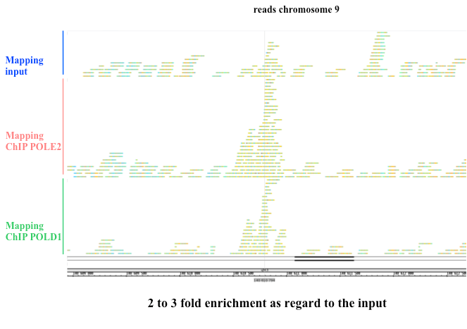

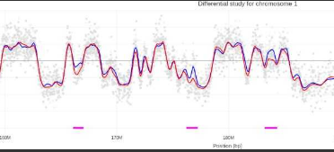
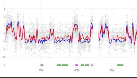


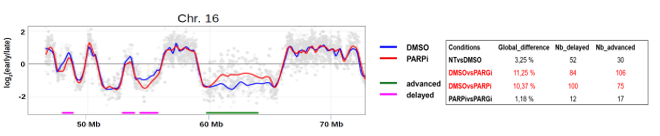

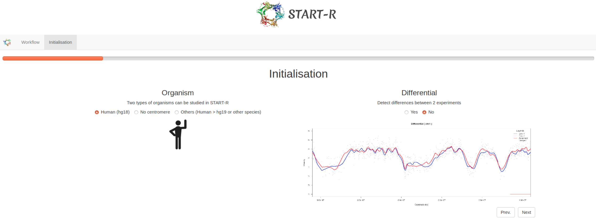

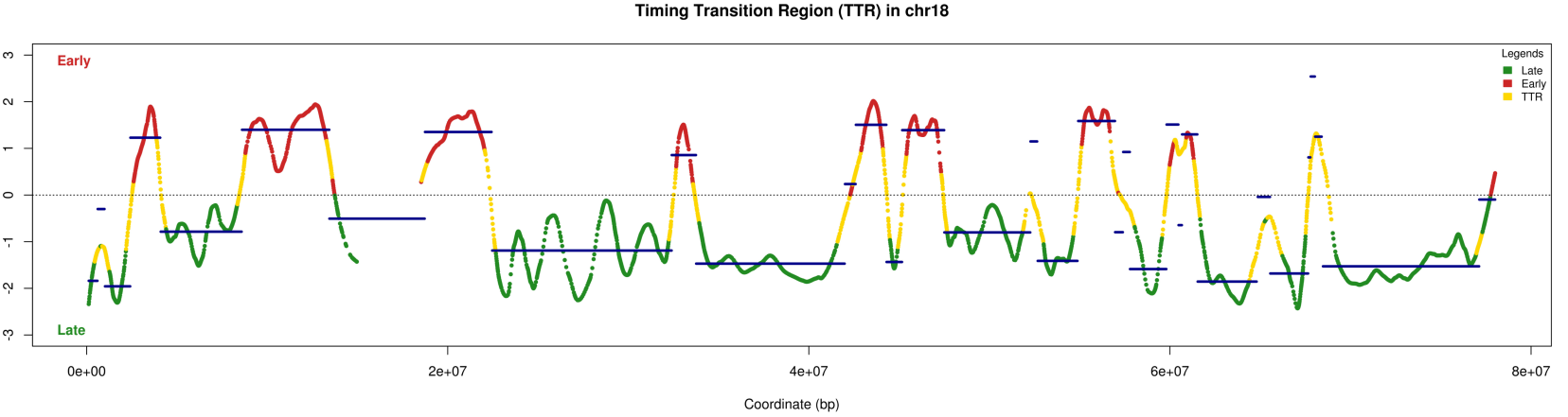


 Giuseppe BALDACCI, Emeritus research professor, CADORET LAB+33 (0)1 57 27 80 73, room 254B
 Chrystelle MARIC ANTOINAT, Researcher, CADORET LAB+33 (0)1 57 27 80 73, room 254B